
Principles of Motifs and Folds
1. Introduction to Protein Motifs and Folds
Proteins are highly organized biological macromolecules whose functions depend on their ability to adopt specific three-dimensional structures. Although a protein is synthesized as a linear sequence of amino acids, its biological activity is determined by the way this sequence folds into a particular conformation. The process of protein folding produces different levels of structural organization, beginning with local secondary structures and progressing toward larger structural arrangements such as motifs, folds, domains, and complete tertiary structures.
The concepts of motifs and folds are particularly important because they explain how relatively simple structural elements can be combined to generate the enormous structural diversity observed among proteins. An α-helix or β-strand by itself represents only a local structural element. When several such elements are arranged in a recurring pattern, they can form a structural motif. When multiple secondary-structure elements and motifs are packed together into a characteristic three-dimensional arrangement, they can generate a protein fold.
The structural hierarchy can therefore be represented as:
Amino acid sequence → Secondary structures → Structural motifs → Protein folds → Domains → Tertiary structure → Biological function
This hierarchy should not be considered a completely rigid sequence of events. Protein folding is a dynamic and cooperative process in which local interactions, long-range interactions, hydrophobic collapse, secondary-structure formation, and tertiary interactions influence one another.
The importance of motifs and folds extends beyond structural biology. They are directly connected with enzyme catalysis, molecular recognition, DNA and RNA binding, protein–protein interactions, signal transduction, protein evolution, structural bioinformatics, protein engineering, and disease-associated protein misfolding.
1.1 Meaning of a Structural Motif
A structural motif is a recognizable and recurring arrangement of two or more secondary-structure elements within a protein.
For example, two β-strands connected by a short turn can form a β-hairpin, whereas two β-strands connected through an α-helix can form a β–α–β motif.
A motif can therefore be viewed as an intermediate level of organization between individual secondary structures and larger structural units such as domains.
The general relationship is:
Secondary structures → Structural motif → Larger structural organization
Motifs are sometimes referred to as supersecondary structures because they contain more than one secondary-structure element but are generally smaller than a complete protein domain.
A structural motif does not necessarily possess independent stability or function. Its significance often depends on its position within the complete protein structure.
1.2 Meaning of a Protein Fold
A protein fold refers to the characteristic three-dimensional arrangement of secondary-structure elements within a protein or protein domain.
A fold describes how α-helices, β-sheets, loops, turns, and other structural elements are arranged relative to one another in three-dimensional space. It also includes information about their connectivity and topology.
For example, two proteins may both contain several α-helices and β-strands, but if these elements are connected and packed differently, the proteins may possess different folds.
A fold therefore represents a larger structural concept than a motif.
The relationship can be expressed as:
Motifs + secondary structures + three-dimensional packing → characteristic fold
Protein folds are particularly important in evolutionary studies because a similar fold can sometimes be retained even after considerable sequence divergence.
1.3 Motif and Fold Are Not Synonymous

The terms motif and fold should not be used interchangeably.
A motif is generally a relatively small recurring structural pattern.
A fold represents a much larger three-dimensional organization.
For example:
β-strand → α-helix → β-strand
may represent a β–α–β motif.
A complete protein fold may contain several such motifs together with additional helices, sheets, loops, and turns.
Thus:
Motif = local structural pattern
Fold = overall structural arrangement
This distinction becomes particularly important when studying protein classification and structural evolution.
2. Hierarchy of Protein Structural Organization
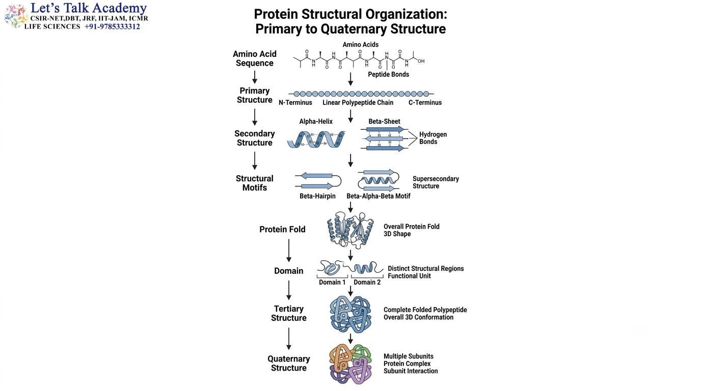
Protein structure is organized at several interconnected levels.
The primary structure is the amino acid sequence. The secondary structure describes local arrangements such as α-helices and β-sheets. These secondary structures can combine into structural motifs. Several motifs and secondary structures can then pack together into a larger fold, often forming a structural domain. The complete three-dimensional organization of one polypeptide chain constitutes its tertiary structure.
When multiple polypeptide chains associate, they form a quaternary structure.
The complete hierarchy can be represented as:
Primary structure
↓
Secondary structure
↓
Structural motifs
↓
Protein fold
↓
Domain
↓
Tertiary structure
↓
Quaternary structure
Each level depends on the preceding structural organization, but interactions can also occur across levels. For example, a mutation in the primary sequence may alter an α-helix, which can change a motif, disturb the fold, and ultimately affect protein function.
2.1 Primary Structure
The primary structure is the linear sequence of amino acids connected by peptide bonds.
For example:
N-terminus → Ala–Gly–Val–Leu–Ser–Lys–Asp–Leu → C-terminus
Although this structure appears simple, the chemical characteristics of the amino acids determine the possible conformations of the chain.
Hydrophobic residues, charged residues, polar residues, aromatic residues, and residues with unusual conformational properties influence how the chain folds.
Thus, the primary structure provides the fundamental information from which higher-order protein structure develops.
2.2 Secondary Structure
The secondary structure consists mainly of recurring local arrangements of the peptide backbone.
The two major regular secondary structures are:
α-helix
β-sheet
Other important structural elements include β-turns and loops.
The α-helix is stabilized mainly by hydrogen bonds between backbone carbonyl and amide groups. β-sheets are stabilized by hydrogen bonds between neighboring β-strands.
These secondary structures serve as the basic building blocks from which motifs and folds are constructed.
2.3 From Secondary Structures to Motifs
Secondary structures rarely function as isolated elements.
They are commonly connected and organized into recurring patterns.
For example:
β-strand → turn → β-strand
produces a β-hairpin.
Similarly:
β-strand → α-helix → β-strand
produces a β–α–β motif.
These arrangements provide structural solutions that are repeatedly used in proteins.
2.4 From Motifs to Folds
Several motifs can combine with additional secondary structures and loops to produce a stable three-dimensional fold.
For example:
β–α–β motif + β-hairpin + α-helical region → larger structural fold
The fold determines the spatial arrangement of structural elements and creates the three-dimensional environment required for biological activity.
3. Structural Motifs as Building Blocks of Protein Architecture

Structural motifs are important because they represent recurring solutions to the problem of organizing a polypeptide chain in three-dimensional space.
Protein folding is constrained by geometry, chemical interactions, and steric limitations. Consequently, only certain combinations of secondary structures are particularly favorable.
Over evolutionary time, these favorable arrangements can appear repeatedly in unrelated or distantly related proteins.
This explains why motifs such as β-hairpins, β–α–β units, helix-turn-helix structures, and Greek-key arrangements occur in many different protein structures.
A motif should therefore not be considered simply as a visual pattern. It represents a recurring structural solution generated by the physical and chemical properties of proteins.
3.1 β-Hairpin Motif
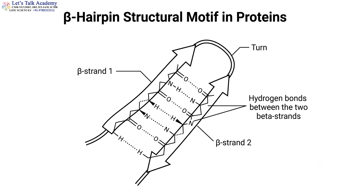
A β-hairpin consists of two β-strands connected by a relatively short turn or loop.
The two strands commonly run in opposite directions and are stabilized by hydrogen bonding between their peptide backbones.
The arrangement can be represented as:
β-strand → turn → β-strand
β-hairpins are frequently observed in β-sheet-rich proteins.
They are important not only for stabilizing β-sheet structures but also for creating loops and surfaces involved in molecular recognition.
3.2 β–α–β Motif
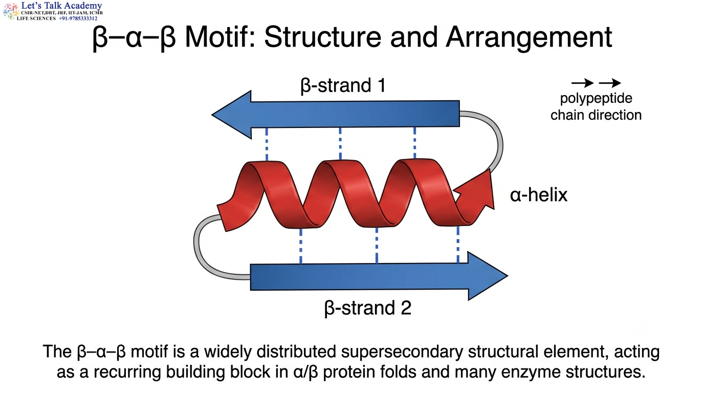
The β–α–β motif contains two β-strands connected by an α-helix.
Its simplified arrangement is:
β-strand → α-helix → β-strand
This motif is particularly common in α/β proteins.
Several β–α–β units can occur together and contribute to the formation of larger β-sheets.
Because α-helices and β-strands can pack efficiently, β–α–β arrangements are found in many enzyme folds.
3.3 Helix-Turn-Helix Motif
The helix-turn-helix (HTH) motif consists of two α-helices separated by a short turn.
It is especially important in DNA-binding proteins.
The structural arrangement positions one of the helices so that specific amino acid side chains can interact with DNA.
The HTH motif demonstrates how a structural pattern can simultaneously contribute to both protein architecture and molecular recognition.
3.4 Greek-Key Motif
The Greek-key motif is a characteristic arrangement of β-strands in which the strands form a specific connectivity and spatial organization.
Greek-key arrangements occur in several β-rich protein structures.
The motif contributes to larger β-sheet architectures and can therefore form part of a complete protein fold.
3.5 Helix-Loop-Helix Motif
The helix-loop-helix arrangement contains two α-helices connected by a flexible loop.
Some helix-loop-helix structures are involved in DNA recognition and protein–protein interactions.
The biological function depends on the precise sequence and three-dimensional context surrounding the motif.
4. Structural Motifs and Biological Function
The presence of a motif does not automatically determine the complete function of a protein. The same motif can occur in different structural environments and participate in different biological processes.
The surrounding fold determines how the motif is positioned and what other residues are brought into its vicinity.
For example, a structural motif may contribute to an active site by positioning one catalytic residue, while another residue from a different structural region completes the catalytic environment.
Thus:
Motif + surrounding structural environment → functional activity
This principle explains why the biological function of a protein cannot usually be predicted from a single short structural motif alone.
5. Protein Folds and Three-Dimensional Organization
A protein fold represents the overall three-dimensional arrangement of the structural elements within a domain.
The fold depends on:
- Secondary-structure composition
- Connectivity
- Topology
- Three-dimensional packing
- Hydrophobic core formation
- Long-range interactions
The fold determines which residues become spatially close to one another.
This is particularly important for enzymes. Two residues may be separated by dozens of amino acids in the primary sequence but become adjacent after folding, allowing them to participate together in catalysis.
5.1 Topology of Protein Folds
Protein topology describes the connectivity and arrangement of structural elements.
Consider two hypothetical structures:
β₁ → α₁ → β₂ → α₂
and:
β₁ → β₂ → α₁ → α₂
Both contain the same basic types of secondary structures, but their connectivity is different.
This difference can result in different folds.
Therefore, protein classification considers not only which secondary structures are present but also how they are connected and arranged.
5.2 Three-Dimensional Packing
The packing of secondary structures is central to fold formation.
Hydrophobic residues tend to become buried within the protein interior.
Polar and charged residues are frequently exposed to solvent, although some can be buried when their interactions provide sufficient stabilization.
The packing must be sufficiently tight to stabilize the structure but must also avoid severe steric clashes.
A well-packed hydrophobic core is therefore one of the important features of many globular protein folds.
6. Forces Responsible for Motif and Fold Stability

Protein motifs and folds are stabilized by the combined effect of several molecular forces.
The major contributions include:
- Hydrophobic interactions
- Hydrogen bonds
- Electrostatic interactions
- Van der Waals interactions
- Disulfide bonds
No single interaction generally determines the complete protein structure. Instead, thousands of interactions collectively produce the native conformation.
6.1 Hydrophobic Effect
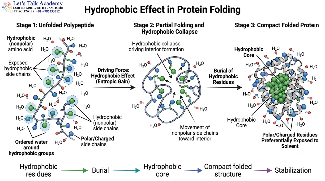
The hydrophobic effect is one of the major driving forces in the folding of soluble proteins.
Nonpolar side chains tend to become buried within the interior of the protein.
This reduces their unfavorable exposure to water and promotes formation of a compact structure.
The process can be summarized as:
Hydrophobic residues → burial → compact core → stabilization of folded structure
The hydrophobic effect is especially important during the early stages of protein folding.
6.2 Hydrogen Bonds
Hydrogen bonds stabilize secondary structures and tertiary interactions.
In an α-helix, hydrogen bonds form between backbone groups within the same chain.
In a β-sheet, hydrogen bonds occur between neighboring β-strands.
Hydrogen bonds can also occur between side chains and between side chains and backbone atoms.
Therefore:
Hydrogen bonding → secondary-structure stabilization → motif stabilization → fold stabilization
6.3 Electrostatic Interactions
Charged amino acid side chains can form favorable electrostatic interactions.
Examples include:
Lys⁺ ↔ Asp⁻
Arg⁺ ↔ Glu⁻
These interactions may stabilize particular structural arrangements.
However, their energetic contribution depends on the local environment, solvent accessibility, and dielectric properties of the protein.
6.4 Van der Waals Interactions
Van der Waals interactions occur when atoms are positioned at appropriate distances.
Each interaction is relatively weak, but the large number of contacts within a well-packed protein can contribute significantly to the stability of the folded state.
6.5 Disulfide Bonds
Disulfide bonds are covalent bonds formed between cysteine residues.
The reaction can be represented as:
2 R–SH → R–S–S–R + 2H⁺ + 2e⁻
Disulfide bonds are particularly important in many extracellular proteins.
They can stabilize a fold by linking two distant regions of the polypeptide chain and restricting conformational freedom.
7. Thermodynamic Basis of Protein Folding
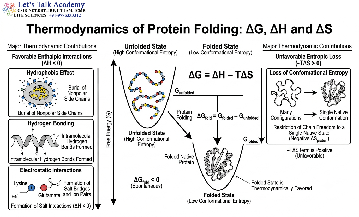
Protein folding is fundamentally a thermodynamic process.
The folded and unfolded states exist in equilibrium, and the relative stability of these states depends on their free energies.
The free-energy difference can be represented as:
ΔG_fold = G_folded − G_unfolded
When the folded state has lower free energy:
ΔG_fold < 0
the folded state is thermodynamically favored.
The general relationship between free energy, enthalpy, and entropy is:
ΔG = ΔH − TΔS
Here, ΔH represents enthalpic contributions, T represents absolute temperature, and ΔS represents the entropy change.
Protein folding often involves favorable enthalpic interactions but an unfavorable loss of conformational entropy. The native state becomes favored when the overall free-energy balance supports folding.
7.1 Cooperative Nature of Folding
Protein folding can be cooperative.
The formation of one structural interaction can influence the formation of other interactions.
As a result, a protein does not necessarily fold by independently constructing one secondary structure after another.
Instead, local and long-range interactions can develop together.
This cooperative behavior helps explain why some proteins show relatively sharp transitions between folded and unfolded states.
7.2 Protein Folding Is Dynamic
A folded protein should not be regarded as a completely rigid structure.
Even when a protein is correctly folded, its atoms continuously undergo thermal motion.
Side chains rotate, loops fluctuate, and entire domains may move relative to one another.
Therefore, the native state should be viewed as a dynamic ensemble of closely related conformations rather than a single absolutely fixed structure.
8. Protein Folding and Energy Landscapes
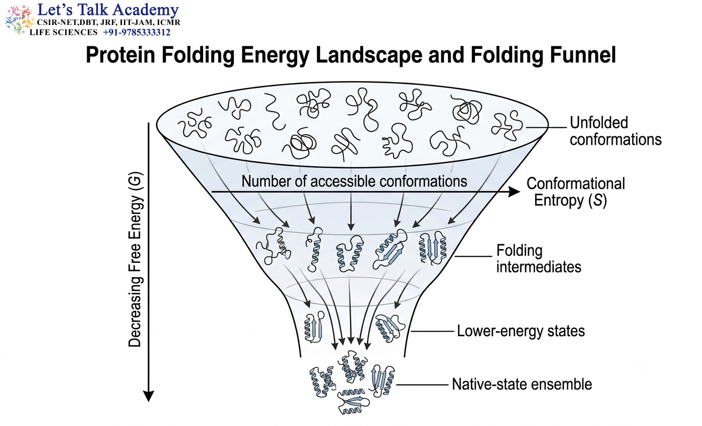
The folding of a protein can be represented conceptually using an energy landscape.
An unfolded polypeptide can exist in a huge number of conformations.
As the protein folds, the number of accessible conformations decreases and the molecule generally moves toward lower-energy regions.
A simplified representation is:
Many unfolded conformations
↓
Folding intermediates
↓
Lower-energy conformational states
↓
Native-state ensemble
The energy landscape is often represented as a funnel because many possible unfolded conformations can converge toward a smaller set of native-like structures.
This model helps explain why proteins can efficiently reach their functional conformations despite having enormous numbers of theoretically possible conformations.
9. Protein Folds and Structural Classes
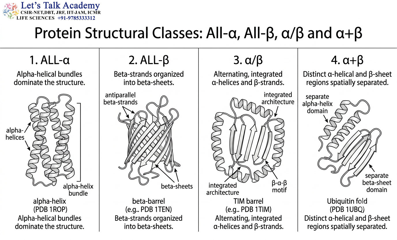
Protein folds can be broadly organized according to the types and arrangements of secondary structures.
The major broad structural classes include:
- All-α
- All-β
- α/β
- α+β
These categories provide a general description of protein architecture.
They do not capture every detail of a protein fold, but they provide a useful starting point for structural classification.
9.1 All-α Structures
All-α structures are dominated by α-helices.
The helices may form compact bundles, hairpins, or larger helical arrangements.
Hydrophobic residues frequently occur at the interfaces between helices, producing a stable internal core.
All-α architectures are common in many regulatory proteins, DNA-binding proteins, and structural proteins.
9.2 All-β Structures
All-β structures are dominated by β-strands.
The β-strands form β-sheets that can be organized into β-sandwiches, barrels, propellers, and other architectures.
Hydrogen bonding between β-strands provides an important stabilizing force.
9.3 α/β Structures
α/β structures contain α-helices and β-strands that are closely integrated.
Many enzymes belong to this broad structural category.
The β–α–β motif is particularly common in these proteins.
The arrangement allows β-sheets to form while α-helices contribute to packing and structural stability.
9.4 α+β Structures
In α+β structures, α-helical and β-sheet regions tend to be more spatially separated.
The distinction between α/β and α+β depends on the topology and organization of the secondary structures rather than simply on the presence of both types.
10. Important Protein Folds

Certain protein folds occur repeatedly in nature because they provide stable and useful structural frameworks.
Examples include:
- Rossmann-like fold
- TIM barrel
- Immunoglobulin fold
- β-propeller
- α-helical bundles
These folds are found in proteins with a wide range of functions.
10.1 Rossmann Fold
The Rossmann fold is a common nucleotide-binding architecture.
It generally contains a repeating α/β arrangement in which β-strands are surrounded by α-helices.
The fold occurs in many proteins that bind cofactors such as NAD⁺ and NADP⁺.
The recurrence of the Rossmann fold demonstrates how one structural solution can be used repeatedly for nucleotide recognition and related biochemical functions.
10.2 TIM Barrel

The TIM barrel, also known as the (β/α)₈ barrel, consists of eight β-strands and eight α-helices arranged in an alternating pattern.
The β-strands form the central barrel-like core, while the α-helices generally surround it.
The repeating pattern can be represented as:
(β–α)₈
Many enzymes use this architecture.
The active site is frequently located near one end of the barrel, where loops connecting β-strands and α-helices can position catalytic residues.
10.3 Immunoglobulin Fold
The immunoglobulin fold is a β-rich architecture containing β-strands arranged into a characteristic β-sandwich.
It is found in antibodies and numerous proteins involved in cell adhesion, recognition, and immune-related functions.
The same general fold can therefore support different biological activities.
10.4 β-Propeller Fold
A β-propeller contains repeated β-sheet units arranged around a central axis.
The individual repeating units are often called blades.
The complete structure resembles a propeller when viewed from above.
β-propeller structures participate in molecular recognition, protein interactions, and other cellular processes.
11. Protein Fold Versus Domain
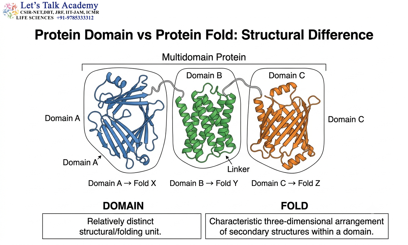
A protein domain is a structurally distinct region of a polypeptide that often behaves as a relatively independent folding unit.
A fold describes the characteristic three-dimensional organization adopted by the structural elements within that region.
Thus:
Domain = structural unit
Fold = three-dimensional architecture of that unit
A multidomain protein may therefore contain:
Domain A → Fold X
Domain B → Fold Y
Domain C → Fold Z
The domains can interact with one another to produce the complete tertiary structure.
12. Motifs Within Protein Folds
A protein fold commonly contains multiple motifs.
For example, an α/β fold may contain several β–α–β motifs.
A β-rich fold may contain several β-hairpins.
The motifs contribute to the overall architecture, while the complete fold determines the global organization.
The relationship can be represented as:
Motif A + Motif B + Motif C + additional structural elements → Fold
This modular organization explains why similar motifs can occur in many different proteins.
13. Motifs and Active-Site Formation

Enzyme active sites are often created by the three-dimensional arrangement of amino acid residues that are distant from one another in the primary sequence.
Protein folding brings these residues together.
For example, one catalytic residue may be located in an α-helix, while another catalytic residue may occur in a loop connected to a β-strand.
After folding, the residues can become adjacent and form a functional active site.
Thus:
Primary sequence → folding → motif organization → active-site geometry → catalysis
This is one of the clearest examples of the relationship between protein conformation and biological function.
14. Motifs and Molecular Recognition
Structural motifs can contribute to recognition of DNA, RNA, proteins, lipids, and small molecules.
Recognition depends on the three-dimensional arrangement of chemical groups.
The interacting molecules must have complementary:
- Shape
- Charge
- Hydrophobicity
- Hydrogen-bonding patterns
For example, DNA-binding motifs often position positively charged residues near the negatively charged phosphate backbone of DNA.
Specific residues may also recognize individual bases through hydrogen bonding and other interactions.
15. Protein Folds and Molecular Recognition
The fold creates the complete three-dimensional surface required for molecular recognition.
A binding pocket may contain residues that are widely separated in the primary sequence but become adjacent after folding.
Therefore:
Correct fold → correct spatial arrangement of residues → specific molecular recognition
If the fold changes, the shape and chemistry of the binding site may also change.
This explains why denaturation often causes loss of biological activity.
16. Motifs, Folds, and Enzyme Catalysis
Enzyme catalysis depends on precise structural organization.
The fold positions catalytic residues in a specific three-dimensional environment.
Structural motifs can contribute to substrate binding, catalytic chemistry, or stabilization of the transition state.
The overall process can be represented as:
Protein sequence → fold formation → active-site organization → substrate recognition → catalysis
A mutation in a motif may therefore affect enzyme activity even if it does not directly alter a catalytic residue.
17. Protein Folds and Allosteric Regulation
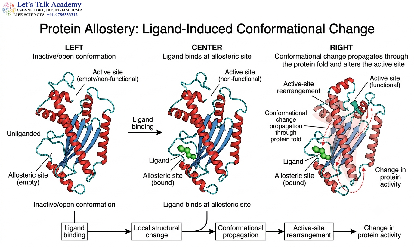
Allosteric regulation occurs when binding or conformational changes at one region of a protein influence another region.
A protein fold provides the structural pathway through which this information can be transmitted.
The process may be represented as:
Ligand binding → local structural change → conformational propagation → altered active site → change in activity
In multidomain proteins, the communication may occur between domains.
In single-domain proteins, the communication can occur through networks of interactions within the fold.
18. Protein Folds and Conformational Dynamics
Protein folds are dynamic structures.
A protein may move between multiple conformational states without losing its overall fold.
For example:
Open ↔ Closed
Active ↔ Inactive
Ligand-free ↔ Ligand-bound
These conformational changes are often essential for function.
Enzymes may close over their substrates, receptors may change conformation after ligand binding, and molecular motors may change shape during nucleotide hydrolysis.
Thus, the functional state of a protein is often determined by its conformational dynamics rather than by a single static structure.
19. Loops and Turns in Protein Folds
Loops and turns connect regular secondary structures.
Although they may not display the regular hydrogen-bonding patterns characteristic of α-helices and β-sheets, they are extremely important.
Loops frequently participate in:
Active-site formation
Ligand binding
Protein–protein interactions
Molecular recognition
Conformational changes
Loops are often more flexible than regular secondary structures.
This flexibility allows proteins to respond to changes in their molecular environment.
20. Motifs and Protein Dynamics
Structural motifs can undergo small or large conformational changes.
A loop within a motif may move when a ligand binds.
An α-helix may rotate relative to another structural element.
A β-sheet may change its orientation as a domain undergoes conformational rearrangement.
These changes allow motifs to participate actively in molecular recognition and regulation.
Therefore, motifs should be understood as dynamic structural components rather than completely rigid patterns.
21. Evolutionary Conservation of Protein Folds
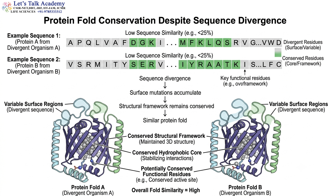
Protein folds are often more conserved than amino acid sequences.
During evolution, mutations can accumulate at surface positions while the fundamental architecture remains preserved.
However, residues that are essential for hydrophobic packing, structural stability, or catalytic activity are usually under stronger evolutionary constraints.
This produces a common pattern:
Conserved structural framework + variable surface regions
Consequently, two proteins can have relatively low sequence identity while retaining a similar three-dimensional fold.
22. Divergence and Conservation of Motifs
Some structural motifs remain highly conserved because they are essential for protein stability or function.
For example, a motif involved in nucleotide binding or catalytic activity may tolerate only limited changes.
Other motifs may evolve more rapidly if they occur on exposed surfaces or participate in species-specific interactions.
Thus, the degree of conservation depends on the functional importance of the motif.
23. Structural Convergence
Similar structural arrangements can sometimes evolve independently.
This phenomenon is associated with convergent evolution.
If a particular fold provides an especially effective structural solution to a biochemical problem, unrelated proteins may evolve similar architectures.
Therefore:
Structural similarity does not automatically establish evolutionary relatedness.
Evidence for common ancestry requires additional information, including sequence, structural, functional, and evolutionary relationships.
24. Structural Classification of Protein Folds

Because proteins display enormous structural diversity, classification systems have been developed to organize their structures.
Two important systems are:
- SCOP
- CATH
These systems classify protein domains based on structural characteristics and, at deeper levels, evolutionary relationships.
They are widely used in structural bioinformatics and protein evolution studies.
24.1 SCOP Classification
The Structural Classification of Proteins (SCOP) system organizes protein domains hierarchically.
A simplified hierarchy is:
Class → Fold → Superfamily → Family
At the class level, proteins are grouped according to broad structural composition.
At the fold level, proteins have similar arrangements and topology of secondary structures.
At the superfamily level, proteins show stronger evidence of common evolutionary origin despite sequence divergence.
At the family level, proteins are more closely related in sequence and function.
This hierarchy allows structural similarity to be distinguished from stronger evolutionary relationships.
24.2 CATH Classification
The CATH system classifies protein structures using four major levels:
Class → Architecture → Topology → Homologous Superfamily
The class describes broad secondary-structure composition.
Architecture describes the overall three-dimensional arrangement of secondary structures without necessarily considering their connectivity.
Topology describes the connectivity and arrangement of secondary structures.
Homologous superfamily groups proteins that show evidence of common ancestry.
25. Protein Motifs and Structural Bioinformatics
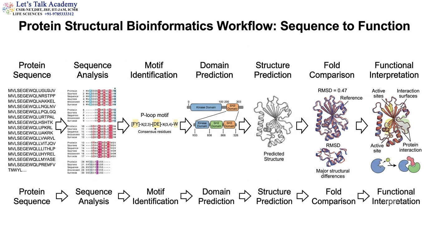
Motif and fold analysis is a major component of structural bioinformatics.
When a protein sequence is obtained, researchers can search for conserved motifs and compare the predicted or experimentally determined structure with known folds.
A general workflow is:
Protein sequence
↓
Sequence analysis
↓
Motif identification
↓
Domain prediction
↓
Structure prediction
↓
Fold comparison
↓
Functional interpretation
This approach is particularly useful when studying proteins for which experimental biochemical information is limited.
25.1 Sequence Motif Versus Structural Motif

A sequence motif is a recurring pattern of amino acids.
A structural motif is a recurring three-dimensional arrangement of structural elements.
These concepts are related but should not be confused.
A short conserved sequence may contribute to a structural motif, but a structural motif generally involves multiple amino acids arranged in three-dimensional space.
Therefore:
Sequence motif ≠ Structural motif
although sequence information can be used to predict structural motifs.
25.2 Structural Alignment
Structural alignment compares three-dimensional structures to identify corresponding regions.
It can reveal:
- Conserved α-helices
- Conserved β-sheets
- Similar motifs
- Similar active-site geometry
- Similar folds
Structural alignment is particularly valuable when sequence similarity is low.
26. Protein Folds and Structural Prediction
Modern computational methods can predict the three-dimensional structures of proteins and help identify potential folds.
A predicted structure can provide information about:
- Secondary structures
- Domain boundaries
- Potential motifs
- Folding patterns
- Binding pockets
- Interdomain interfaces
However, a predicted structure should not automatically be interpreted as experimental confirmation of biological function.
Functional interpretation requires additional biochemical, genetic, or structural evidence.
27. Protein Folds and Molecular Evolution
Protein folds provide stable frameworks upon which evolutionary changes can act.
Instead of creating a completely new protein structure, evolution can modify an existing fold.
Changes in loops and surface residues can alter:
- Ligand specificity
- Protein interactions
- Cellular localization
- Regulation
while the central fold remains conserved.
This provides a molecular explanation for how proteins can diversify while retaining structural stability.
28. Domain Fusion and Fold Combination
Evolution can combine different domains with different folds into a single protein.
For example:
Binding domain → Fold A
Catalytic domain → Fold B
↓
Multidomain protein with integrated function
Such fusion can improve biochemical efficiency by bringing two related activities into close proximity.
This mechanism is particularly important in metabolic enzymes, signaling proteins, and regulatory proteins.
29. Protein Engineering Based on Motifs and Folds
Understanding motifs and folds allows scientists to modify proteins rationally.
A structural motif can be altered to change binding specificity.
An active-site loop can be modified to alter substrate preference.
A hydrophobic core can be redesigned to increase stability.
Domains can be combined to create proteins with new activities.
However, protein engineering must consider the possibility that modifying one region may indirectly alter the entire fold.
A successful engineered protein must maintain an appropriate balance between:
- Structure
- Stability
- Flexibility
- Activity
- Specificity
30. Motifs, Folds, and Protein Misfolding
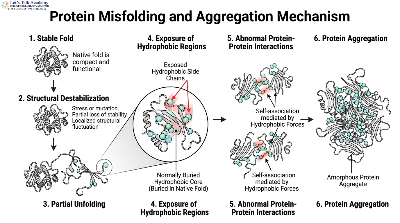
The same structural principles that allow proteins to fold correctly can also help explain protein misfolding.
If a mutation disrupts a motif or destabilizes the hydrophobic core, the protein may partially unfold.
Exposed hydrophobic regions can interact with other protein molecules and promote aggregation.
The process can be represented as:
Stable fold → structural destabilization → partial unfolding → exposed hydrophobic regions → aggregation
Protein misfolding is associated with several pathological conditions, making the study of protein folds important not only for basic biology but also for biomedical research.
31. Motifs, Folds, and Protein Aggregation
Protein aggregation can occur when unfolded or partially folded proteins form abnormal intermolecular interactions.
In some cases, aggregation is associated with formation of highly ordered β-sheet-rich structures.
This illustrates that a change in structural organization can dramatically alter protein behavior.
The biological activity of a protein therefore depends not only on its amino acid sequence but also on maintaining the correct structural state.
32. Motifs and Folds in Protein–Protein Interactions
Protein–protein interactions often involve specific structural surfaces created by folds and motifs.
The interacting surfaces may contain complementary:
Hydrophobic regions
Charged residues
Hydrogen-bonding groups
Aromatic residues
The strength and specificity of the interaction depend on the combined contribution of these contacts.
Some interaction motifs are highly conserved because they recognize specific protein partners.
33. Motifs and Folds in DNA-Binding Proteins
DNA-binding proteins provide excellent examples of the relationship between motifs, folds, and biological function.
A DNA-binding protein may contain a structural motif that positions particular residues within the major groove of DNA.
The complete protein fold determines the orientation of the motif and the overall DNA-binding surface.
Therefore:
Fold → motif positioning → DNA recognition → biological regulation
This organization is particularly important in transcription factors and DNA-binding regulatory proteins.
34. Motifs and Folds in RNA-Binding Proteins
RNA-binding proteins also use structural motifs and folds to recognize RNA molecules.
RNA-binding regions can interact with:
- RNA bases
- Phosphate groups
- Ribose groups
Recognition may depend on both sequence-specific and shape-specific interactions.
The three-dimensional fold positions the RNA-binding residues correctly and provides the structural framework for recognition.
35. Motifs and Folds in Membrane Proteins
Membrane proteins contain specialized structural arrangements that allow them to interact with lipid bilayers.
Transmembrane α-helices can form bundles.
β-strands can form membrane-associated β-barrels.
These structural arrangements represent specialized solutions to the problem of stabilizing proteins within a hydrophobic membrane environment.
The surrounding protein architecture can then create channels, receptors, transporters, and signaling structures.
36. Motifs and Folds in Enzyme Evolution

Enzymes belonging to the same structural fold can evolve different substrate specificities.
The central fold may remain conserved, while loops surrounding the active site undergo evolutionary changes.
This can alter the size, shape, charge, and chemical properties of the substrate-binding pocket.
Therefore:
Conserved fold + variable active-site loops → functional diversification
This mechanism is particularly important in enzyme evolution.
37. Protein Fold and Functional Diversity
One fold can support multiple biological functions.
The functional differences may result from changes in:
- Active-site residues
- Binding pockets
- Surface loops
- Domain interactions
- Regulatory regions
Thus, the fold provides a structural framework, while specific sequence features determine much of the detailed molecular behavior.
38. Motifs and Folds as Evolutionary Modules
Protein motifs and folds can act as reusable structural modules.
A successful structural arrangement can be retained and incorporated into different proteins.
Evolution can modify the surrounding sequence or combine the structural module with other domains.
This produces:
Conserved structural module + new molecular context → new functional possibility
This modular principle is one of the major sources of protein diversity.
39. Integrated Relationship Between Motifs, Folds, and Domains
The relationship among the major structural concepts can be summarized as:
Amino acid sequence
↓
Secondary structures
↓
Structural motifs
↓
Motif organization
↓
Protein fold
↓
Domain
↓
Interdomain interactions
↓
Tertiary structure
↓
Quaternary structure, when present
↓
Biological function
A motif therefore represents a relatively local structural organization, a fold represents a larger three-dimensional architecture, and a domain represents a structurally distinct unit that commonly contains a characteristic fold.


