
Conformation of Proteins: Domains
1. Introduction to Protein Domains

Proteins are highly organized biological macromolecules in which structure and function are closely interconnected. A protein is synthesized as a linear chain of amino acids, but its biological activity does not arise from the linear sequence alone. The polypeptide chain folds into a specific three-dimensional arrangement in which particular groups of amino acids are brought together to form structural and functional regions. One of the most important organizational units within this three-dimensional structure is the protein domain.
A protein domain is a distinct region of a polypeptide chain that forms a characteristic three-dimensional structure and, in many cases, can fold relatively independently from the rest of the protein. A domain may also possess a specific biological function, such as binding a ligand, interacting with another protein, recognizing DNA or RNA, catalyzing a chemical reaction, or regulating the activity of another region of the same protein.
The concept of a domain is particularly useful because proteins are not always single, uninterrupted structural units. Many proteins contain two or more domains, and each domain can contribute a different structural or functional property. A multidomain protein can therefore be considered a molecular assembly in which different structural modules cooperate to perform a complex biological task.
A protein domain is larger and more complex than an individual secondary-structure element. An α-helix or β-strand represents a local structural conformation of the polypeptide backbone, whereas a domain contains several secondary-structure elements arranged into a stable three-dimensional fold.
The structural hierarchy can therefore be represented as:
Amino acid sequence → Secondary structures → Structural motifs → Domains → Tertiary structure → Functional protein
Domains provide an important connection between the local organization of a polypeptide chain and the overall architecture and biological function of the protein.
1.1 Definition of a Protein Domain
A protein domain can be defined as a compact, structurally distinct region of a protein that possesses a characteristic fold and often behaves as an independent folding unit.
The word “often” is important because not every domain folds completely independently. Some domains require interactions with neighboring domains for proper stability or function. Similarly, a domain may have a recognizable structural identity but may not perform an independent biological function.
A domain generally contains a combination of α-helices, β-sheets, turns, loops, and other structural elements. These components are arranged in a particular topology and stabilized by numerous molecular interactions.
For example:
α-helices + β-sheets + loops + turns → specific three-dimensional fold → protein domain
The sequence of amino acids determines the possible structural conformations, while the physical and chemical properties of the residues determine which conformations are energetically favorable.
1.2 Domains as Structural Units
A domain is primarily recognized by its three-dimensional organization.
A folded domain usually contains a relatively compact core in which hydrophobic residues are efficiently packed. The outer surface frequently contains polar and charged residues that interact with the aqueous environment or with other biological molecules.
The domain therefore has an internal organization that is not random. The secondary-structure elements are positioned relative to one another in a reproducible manner.
For example, one domain may contain a central β-sheet surrounded by α-helices, whereas another may consist mainly of α-helices packed together into a helical bundle.
The particular arrangement of secondary structures is called the fold of the domain.
1.3 Domains as Functional Units
Many domains also perform recognizable biological functions.
A domain may form a binding pocket for ATP, recognize a specific DNA sequence, interact with another protein, bind a membrane lipid, or provide catalytic residues for an enzymatic reaction.
In a multidomain protein, different domains may perform complementary functions.
For example:
Binding domain + catalytic domain → substrate recognition and catalysis
or:
Regulatory domain + catalytic domain → controlled enzymatic activity
or:
DNA-binding domain + regulatory domain → controlled gene expression
Thus, domain organization provides a structural basis for the functional complexity of proteins.
2. Position of Domains in the Hierarchy of Protein Structure
Protein structure is commonly described at four major levels: primary, secondary, tertiary, and quaternary structure.
The primary structure represents the linear sequence of amino acids.
The secondary structure represents local recurring conformations such as α-helices, β-sheets, turns, and related structures.
The tertiary structure represents the complete three-dimensional arrangement of a single polypeptide chain.
The quaternary structure describes the association of multiple polypeptide chains.
The concept of a domain occurs within the tertiary organization of a protein. A single polypeptide chain may contain one domain or several domains, and these domains together contribute to the overall tertiary architecture.
A useful representation is:
Primary structure
↓
Secondary structure
↓
Structural motifs
↓
Protein domains
↓
Tertiary structure
↓
Quaternary structure, when present
The hierarchy is not simply a sequence of independent steps. Structural elements at one level influence and stabilize those at other levels.
2.1 Secondary Structure Versus Domain
Secondary structure describes local backbone conformations.
The major examples are:
α-helix
β-sheet
β-turn
Loop
A domain, in contrast, contains several such structural elements arranged into a characteristic three-dimensional structure.
For example:
α-helix + β-strand + α-helix + loops → domain fold
Therefore, a domain should never be equated with a single α-helix or β-sheet.
2.2 Motif Versus Domain
A structural motif is a recurring combination of secondary-structure elements.
Examples include the β-hairpin, β–α–β motif, helix-turn-helix motif, and Greek-key motif.
A domain is generally larger and can contain several motifs.
The relationship can be represented as:
Secondary structures → motifs → domains
A motif may occur in many different domains, whereas a domain represents a more extensive structural and often evolutionary unit.
2.3 Domain Versus Protein
A protein can consist of a single domain or multiple domains.
A small protein may essentially correspond to one structural domain:
Protein = Domain A
A larger multidomain protein may have:
Protein = Domain A + Domain B + Domain C
Therefore, the terms protein and domain cannot be used interchangeably.
2.4 Domain Versus Subunit
A domain is a structural region within a polypeptide chain.
A subunit is an individual polypeptide chain within a multimeric protein.
For example, a protein may contain:
One polypeptide chain → three domains
or:
Three polypeptide chains → three subunits
Each subunit can itself contain one or several domains.
This distinction is important when interpreting protein architecture.
3. Structural Organization of a Protein Domain
The three-dimensional structure of a domain results from the folding of its amino acid sequence.
The amino acid sequence determines the chemical groups available for interaction. During folding, residues that may be far apart in the primary sequence can become close in three-dimensional space.
This is particularly important for functional sites. Catalytic residues, for example, may be separated by many amino acids in the sequence but become neighbors after the domain folds.
The organization can therefore be represented as:
Linear sequence → folding → three-dimensional proximity → functional site
This is one of the central principles of structural biology.
3.1 Hydrophobic Core
Most soluble protein domains contain a hydrophobic core.
Nonpolar amino acid side chains tend to become buried within the interior of the folded protein, reducing their exposure to water.
The hydrophobic effect is a major driving force in protein folding.
The formation of a compact hydrophobic core also allows efficient packing of side chains and contributes to the stability of the domain.
The core must be packed carefully. Large cavities can destabilize a protein, whereas excessive steric crowding can also be unfavorable.
Thus, domain stability depends on an appropriate balance between packing efficiency and conformational flexibility.
3.2 Protein Domain Surface
The surface of a soluble domain is frequently enriched in polar and charged residues because these groups can interact favorably with water.
However, the surface can also contain hydrophobic patches involved in protein–protein interactions, membrane binding, or ligand recognition.
The chemical properties of the domain surface therefore depend strongly on its biological role.
For example, a DNA-binding domain may contain positively charged residues that interact with the negatively charged phosphate backbone of DNA.
3.3 Core-Surface Relationship
The core and surface of a domain are structurally interconnected.
A mutation within the hydrophobic core can change the shape of the entire domain and indirectly affect the surface.
Conversely, a surface mutation can sometimes influence internal packing or domain stability.
Therefore, the functional consequences of a mutation cannot always be predicted simply by examining whether the altered residue is located at an active site.
4. Domain Folding

The folding of a domain involves the conversion of a flexible polypeptide chain into a specific three-dimensional structure.
The unfolded chain can adopt a very large number of possible conformations. Folding restricts this conformational freedom and favors a smaller set of energetically favorable structures.
During folding, local secondary structures can develop, hydrophobic residues can become buried, and long-range interactions can bring distant regions of the sequence together.
A simplified representation is:
Unfolded polypeptide
↓
Formation of local conformations
↓
Secondary-structure formation
↓
Hydrophobic collapse and long-range interactions
↓
Packing of structural elements
↓
Folded domain
The actual folding pathway is more complex and may involve multiple intermediates.
4.1 Role of the Amino Acid Sequence
The amino acid sequence contains the information required to establish the native structure of the domain.
Different amino acids have different:
- side-chain sizes,
- charges,
- hydrophobicities,
- hydrogen-bonding properties,
- conformational preferences.
These properties determine which backbone conformations and side-chain arrangements are energetically favorable.
Consequently, even a single amino acid substitution can sometimes significantly alter domain stability or function.
4.2 Role of Secondary Structures
Secondary structures provide the basic framework from which domains are constructed.
A domain can contain several helices and sheets that pack together in a specific orientation.
For example, a domain may have a central β-sheet surrounded by α-helices.
Another domain may consist almost entirely of α-helices.
The exact arrangement is determined by the amino acid sequence and the energetics of folding.
4.3 Long-Range Interactions
An important characteristic of domain formation is the development of interactions between residues that are distant in the primary sequence.
For example, residues separated by dozens of amino acids can become adjacent after folding.
These long-range interactions are essential for establishing the final three-dimensional fold.
They include hydrophobic contacts, hydrogen bonds, salt bridges, aromatic interactions, and van der Waals interactions.
5. Forces Stabilizing Protein Domains
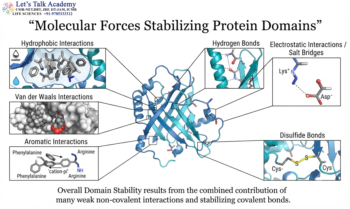
No single interaction is responsible for maintaining the entire domain structure.
Domain stability results from the combined contribution of many weak and strong interactions.
The major contributors include hydrophobic interactions, hydrogen bonds, electrostatic interactions, van der Waals interactions, aromatic interactions, and, in appropriate proteins, covalent disulfide bonds.
The overall stability of a domain therefore reflects the total free-energy balance of its folded and unfolded states.
5.1 Hydrophobic Interactions
Hydrophobic interactions are especially important for soluble proteins.
Nonpolar side chains tend to become buried within the protein interior, reducing their unfavorable exposure to water.
This promotes compact folding and contributes substantially to the stability of the domain.
5.2 Hydrogen Bonding
Hydrogen bonds occur throughout folded domains.
They can form between backbone groups as well as between side chains.
Backbone hydrogen bonding contributes strongly to α-helices and β-sheets, while side-chain hydrogen bonds can stabilize tertiary interactions.
Hydrogen bonds can also participate directly in ligand recognition and catalysis.
5.3 Electrostatic Interactions
Charged side chains can form favorable interactions with oppositely charged groups.
For example:
Lys⁺ ↔ Asp⁻
Arg⁺ ↔ Glu⁻
Such interactions are often called salt bridges when they occur between oppositely charged groups.
Their contribution to protein stability depends strongly on the surrounding environment and whether the interacting groups are buried or exposed.
5.4 Van der Waals Interactions
When atoms are packed closely and appropriately, van der Waals interactions contribute to the stability of the folded domain.
Although each interaction is weak, the large number of contacts within a well-packed protein core can produce a significant cumulative effect.
5.5 Disulfide Bonds
Disulfide bonds are covalent interactions formed between two cysteine residues.
The reaction can be represented as:
2 R–SH → R–S–S–R + 2H⁺ + 2e⁻
Disulfide bonds are particularly important in many extracellular proteins because the extracellular environment is generally more oxidizing than the cytosol.
A disulfide bond can stabilize a domain by linking two regions of the polypeptide chain that would otherwise have greater conformational freedom.
6. Thermodynamics of Domain Folding
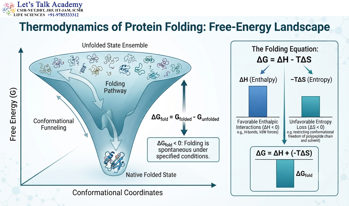
Protein-domain folding is governed by thermodynamics.
The free-energy change associated with folding can be represented as:
ΔG_fold = G_folded − G_unfolded
If:
ΔG_fold < 0
the folded state is thermodynamically favored under the specified conditions.
The relationship between free energy and equilibrium is:
ΔG = −RT ln K
where R is the gas constant, T is absolute temperature, and K is the equilibrium constant.
The folded state is therefore determined by a balance of favorable and unfavorable energetic contributions.
6.1 Enthalpy and Entropy
The relationship:
ΔG = ΔH − TΔS
illustrates the balance between enthalpy and entropy.
Formation of favorable interactions such as hydrogen bonds and electrostatic contacts can contribute favorably to enthalpy.
However, folding restricts the conformational freedom of the polypeptide, creating an unfavorable entropy contribution.
The final structure is determined by the overall free-energy balance.
6.2 Cooperative Folding
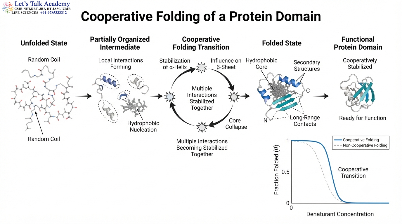
Many protein domains exhibit cooperative folding.
This means that the stabilization of one part of the domain can influence the stabilization of other regions.
Instead of every residue folding independently, many interactions become organized together during the folding transition.
Cooperative folding helps explain why some domains show relatively sharp transitions between folded and unfolded states.
6.3 Domain Stability Is Context Dependent
A domain that is stable under one condition may become unstable under another.
Factors such as:
Temperature
pH
Ionic strength
Solvent composition
Ligand concentration
Redox conditions
can alter the stability of the folded state.
Thus, protein-domain structure must always be considered in relation to its molecular environment.
7. Independent Folding of Domains
Many domains can fold independently when isolated from the rest of the protein.
This property is one of the reasons domains are considered important units of protein organization.
For example:
Domain A — linker — Domain B
may allow each domain to form its characteristic structure while the linker provides flexibility.
However, independent folding is not universal.
Some domains depend on neighboring regions for proper folding, stability, or function.
7.1 Folding Unit Versus Functional Unit
A domain may be an independent folding unit without being an entirely independent functional unit.
Two domains may each form stable structures but need to interact to produce a functional active site.
Thus:
Folding independence does not necessarily mean functional independence.
This distinction is particularly important for multidomain enzymes and regulatory proteins.
7.2 Domain Boundaries
Domain boundaries are often found near flexible loops or linker regions.
Such regions may be relatively exposed to solvent and can connect two compact structural regions.
A domain boundary is not necessarily a sharp physical boundary. It represents a transition between regions with different structural organization.
Different computational algorithms may predict slightly different boundaries for the same protein.
8. Multidomain Proteins
Many biologically important proteins contain multiple domains.
A typical multidomain protein may be represented as:
Domain A — Linker — Domain B — Linker — Domain C
Each domain can contribute a different structural or functional property.
One domain may bind a substrate, another may catalyze the reaction, and another may regulate activity.
The combination of these modules allows a single protein to perform complex functions.
8.1 Functional Advantages of Multidomain Organization
Multidomain organization allows different activities to be physically connected.
This can increase the efficiency of sequential biochemical reactions because products generated by one domain can be transferred directly to another domain.
It can also facilitate regulation because one domain can control the conformation of another.
Furthermore, multidomain organization allows evolution to modify individual modules without completely redesigning the entire protein.
8.2 Linkers Between Domains

Domains are frequently connected by flexible or semi-flexible linker regions.
The properties of the linker influence the relative movement of the domains.
A short rigid linker may restrict domain movement, whereas a longer flexible linker may allow considerable conformational freedom.
Thus, linkers can act as mechanical elements within multidomain proteins.
8.3 Linkers as Functional Elements
A linker is not necessarily an inactive connector.
It can participate in:
Conformational changes
Allosteric communication
Regulation
Proteolytic processing
Substrate positioning
Domain orientation
Consequently, the properties of the linker can strongly influence the biological behavior of the complete protein.
9. Domain Architecture
The term domain architecture describes the number, identity, order, and arrangement of domains within a protein.
For example:
Domain A — Domain B — Domain C
represents a different architecture from:
Domain B — Domain A — Domain C
even if the same domains are present.
The order of domains can influence protein folding, localization, molecular interactions, and function.
9.1 Single-Domain Architecture
A single-domain protein contains one principal folded domain.
In such proteins, most of the polypeptide chain contributes to a single structural unit.
The relationship can be represented as:
Protein → Domain A
Many small globular proteins have architectures of this type.
9.2 Two-Domain Architecture
A two-domain protein can be represented as:
Domain A — Linker — Domain B
The two domains may perform complementary functions or may participate in regulatory interactions.
Changes in the relative orientation of the domains can sometimes control activity.
9.3 Repeated Domain Architecture
Some proteins contain repeated copies of related domains:
Domain A — Domain A′ — Domain A″
Such repetition can provide multiple binding sites or increase structural stability.
Repeated domains often arise through gene duplication followed by evolutionary divergence.
10. Structural Classes of Protein Domains
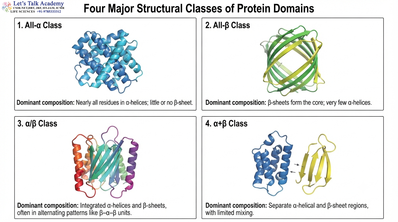
Protein domains can be broadly classified according to their secondary-structure composition.
Four broad categories are:
- All-α
- All-β
- α/β
- α+β
These categories describe the overall organization rather than defining every detail of a domain.
10.1 All-α Domains
All-α domains are dominated by α-helices.
The helices can pack together to form helical bundles and other architectures.
Hydrophobic residues often form the internal interfaces between neighboring helices.
The resulting structure can be rigid enough to provide mechanical support while retaining functional flexibility.
10.2 All-β Domains
All-β domains are dominated by β-strands organized into β-sheets.
The sheets can form structures such as β-sandwiches and β-barrels.
Backbone hydrogen bonds between β-strands are central to their structural organization.
10.3 α/β Domains
α/β domains contain α-helices and β-strands that are closely integrated.
Many metabolic enzymes contain α/β architectures.
The repeated β–α–β motif is an important building block of several α/β folds.
10.4 α+β Domains
In α+β domains, α-helical and β-sheet regions are often more structurally separated.
The distinction between α/β and α+β is therefore based mainly on the topology and organization of the secondary structures.
11. Protein Folds and Domains
The term fold describes the overall three-dimensional arrangement of secondary-structure elements within a domain.
Two proteins may have similar folds even if their amino acid sequences are considerably different.
A common fold may therefore be conserved over evolutionary time while individual sequences undergo substantial divergence.
This is one reason structural comparison can reveal evolutionary relationships that are difficult to detect through direct sequence comparison.
11.1 Fold Conservation
Protein folds can be remarkably conserved.
This occurs because the physical constraints required to maintain a stable three-dimensional structure can limit the range of acceptable mutations.
Hydrophobic core residues, important structural contacts, and residues involved in functional sites may be conserved strongly.
Other surface residues can evolve more rapidly.
11.2 Structural Conservation Versus Sequence Conservation
Two homologous proteins can retain similar domain structures despite substantial sequence differences.
The reason is that the exact amino acid identity is not always essential at every position.
A conservative substitution may preserve the chemical characteristics required for the fold.
Thus, structural conservation can persist even when sequence identity becomes low.
12. Domain Evolution

Protein domains are important units of molecular evolution.
Evolution can modify protein architecture through duplication, fusion, fission, insertion, deletion, and rearrangement of domains.
This modular evolutionary strategy allows new proteins to emerge by modifying existing structural units.
12.1 Gene Duplication
Gene duplication produces an additional copy of a gene or domain-coding region.
The duplicated copy can accumulate mutations.
Over time, the two copies may retain similar functions, divide their original functions, or acquire new functions.
The general pattern can be represented as:
Duplication → divergence → functional specialization
12.2 Domain Fusion
Domain fusion occurs when two previously separate coding regions become combined into a single gene.
The resulting protein may contain two domains that function together.
For example:
Catalytic domain + binding domain → integrated multifunctional protein
Fusion can improve functional coordination by bringing activities into the same polypeptide.
12.3 Domain Fission
Domain fission is conceptually the reverse process.
A previously integrated protein region can become separated into distinct protein products through evolutionary changes.
This can alter regulation, localization, and functional specialization.
12.4 Domain Shuffling
Domain shuffling refers to the evolutionary rearrangement of domains among proteins.
A domain that performs one function in one protein can become associated with a different domain in another protein.
This mechanism can generate new combinations of functions.
For example:
Protein 1: Domain A + Domain B
Protein 2: Domain A + Domain C
The same Domain A can therefore participate in different functional contexts.
12.5 Domain Insertion and Deletion
Evolution can also insert a domain into an existing protein architecture or remove a domain from it.
Insertion can provide a new regulatory or binding capability.
Deletion can simplify a protein or eliminate a function that is no longer required.
These changes can have major consequences for protein architecture.
13. Domains as Evolutionary Modules
Domains are often described as evolutionary modules because they can be conserved, duplicated, rearranged, and recombined.
This modularity provides an efficient mechanism for generating protein diversity.
Instead of evolving an entirely new protein structure from the beginning, evolution can modify existing domains and their combinations.
This is particularly evident in signaling proteins, transcription factors, extracellular proteins, and regulatory proteins.
13.1 Domain Duplication and Functional Divergence
Duplicated domains can accumulate mutations.
Some mutations may occur at positions involved in molecular recognition.
As a result, one copy may develop a different binding specificity while retaining the same general structural fold.
Thus:
Common ancestral domain → duplication → sequence divergence → different functional specificity
13.2 Subfunctionalization
Subfunctionalization occurs when duplicated genes or domains divide the ancestral function.
One copy may become specialized for one cellular context, while another performs a different part of the original function.
13.3 Neofunctionalization
Neofunctionalization occurs when a duplicated gene or domain acquires a novel function.
Because the existing fold provides a stable structural framework, mutations can modify surface chemistry or active-site properties without necessarily destroying the overall architecture.
14. Functional Domains
The biological roles of domains are extremely diverse.
Domains can participate in:
Catalysis
Ligand binding
DNA recognition
RNA recognition
Protein–protein interaction
Regulation
Membrane association
Localization
A single protein can combine several of these functions through different domains.
14.1 Catalytic Domains
A catalytic domain provides a three-dimensional environment in which a chemical reaction can occur efficiently.
Catalytic residues that are separated in the primary sequence can become spatially adjacent after folding.
The catalytic domain can bind substrates, orient reactive groups, stabilize transition states, and facilitate proton transfer or other chemical processes.
This is one reason why protein conformation is essential for enzyme activity.
14.2 Binding Domains
Binding domains recognize particular molecules through complementary chemical and structural features.
A binding pocket may contain hydrophobic residues, charged residues, hydrogen-bond donors and acceptors, and aromatic groups arranged in a specific three-dimensional configuration.
The affinity and specificity of the interaction depend on the combined contribution of these features.
14.3 Regulatory Domains
Regulatory domains modulate the activity of other regions of a protein.
A regulatory domain may bind a ligand and undergo a conformational change.
This change can alter the position or accessibility of a catalytic domain.
Thus:
Ligand binding → regulatory-domain change → interdomain rearrangement → altered protein activity
This provides a structural basis for allosteric regulation.
15. Domain–Domain Interactions
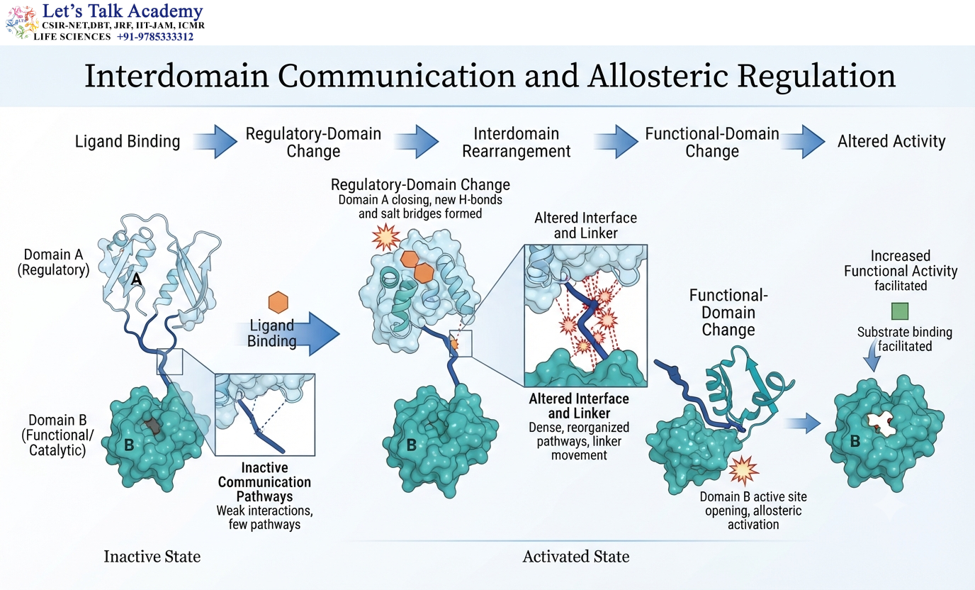
Domains within a multidomain protein can interact through specific interfaces.
These interfaces may involve hydrophobic contacts, hydrogen bonds, salt bridges, van der Waals interactions, and aromatic interactions.
The strength and nature of these interactions determine how the domains are positioned relative to one another.
In some proteins, domain interactions remain relatively stable.
In others, domains move significantly during the functional cycle.
15.1 Interdomain Communication
A change in one domain can influence another domain even when the two domains are not directly involved in the same chemical reaction.
This is particularly important in allosteric proteins.
The communication may occur through changes in:
- Domain orientation
- Interface contacts
- Linker conformation
- Electrostatic networks
- Hydrogen-bonding networks
Such communication allows local molecular events to produce larger structural and functional changes.
15.2 Domain Dynamics
Domains are not perfectly rigid structures.
Even in a well-folded protein, atoms and side chains undergo continuous thermal motion.
Loops can move, side chains can rotate, and domains can undergo larger conformational changes.
These dynamics are often essential for biological activity.
A catalytic protein may alternate between open and closed conformations, while a signaling protein may shift between inactive and active states.
16. Domains and Allosteric Regulation
Allosteric regulation occurs when binding or structural changes at one region influence the behavior of another region.
Multidomain architecture provides an effective structural basis for this phenomenon.
A simplified sequence is:
Regulatory ligand binds Domain A
↓
Domain A changes conformation
↓
Interdomain contacts change
↓
Domain B changes conformation
↓
Functional activity changes
Allosteric communication is therefore an example of how domains function as integrated molecular units rather than isolated structures.
17. Domains in Enzymes
Many enzymes contain multiple domains.
Different domains can contribute to substrate recognition, catalytic chemistry, cofactor binding, or regulation.
For example, one domain may bind the substrate while another domain closes over the active site.
This arrangement can increase catalytic efficiency by positioning the substrate correctly and shielding the reaction site from solvent.
17.1 Open and Closed Domain Conformations

Some enzymes undergo domain movements during catalysis.
A simplified cycle is:
Open enzyme → substrate binding → domain closure → catalysis → product release → open enzyme
Domain closure can bring catalytic residues into the appropriate position and reduce unwanted solvent exposure.
The movement may involve rotation of one domain relative to another rather than major unfolding of the protein.
17.2 Functional Significance of Domain Movement
Domain movement can influence:
Substrate affinity
Catalytic efficiency
Transition-state stabilization
Product release
Allosteric regulation
Therefore, domain architecture must be understood dynamically rather than only as a static structural arrangement.
18. Domains in DNA-Binding Proteins
DNA-binding proteins frequently contain domains specialized for recognizing DNA sequences.
Examples include helix-turn-helix domains, zinc-finger domains, homeodomains, and other DNA-recognition architectures.
The DNA-binding domain positions specific amino acid side chains near the DNA bases and phosphate backbone.
The interaction is determined by shape complementarity, hydrogen bonding, electrostatic interactions, and other molecular forces.
18.1 Zinc-Finger Domains
Zinc-finger domains contain zinc ions that help stabilize the three-dimensional structure.
Cysteine and histidine residues commonly participate in zinc coordination, depending on the particular zinc-finger type.
The general principle can be represented as:
Cys/His residues + Zn²⁺ → stabilized structural fold
The stabilized domain can then participate in DNA, RNA, protein, or other molecular interactions.
18.2 Protein–Protein Interaction Domains
Many signaling and regulatory proteins contain specialized interaction domains.
Examples include SH2, SH3, PDZ, and WW domains.
These domains recognize particular sequence or structural features in partner proteins.
Such interaction modules allow proteins to assemble into larger signaling and regulatory complexes.
19. Domains in Signal Transduction
Signal-transduction proteins often contain multiple domains because signaling requires several coordinated activities.
A protein may contain a ligand-recognition region, regulatory domain, catalytic domain, and protein-interaction domain.
This allows the protein to:
Receive a signal → undergo conformational change → interact with another protein → catalyze a reaction → transmit the signal
The modular organization of signaling proteins is therefore directly related to their ability to process cellular information.
19.1 SH2 Domains

SH2 domains commonly recognize phosphorylated tyrosine-containing sequences.
They are important components of signaling pathways regulated by tyrosine phosphorylation.
The domain recognizes the phosphorylated tyrosine together with neighboring sequence information, allowing selective recruitment of signaling proteins.
19.2 SH3 Domains
SH3 domains commonly recognize proline-rich sequence motifs.
They are frequently involved in protein–protein interactions associated with signaling and cytoskeletal organization.
Their recognition illustrates how a domain can translate a short sequence feature into a specific molecular interaction.
19.3 PDZ Domains
PDZ domains commonly recognize short peptide sequences, often located at the C-terminal ends of target proteins.
They play important roles in organizing membrane-associated signaling complexes and protein assemblies.
20. Domains in Membrane Proteins
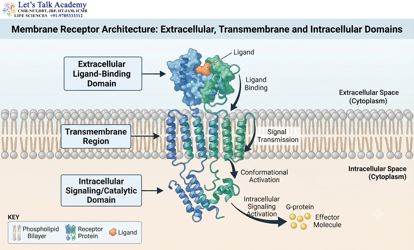
Membrane proteins often contain multiple structural regions with different functions.
A typical receptor architecture may be represented as:
Extracellular domain — transmembrane region — intracellular domain
The extracellular region may recognize a ligand.
The transmembrane region anchors the protein within the lipid bilayer.
The intracellular domain can transmit the signal or perform catalytic activity.
This is an excellent example of how different structural modules cooperate within one protein.
20.1 Membrane-Binding Domains
Some soluble domains associate with membranes by recognizing specific lipid molecules.
The interaction may involve positively charged residues, hydrophobic patches, or specific lipid-binding pockets.
Membrane-binding domains allow proteins to become localized to particular cellular membranes without necessarily containing a transmembrane helix.
21. Domains in Motor Proteins
Motor proteins convert chemical energy, usually derived from nucleotide hydrolysis, into mechanical movement.
A motor protein may contain:
ATPase domain + mechanical region + cargo-binding domain
The ATPase domain performs the chemical reaction, while other regions connect the motor to cellular structures or cargo.
Conformational changes associated with nucleotide binding and hydrolysis can produce mechanical movement.
Thus, domain organization provides a structural mechanism for converting chemical energy into mechanical work.
22. Domains in DNA Repair
DNA-repair proteins frequently contain multiple functional domains.
A protein may combine a DNA-binding domain with a catalytic domain and one or more interaction domains.
This organization allows it to recognize damaged DNA, recruit other proteins, and perform the appropriate chemical reaction.
The modular nature of DNA-repair proteins therefore contributes to the complexity and specificity of DNA maintenance pathways.
23. Domain Swapping
Domain swapping is a structural phenomenon in which equivalent structural elements from two protein molecules exchange with one another.
Instead of each protein containing only its own structural segment, part of one molecule becomes associated with the corresponding region of another molecule.
Domain swapping demonstrates that the interactions maintaining protein structure can sometimes be reorganized between molecules.
It is relevant to protein oligomerization, structural evolution, and in some cases protein aggregation.
23.1 Structural Significance of Domain Swapping
Domain swapping illustrates the balance between:
Intramolecular interactions and Intermolecular interactions
A structural element that normally interacts with another part of the same protein can instead interact with the corresponding region of another protein molecule.
This can produce stable oligomeric assemblies.
24. Domains and Protein Misfolding

Correct domain folding is essential for protein function.
A mutation that destabilizes a domain can cause partial unfolding.
Partial unfolding can expose hydrophobic surfaces that are normally buried inside the protein.
These exposed surfaces can interact with other molecules and promote aggregation.
Therefore:
Domain destabilization → partial unfolding → exposure of hydrophobic surfaces → abnormal protein association
Protein aggregation is an important feature of several pathological conditions.
24.1 Structural Changes During Aggregation
In some aggregation processes, proteins undergo changes in secondary structure and become enriched in β-sheet-containing assemblies.
This demonstrates that protein domains are dynamic structures and that changes in their conformational state can influence biological behavior.
25. Domains and Post-Translational Modifications
Protein domains can contain residues that undergo post-translational modification.
Common modifications include phosphorylation, acetylation, methylation, ubiquitination, and glycosylation.
These modifications can alter:
- Domain stability
- Protein interactions
- Subcellular localization
- Catalytic activity
- Conformational dynamics
For example, phosphorylation can create a recognition site for another protein domain.
Thus, domains can function as platforms for regulated molecular communication.
26. Domain Databases and Classification
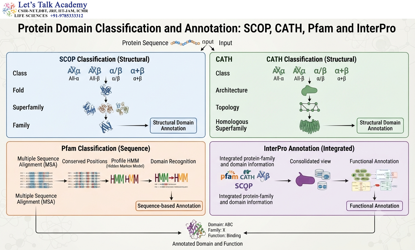
Modern structural biology uses specialized databases to identify, classify, and compare protein domains.
Important resources include:
- SCOP
- CATH
- Pfam
- InterPro
These resources use different approaches but collectively provide valuable information about protein families, domains, structural folds, and evolutionary relationships.
26.1 SCOP
The Structural Classification of Proteins (SCOP) system classifies protein structures according to structural and evolutionary relationships.
Its classical organization includes:
Class → Fold → Superfamily → Family
A class describes broad structural composition.
A fold describes the arrangement of secondary structures.
A superfamily groups proteins with probable evolutionary relationships despite considerable sequence divergence.
A family generally contains more closely related proteins.
26.2 CATH
The CATH system classifies protein domains using:
Class → Architecture → Topology → Homologous superfamily
Class describes broad secondary-structure composition.
Architecture describes the overall spatial arrangement of secondary structures without necessarily considering their connectivity.
Topology describes the connectivity and arrangement of secondary structures.
Homologous superfamily represents proteins with evolutionary relationships.
26.3 Pfam
Pfam is a protein-family database that uses profile hidden Markov models to identify conserved sequence domains.
Profile HMMs are particularly useful for detecting distant homologues because they capture position-specific patterns of conservation.
The general concept is:
Multiple sequence alignment → conserved positions → statistical profile → domain recognition
26.4 InterPro
InterPro integrates information from several protein-family and domain databases.
It helps identify conserved domains and functional regions in protein sequences.
Such resources are widely used for protein annotation and bioinformatics analysis.
27. Domain Detection from Protein Sequences
When the three-dimensional structure of a protein is unknown, domain architecture can often be predicted from its sequence.
A general workflow is:
Protein sequence → sequence analysis → conserved-region identification → domain prediction → structural and functional interpretation
Profile-based methods can detect domains that may be difficult to identify through simple pairwise sequence comparisons.
However, computational domain prediction should be interpreted carefully because a predicted domain does not automatically establish its exact biological function.
27.1 Hidden Markov Models in Domain Identification
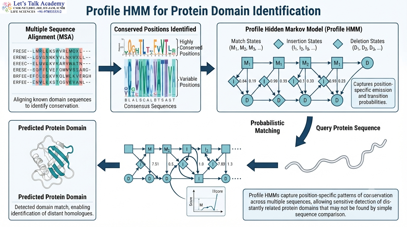
A profile hidden Markov model describes the probability of observing different amino acids at different positions within a conserved protein family.
This allows the model to recognize weak but biologically meaningful sequence similarities.
The approach is particularly valuable when proteins have diverged significantly during evolution.
27.2 Sequence Motif Versus Domain Prediction
A short conserved motif may provide evidence for a particular function, but a motif is not equivalent to an entire domain.
A domain generally contains a much larger sequence and structural region.
Therefore, domain prediction often combines multiple types of evidence rather than relying on a single short sequence pattern.
28. Domain Architecture and Bioinformatics

Domain analysis is an important part of structural bioinformatics.
For an unknown protein, identifying domains can provide clues about its potential function, evolutionary history, and structural organization.
A typical computational analysis may proceed as:
Sequence
↓
Domain identification
↓
Secondary-structure prediction
↓
Structure prediction
↓
Fold comparison
↓
Functional annotation
↓
Experimental hypothesis
This approach is particularly valuable when experimental structures are unavailable.
29. Domain Prediction and Structural Modeling
Modern structure-prediction methods can provide detailed information about folded regions of proteins.
Predicted structures can help identify:
- Compact domains
- Flexible linkers
- Disordered regions
- Interdomain interfaces
- Potential ligand-binding sites
However, structural prediction should not be interpreted as complete experimental proof of biological function.
Experimental validation remains important for determining activity, dynamics, ligand interactions, and cellular behavior.
30. Domains and Protein Engineering
Protein domains are extremely useful in protein engineering because they can function as modular components.
Researchers can combine or modify domains to create proteins with new properties.
For example:
DNA-binding domain + activation domain
can produce a transcriptional regulatory system.
Similarly:
Targeting domain + catalytic domain
can direct enzymatic activity toward a specific cellular location.
The success of domain engineering depends on proper folding, linker design, domain orientation, and compatibility between the domains.
30.1 Domain Deletion
A domain can be experimentally deleted to investigate its contribution to protein function.
If removal of a domain eliminates activity, the domain may be directly involved in the function.
However, loss of activity may also result from destabilization of the complete protein.
Therefore, domain deletion experiments must be interpreted carefully.
30.2 Domain Replacement
One domain can sometimes be replaced by another related domain.
Comparison of the resulting protein with the original can reveal which properties are controlled by the exchanged domain.
This approach is useful for studying:
- Specificity
- Regulation
- Localization
- Protein interactions
- Catalytic activity
31. Domain Architecture and Synthetic Biology
Synthetic biology frequently exploits natural domain organization.
Protein components can be assembled into new combinations to create engineered molecular systems.
A simplified design principle is:
Recognition module + regulatory module + functional module
The recognition module determines what the engineered protein interacts with.
The regulatory module controls when the protein becomes active.
The functional module performs the desired biological activity.
This modular strategy is based directly on the natural organization of proteins.
32. Domains and Protein–Ligand Recognition
A domain can contain a binding pocket that recognizes a ligand with high specificity.
The pocket is formed by residues that may be far apart in the primary sequence but close together in three-dimensional space.
Binding depends on:
- Shape complementarity
- Electrostatic complementarity
- Hydrogen bonding
- Hydrophobic interactions
- van der Waals interactions
The folded domain therefore creates a specific chemical environment that cannot be reproduced by the linear sequence alone.
32.1 Induced Fit
In the induced-fit model, ligand binding induces a conformational change in the protein.
A simplified representation is:
Protein + ligand → initial complex → conformational rearrangement → optimized complex
The structural change can involve movement of a loop, rotation of a domain, or rearrangement of side chains.
32.2 Conformational Selection
In conformational selection, the protein already exists as an ensemble of conformations.
The ligand preferentially binds one of those conformations and shifts the equilibrium toward it.
The two models are not mutually exclusive and can operate together in real protein–ligand interactions.
33. Domains and Intrinsically Disordered Regions
Not every region of a protein forms a stable folded domain.
Some proteins contain intrinsically disordered regions, which lack a single stable three-dimensional structure under particular physiological conditions.
A protein can therefore contain:
Folded domain — disordered region — folded domain
Disordered regions often contain regulatory sites and flexible interaction regions.
This demonstrates that protein architecture can combine highly ordered domains with structurally flexible segments.
34. Domains and Conformational Flexibility
A folded domain is not completely rigid.
Thermal motion continuously causes small fluctuations in the structure.
Side chains rotate, loops move, and backbone atoms undergo small displacements.
Some domains undergo larger movements during function.
This flexibility is particularly important in enzymes and signaling proteins.
A protein can therefore be understood as a dynamic molecular machine rather than a static structure.
35. Domains and Protein Stability
Domain stability is determined by the balance between the energetic benefits of folding and the energetic costs of restricting the polypeptide.
The folded state gains stabilization from favorable interactions, while folding reduces conformational entropy.
A stable domain therefore represents a favorable overall free-energy state.
However, the stability margin of many proteins is relatively small, meaning that modest changes in temperature, pH, mutation, or solvent conditions can substantially influence folding.
35.1 Effects of Temperature
Increasing temperature increases molecular motion and can eventually destabilize the interactions maintaining the domain.
At sufficiently high temperatures, the folded structure may undergo thermal denaturation.
The temperature at which a protein undergoes a major unfolding transition is commonly associated with its melting temperature, Tₘ.
35.2 Effects of pH
Changes in pH alter the protonation state of ionizable amino acid side chains.
This can disrupt salt bridges, hydrogen-bonding networks, and other interactions.
As a result, extreme pH conditions can destabilize domains.
35.3 Effects of Chemical Denaturants
Chemical denaturants such as urea or guanidinium-based compounds can disrupt the interactions responsible for protein folding.
They can promote exposure of hydrophobic regions and shift the equilibrium toward unfolded states.
36. Domains and Molecular Chaperones
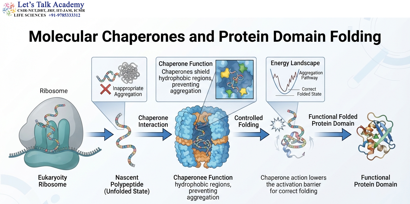
Some proteins require molecular chaperones to fold efficiently inside cells.
Chaperones do not usually provide the structural information necessary to determine the final fold. Instead, they help prevent inappropriate interactions and aggregation while the protein folds.
A simplified process is:
Nascent polypeptide → chaperone interaction → controlled folding → functional protein
Chaperones are especially important for proteins with complex multidomain architectures.
37. Domains and Cellular Crowding
Protein domains fold inside cells rather than in an empty solution.
The cellular environment contains high concentrations of proteins, nucleic acids, metabolites, ions, and membranes.
This molecular crowding can influence protein folding, stability, association, and aggregation.
Therefore, the behavior of an isolated protein domain in a laboratory solution may not always perfectly represent its behavior inside the cell.
38. Domain Architecture in Receptors
Many receptors contain clearly differentiated domains.
A typical receptor can have:
- Extracellular ligand-binding domain
- Transmembrane region
- Intracellular signaling domain
The extracellular region interacts with an external signal.
The transmembrane region anchors the receptor.
The intracellular domain transmits the information into the cell.
This architecture converts an extracellular chemical signal into an intracellular molecular response.
39. Domains in Protein Kinases
Protein kinases contain catalytic domains specialized for transferring phosphate groups from ATP to target molecules.
The catalytic domain contains structural elements that position:
- ATP
- Metal ions
- Substrate
- Catalytic residues
in the correct three-dimensional arrangement.
Additional domains can regulate kinase activity or determine substrate specificity.
Thus, the catalytic domain performs the chemical reaction while the overall domain architecture determines when and where the kinase functions.
40. Domains in Motor Proteins
Motor proteins use nucleotide hydrolysis to generate mechanical work.
Their domain organization allows chemical energy to be coupled to structural movement.
The general relationship is:
Nucleotide binding/hydrolysis → conformational change → mechanical movement
Different domains can connect the chemical reaction to cellular structures or cargo.
This illustrates the importance of domain movement and interdomain communication.
41. Domains in Structural Proteins
Structural proteins often contain repeated or specialized domains that contribute to mechanical strength and assembly.
Domain organization can determine:
- Elasticity
- Mechanical strength
- Fiber formation
- Protein assembly
Repeated domains can also provide multiple interaction surfaces.
This is particularly important in large cytoskeletal and extracellular proteins.
42. Domains and Post-Translational Regulation
A domain can serve as a platform for post-translational modifications and regulatory interactions.
Phosphorylation can change charge and alter domain interactions.
Ubiquitination can affect protein stability or localization.
Acetylation and methylation can influence molecular recognition.
Glycosylation can alter extracellular domain stability and interactions.
Thus, post-translational modification can regulate the structure and function of domains without changing the amino acid sequence.
43. Domains and Disease
Mutations affecting protein domains can cause disease through several mechanisms.
A mutation may destabilize the domain, destroy a catalytic residue, alter ligand binding, disrupt protein–protein interactions, or interfere with domain communication.
The consequences can be represented as:
Mutation → structural change → altered domain stability/function → altered cellular pathway → disease phenotype
The same amino acid substitution can have very different effects depending on whether the affected residue is located in the hydrophobic core, active site, domain interface, or flexible surface region.
44. Domains and Protein Aggregation
When a domain becomes partially unfolded, normally buried hydrophobic residues can become exposed.
These surfaces can interact with other protein molecules.
Repeated interactions can lead to oligomers and larger aggregates.
In some cases, aggregation is associated with the formation of β-sheet-rich assemblies.
Understanding domain stability and conformational transitions is therefore important for understanding protein aggregation.
45. Domain Swapping and Oligomerization
Domain swapping provides another example of how protein domains can participate in intermolecular organization.
A structural element from one protein molecule can interact with another protein molecule rather than its own corresponding region.
This can generate stable oligomers.
The phenomenon demonstrates that protein architecture is determined by a balance between intra- and intermolecular interactions.
46. Domain Boundaries and Limited Proteolysis
Domain boundaries can sometimes be investigated experimentally using controlled or limited proteolysis.
Flexible, solvent-exposed regions are generally more accessible to proteases than compact domain cores.
Consequently, cleavage patterns can provide clues about domain organization.
However, proteolytic sensitivity alone is not sufficient to establish the exact boundary of a domain.
Structural and sequence information is usually considered together.
47. Domain Architecture and Evolutionary Classification
Protein domains can be classified according to their structural and evolutionary relationships.
Important concepts include:
- Fold
- Superfamily
- Family
A fold represents a recurring arrangement of secondary structures.
A superfamily contains structurally related proteins that may have diverged substantially in sequence.
A family generally contains more closely related proteins with stronger sequence and functional similarities.
This classification helps connect protein structure with evolutionary history.
48. Domain Fold
A domain fold represents the characteristic three-dimensional arrangement of its secondary structures.
For example, different proteins can contain similar arrangements of α-helices and β-sheets even when their amino acid sequences differ.
The existence of common folds suggests that certain structural solutions are particularly favorable or have been conserved through evolution.
49. Structural Superfamilies
A structural superfamily contains proteins that share a common structural framework and generally show evidence of evolutionary relationship.
The sequences may have diverged substantially, but important structural features can remain conserved.
Superfamily analysis can therefore reveal evolutionary relationships that are difficult to detect from sequence alone.
50. Protein Families
A protein family generally consists of proteins with significant sequence and structural similarity and often related functions.
Conserved residues within a family can identify important structural or catalytic regions.
The relationship can be represented as:
Protein family → conserved sequence → conserved structure → conserved function
Although functional divergence can occur within a family, family-level relationships provide important clues for protein annotation.
51. Domain Architecture as a Molecular Blueprint
The arrangement of domains can be considered a molecular blueprint for protein function.
A protein containing:
Binding domain + catalytic domain
has a different functional potential from a protein containing:
Binding domain + regulatory domain
even though both proteins share the same binding module.
Thus, domain combinations can be as important as individual domains.
52. Domain Combinations and Functional Complexity
Multidomain proteins can integrate several molecular activities.
For example:
Recognition → binding → conformational change → catalysis → regulation
can occur within a single polypeptide.
This reduces the need for separate proteins to perform each step and allows efficient communication between activities.
Domain organization is therefore an important structural basis for the complexity of cellular pathways.
53. Domains and Functional Specificity
A domain can determine where a protein binds, what it recognizes, or which molecular partner it interacts with.
Small changes in domain surface residues can alter specificity without necessarily destroying the overall fold.
This is an important mechanism of molecular evolution.
A conserved domain can therefore retain its basic architecture while gradually acquiring a new interaction preference.
54. Domain Engineering and Biotechnology
The modular nature of domains makes them valuable tools in biotechnology.
Researchers can combine domains to construct proteins with specific properties.
For example:
Targeting domain + catalytic domain
can direct an enzyme toward a particular cellular compartment.
Similarly:
DNA-binding domain + regulatory domain
can be used to construct artificial transcriptional regulators.
Domain-based engineering therefore takes advantage of the natural modularity of protein architecture.
55. Domain Architecture and Synthetic Biology
Synthetic biology extends the concept of modular protein architecture by assembling functional modules into engineered systems.
Successful domain fusion requires consideration of:
- Folding compatibility
- Domain orientation
- Linker length
- Linker flexibility
- Steric accessibility
- Interdomain interactions
A domain that folds properly in isolation may not necessarily function correctly after fusion with another domain.
Thus, domain engineering requires understanding both individual domains and their interactions.
56. Experimental Study of Protein Domains
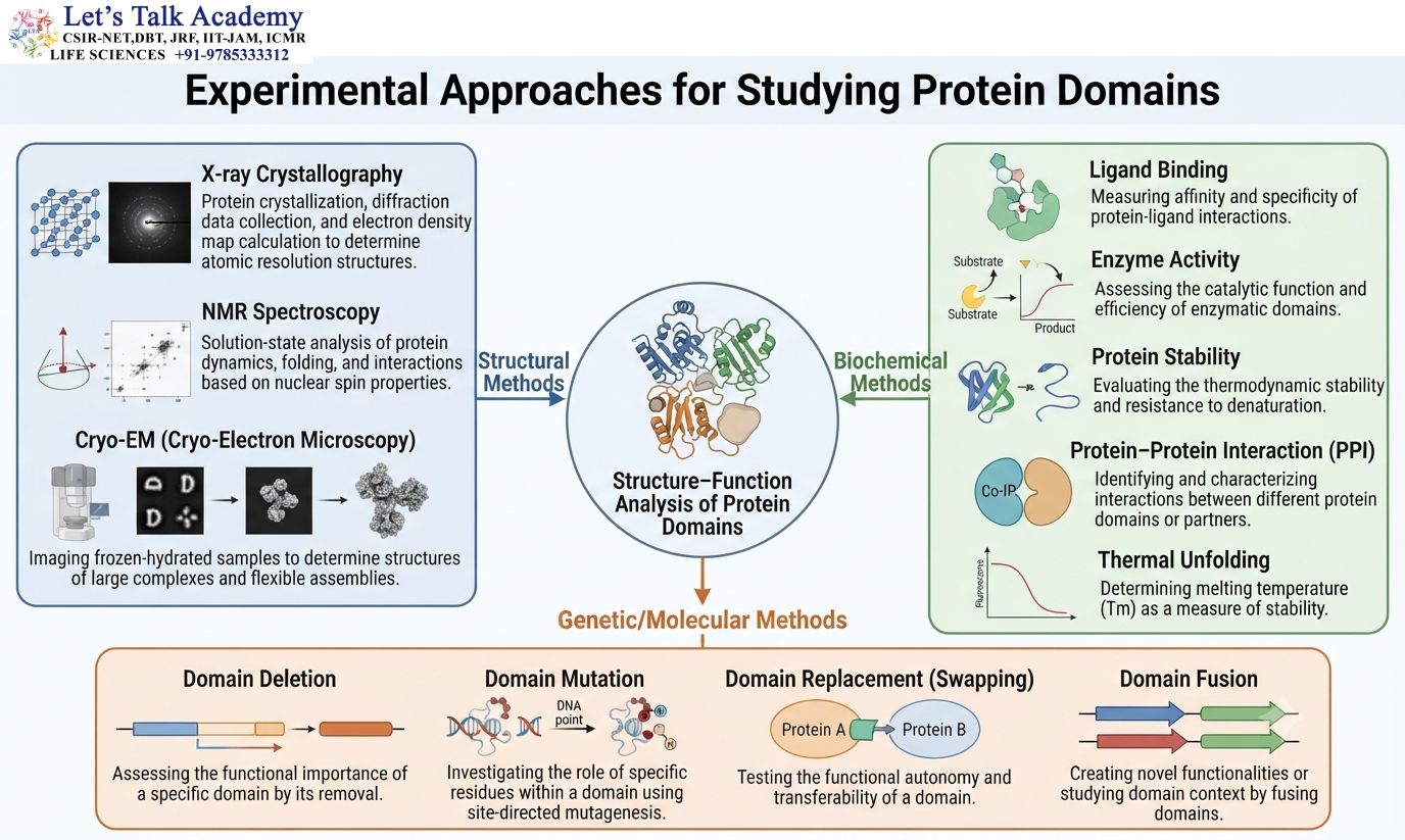
Protein domains can be investigated using several experimental approaches.
Structural methods include:
- X-ray crystallography
- Nuclear magnetic resonance spectroscopy
- Cryo-electron microscopy
Biochemical methods can investigate:
- Ligand binding
- Enzyme activity
- Protein stability
- Protein–protein interactions
- Thermal unfolding
Genetic and molecular approaches can investigate:
- Domain deletion
- Domain mutation
- Domain replacement
- Domain fusion
Combining structural, biochemical, and genetic information provides a more complete understanding of domain function.
57. Domain Structure Determined by X-Ray Crystallography
X-ray crystallography can provide high-resolution structural information for suitable protein crystals.
The resulting atomic coordinates can reveal:
Secondary structures
Domain boundaries
Hydrophobic cores
Active sites
Ligand-binding pockets
Interdomain interfaces
This information is particularly useful for understanding the detailed molecular basis of domain function.
58. Domain Structure Determined by NMR
Nuclear magnetic resonance spectroscopy is particularly useful for studying proteins and domains in solution.
NMR can provide information about:
Three-dimensional structure
- Molecular dynamics
- Residue-level interactions
- Conformational changes
It is especially useful for smaller proteins and domains and for studying dynamic behavior that may not be apparent from a single static structure.
59. Domain Structure Determined by Cryo-EM
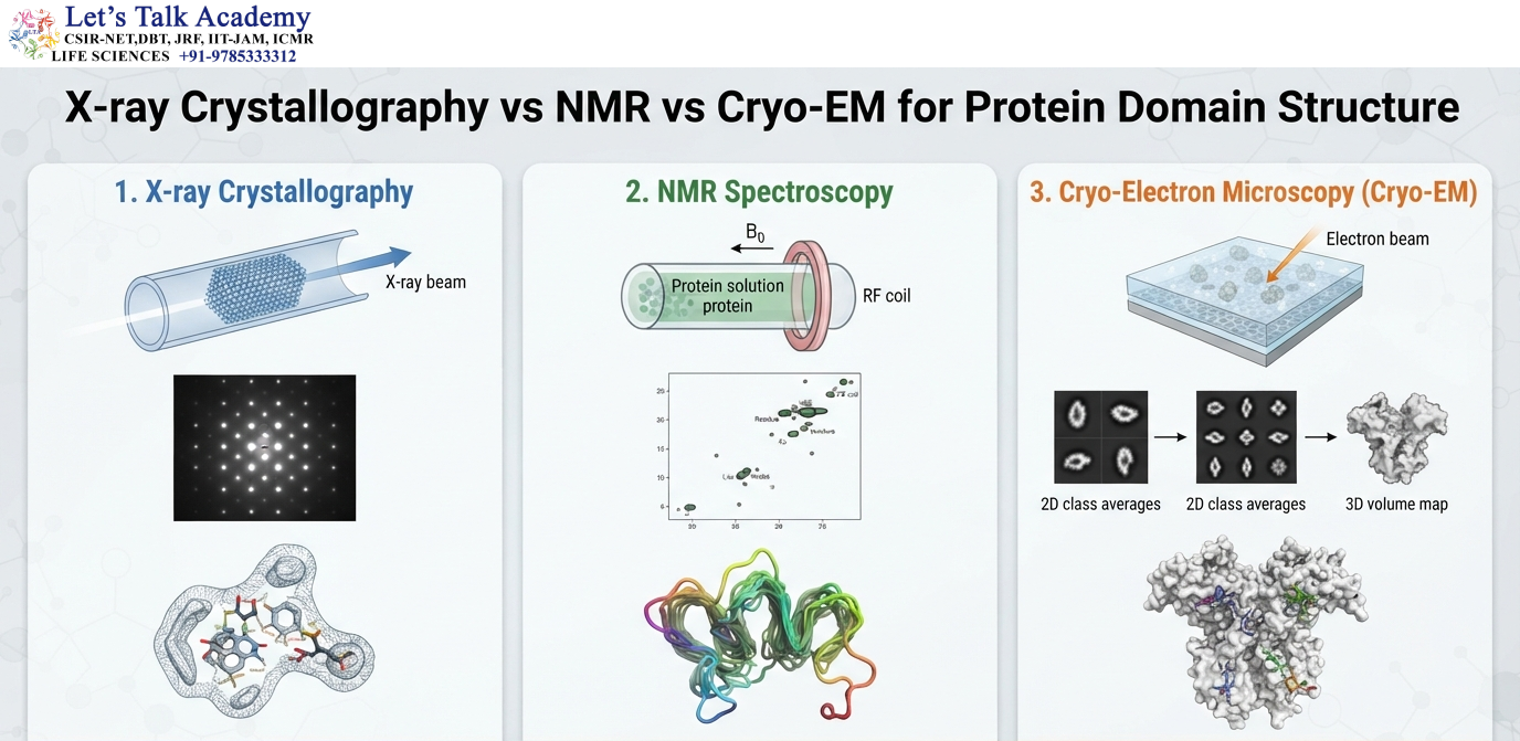
Cryo-electron microscopy has become an important method for determining structures of large protein complexes and multidomain proteins.
It can reveal:
- Domain arrangement
- Interdomain movement
- Ligand binding
- Protein–protein interfaces
- Conformational states
This is particularly useful for large molecular machines and membrane-associated complexes.
60. Domains and Structural Prediction
Modern computational methods can predict protein structures and provide valuable information about domain organization.
A predicted structure may reveal compact regions separated by flexible linkers.
It may also suggest:
- Potential domain boundaries
- Ligand-binding pockets
- Interdomain interfaces
- Disordered regions
Structural prediction has therefore become an important complement to experimental protein characterization.
61. Domain Deletion and Functional Analysis
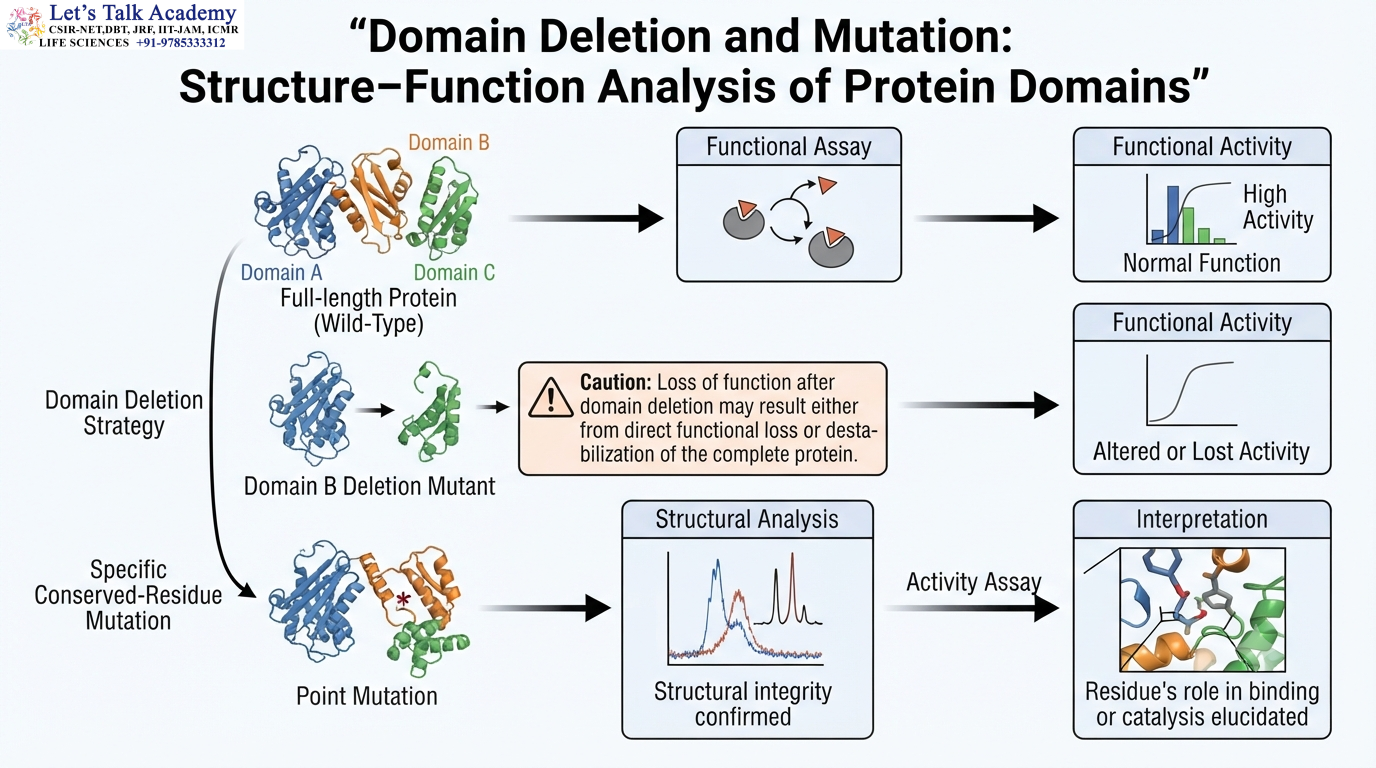
Domain deletion experiments are commonly used to determine the contribution of a domain to protein function.
Suppose:
Full-length protein → functional
and:
Domain-deletion mutant → altered function
The result indicates that the deleted region contributes to the observed behavior.
However, interpretation requires caution because deleting a domain can destabilize the remaining protein.
62. Domain Mutation and Structure–Function Relationships
Specific residues can be mutated to determine their roles.
If mutation of a conserved residue reduces activity, that residue may contribute to catalysis, ligand binding, structural stability, or domain communication.
Structural information helps distinguish these possibilities.
For example, a mutation in the hydrophobic core may reduce activity because the protein becomes unstable rather than because the residue directly participates in catalysis.
63. Domain Architecture and Protein Localization
Some domains determine the cellular location of a protein.
Domains can recognize:
- Membrane lipids
- Protein receptors
- Nucleic acids
- Organellar components
- Cytoskeletal structures
A protein can therefore contain one domain responsible for localization and another responsible for biochemical activity.
This modular arrangement allows the same catalytic activity to be targeted to different cellular environments.
64. Domain Organization in Cellular Signaling
Signaling pathways depend heavily on domain-mediated recognition.
A signaling protein may contain:
- Localization domain
- Interaction domain
- Regulatory domain
- Catalytic domain
The individual modules cooperate to ensure that signaling occurs at the correct location and under the correct conditions.
This organization provides both specificity and regulation.
65. Domain Dynamics and Biological Function
The functional state of a protein is often associated with a particular conformational ensemble rather than one completely fixed structure.
Domains may shift between states such as:
Open ↔ closed
Active ↔ inactive
Bound ↔ unbound
Associated ↔ dissociated
These transitions allow proteins to respond to their molecular environment.
Consequently, understanding domains requires consideration of both structure and dynamics.
66. Domain Architecture and Allosteric Communication
Allosteric communication can occur through networks of interactions extending across a protein.
A conformational change in one domain can alter:
- Domain interfaces
- Hydrogen-bond networks
- Salt bridges
- Hydrophobic contacts
- Linker conformations
These changes can eventually alter the activity of a distant functional site.
Thus, domains can act as interconnected communication modules.
67. Domains and Molecular Recognition
Molecular recognition depends on precise three-dimensional complementarity.
The interacting surfaces of a domain and its ligand must possess compatible:
- Shape
- Charge
- Hydrophobicity
- Hydrogen-bonding patterns
This explains why a protein can distinguish between closely related molecules.
The specificity is generated by the three-dimensional arrangement of residues within the folded domain.
68. Domain Architecture in Protein–Protein Interactions
Protein–protein interactions often involve specialized domain surfaces.
The interface between two domains may contain many weak interactions that collectively provide strong binding.
The total interaction can be expressed conceptually as:
Hydrophobic interactions + hydrogen bonds + electrostatic interactions + van der Waals contacts → stable protein interface
The exact contribution of each interaction depends on the specific protein system.
69. Domain Architecture and Protein Folding Diseases
Diseases associated with protein misfolding can involve domain destabilization, abnormal conformational transitions, or aggregation.
A mutation may shift the equilibrium:
Native state ⇌ partially unfolded state
toward the partially unfolded state.
The exposed regions may then participate in abnormal intermolecular interactions.
Understanding the domain-level structural defect can therefore help explain how a molecular mutation leads to a cellular phenotype.
70. Domain Architecture and Evolutionary Innovation
The modular nature of protein domains provides an efficient mechanism for evolutionary innovation.
Instead of creating an entirely new structural framework, evolution can modify existing modules.
For example:
Existing domain + new partner domain → new protein architecture
The new protein may acquire a novel combination of localization, recognition, regulation, and catalytic properties.
This modular principle is one of the reasons protein evolution can produce enormous functional diversity.
71. Integrated Structural Relationship
The complete organization of a protein can be summarized as:
Amino acid sequence
↓
Backbone conformation
↓
Secondary structures
↓
Structural motifs
↓
Domain formation
↓
Domain packing
↓
Interdomain interactions
↓
Tertiary architecture
↓
Quaternary assembly, when present
↓
Biological function
Each level contributes to the next, while higher-order interactions can also influence local structural behavior.
72. Domain, Motif, Fold, and Protein: Comparison
Structural term |
Description |
|---|---|
| Secondary structure | Local conformation such as α-helix or β-sheet |
| Motif | Recurring arrangement of secondary-structure elements |
| Domain | Compact structural unit with a characteristic fold |
| Fold | Overall three-dimensional arrangement of secondary structures |
| Protein | Complete polypeptide or functional molecular assembly |
| Subunit | Individual polypeptide chain within a multimeric protein |
73. Major Properties of Protein Domains
Protein domains commonly show several characteristic properties. They are structurally organized regions containing multiple secondary-structure elements, and many have a compact hydrophobic core surrounded by a chemically diverse surface. Many domains can fold relatively independently, although this property is not universal. Domains frequently possess recognizable biological functions and may be conserved during evolution even when their amino acid sequences have diverged considerably.
A domain can also serve as an evolutionary module. It may be duplicated, inserted, deleted, fused with another domain, or rearranged within a protein architecture.
These properties make domains fundamental units for understanding protein structure, evolution, and function.
74. Protein Domains and Functional Integration
The true importance of domains becomes apparent when considering the complete function of multidomain proteins.
A protein may need to recognize a molecule, bind it with high specificity, undergo a conformational change, catalyze a chemical reaction, and then interact with another protein.
Rather than requiring separate proteins for each function, a single polypeptide can contain several specialized domains.
For example:
Recognition domain → identifies target
Regulatory domain → controls activity
Catalytic domain → performs reaction
Interaction domain → recruits another protein
The domains work together as an integrated molecular system.
75. Protein Domains as Dynamic Molecular Modules
Although domains are often described as independent structural units, they should not be viewed as rigid blocks.
A domain continuously undergoes small conformational fluctuations, and many domains undergo larger movements during function.
The dynamic behavior of a domain can determine:
- Ligand affinity
- Catalytic activity
- Protein interaction
- Allosteric regulation
- Signal transmission
- Subcellular localization
Therefore, the biological significance of a domain depends on both its static fold and its dynamic conformational behavior.


