
Conformation of Nucleic Acids: Structural Characteristics of A, B and Z DNA
1. Conformation of Nucleic Acids
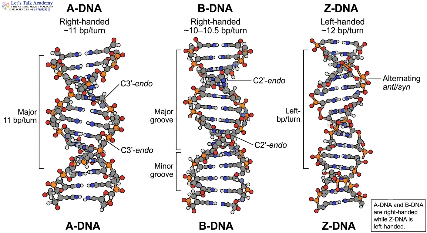
Nucleic acids are not rigid, permanently fixed molecules. Their biological activity depends on their ability to adopt different three-dimensional conformations. DNA is generally represented as a double helix, but the double helix can exist in several structural forms depending on the nucleotide sequence, hydration, ionic conditions, temperature, chemical environment, torsional stress and interactions with proteins.
The term conformation refers to the three-dimensional arrangement of atoms in a molecule that can change without breaking the covalent bonds of the molecule. In DNA, conformational changes mainly involve rotations around bonds in the sugar-phosphate backbone, changes in the orientation of the nitrogenous bases, and changes in the puckering of the deoxyribose sugar.
The most extensively studied double-helical conformations of DNA are A-DNA, B-DNA and Z-DNA. These forms have the same basic chemical components but differ substantially in their three-dimensional organization.
A-DNA and B-DNA are right-handed helices, whereas Z-DNA is a left-handed helix. A-DNA is relatively broad and compact, B-DNA is the classical DNA structure found under many aqueous conditions, and Z-DNA is a distinctive left-handed structure with a characteristic zig-zag phosphate backbone.
The ability of DNA to adopt these different structures is known as DNA structural polymorphism. Structural polymorphism is important because DNA must interact with many proteins, ions and other molecules during replication, transcription, recombination and repair. A conformational change can alter the shape of DNA and consequently change how a protein recognizes or binds to a particular DNA sequence.
The structural form adopted by DNA is determined by the combined effect of several molecular parameters. These include the conformation of the deoxyribose sugar, orientation of the glycosidic bond, arrangement of the phosphate backbone, inclination of base pairs, base stacking, hydration, electrostatic interactions and torsional stress.
Therefore, A-DNA, B-DNA and Z-DNA should not be considered as three completely independent molecules. They are alternative structural states of the same DNA polymer.
1.1 Chemical Organization Underlying DNA Conformation
Each DNA nucleotide consists of a nitrogenous base, a deoxyribose sugar and one or more phosphate groups. The four major nitrogenous bases are adenine, guanine, cytosine and thymine.
Adenine and guanine are purines, containing two fused rings, whereas cytosine and thymine are pyrimidines, containing a single six-membered ring.
In double-stranded DNA, complementary bases interact through hydrogen bonding. Adenine pairs with thymine, while guanine pairs with cytosine.
Although hydrogen bonds are essential for maintaining complementary base pairing, they are not the only source of DNA stability. Base stacking interactions between neighboring aromatic bases contribute substantially to the stability of the double helix. The hydrophobic nature of the bases also favors their burial inside the helix, away from the surrounding aqueous environment.
The sugar-phosphate backbone is negatively charged because of the phosphate groups. This negative charge strongly influences DNA conformation because neighboring phosphate groups repel each other. Water molecules and positively charged ions help reduce this electrostatic repulsion and therefore influence which DNA conformation is energetically favorable.
Consequently, DNA conformation results from a balance among hydrogen bonding, base stacking, sugar geometry, backbone electrostatics, hydration and interactions with surrounding molecules.
1.2 Antiparallel Arrangement of DNA Strands
The two strands of DNA are antiparallel.
One strand runs from 5′ to 3′, while the complementary strand runs from 3′ to 5′.
The antiparallel arrangement is essential for the formation of Watson–Crick base pairs and for the correct geometry of the double helix.
The 5′ and 3′ designations arise from the carbon atoms of the deoxyribose sugar. The phosphate group connects the 5′ carbon of one nucleotide to the 3′ carbon of the next nucleotide through a phosphodiester bond.
Although A-DNA, B-DNA and Z-DNA differ in their three-dimensional geometry, the basic chemical polarity and antiparallel organization of the DNA strands remain fundamental features.
1.3 DNA Structural Parameters
DNA conformation is described using several measurable structural parameters. Understanding these parameters is essential because the differences among A-DNA, B-DNA and Z-DNA are primarily differences in molecular geometry.
Important parameters include:
- Helical handedness
- Helical pitch
- Number of base pairs per turn
- Rise per base pair
- Helix diameter
- Base-pair inclination
- Base-pair displacement
- Sugar-pucker conformation
- Glycosidic bond orientation
- Major-groove dimensions
- Minor-groove dimensions
- Backbone geometry
Each parameter describes a different aspect of the three-dimensional organization of DNA.
2. Helical Handedness of DNA
The term helical handedness describes the direction in which a helix twists around its longitudinal axis.
A DNA helix may be right-handed or left-handed.
A-DNA and B-DNA are right-handed, whereas Z-DNA is left-handed.
This distinction is particularly important because Z-DNA demonstrates that DNA can reverse its overall helical direction while retaining complementary base pairing between its two strands.
2.1 Right-Handed DNA
In a right-handed helix, the DNA strands wind around the central axis in a right-handed direction.
Both A-DNA and B-DNA are right-handed structures.
The fact that A-DNA and B-DNA have the same handedness does not mean that they have the same geometry. They differ in diameter, pitch, sugar pucker, base-pair inclination and groove architecture.
2.2 Left-Handed Z-DNA
Z-DNA is a left-handed double helix.
The change from a right-handed B-form to a left-handed Z-form requires substantial rearrangement of the nucleotide backbone.
The left-handed structure is associated with changes in glycosidic bond orientation and sugar-pucker geometry. In canonical alternating GC sequences, guanine residues adopt the syn conformation while cytosine residues generally remain anti.
This alternating molecular geometry generates the characteristic zig-zag backbone of Z-DNA.
3. Sugar Pucker and DNA Conformation
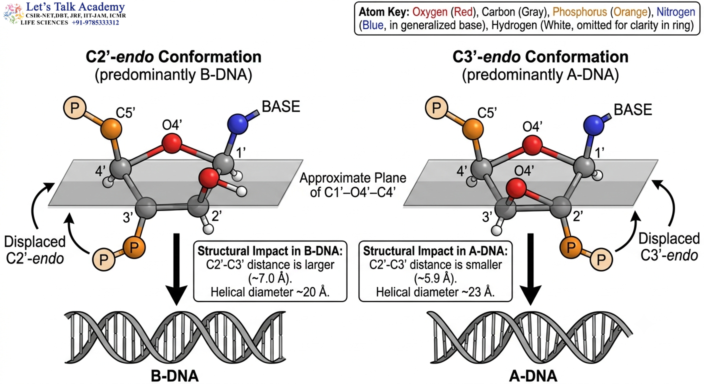
The deoxyribose sugar is not completely flat. It adopts a non-planar three-dimensional conformation called a sugar pucker.
Sugar pucker is one of the most important factors determining the overall geometry of nucleic acids.
The five-membered sugar ring can adopt several conformations. Two particularly important conformations in DNA structural biology are C2′-endo and C3′-endo.
B-DNA predominantly contains C2′-endo sugars, whereas A-DNA predominantly contains C3′-endo sugars.
Z-DNA displays alternating sugar conformations.
3.1 C2′-Endo Conformation
The C2′-endo sugar pucker is characteristic of B-DNA.
In this arrangement, the C2′ carbon is displaced relative to the approximate plane of the remaining sugar atoms.
This geometry affects the orientation of the phosphodiester backbone and contributes to the characteristic dimensions of B-DNA.
Because the sugar is connected to both the phosphate backbone and the nitrogenous base, even a relatively small change in sugar geometry can produce significant changes in the overall DNA helix.
3.2 C3′-Endo Conformation
A-DNA predominantly contains the C3′-endo sugar pucker.
The C3′-endo geometry changes the orientation of the backbone and moves successive base pairs closer together along the helical axis.
This contributes to the smaller rise per base pair characteristic of A-DNA.
The C3′-endo geometry is also strongly associated with RNA duplexes. The presence of the 2′-OH group in ribose influences sugar conformation and makes A-form geometry particularly favorable for RNA.
3.3 Alternating Sugar Geometry in Z-DNA
Z-DNA has an unusual alternating pattern of nucleotide conformations.
In canonical alternating GC Z-DNA, cytosine and guanine residues adopt different conformational states. Cytosine generally adopts an anti glycosidic orientation with C2′-endo-like sugar geometry, whereas guanine adopts a syn orientation with C3′-endo-like geometry.
This alternating pattern contributes to the repeating two-nucleotide structure and zig-zag backbone of Z-DNA.
4. Glycosidic Bond Orientation
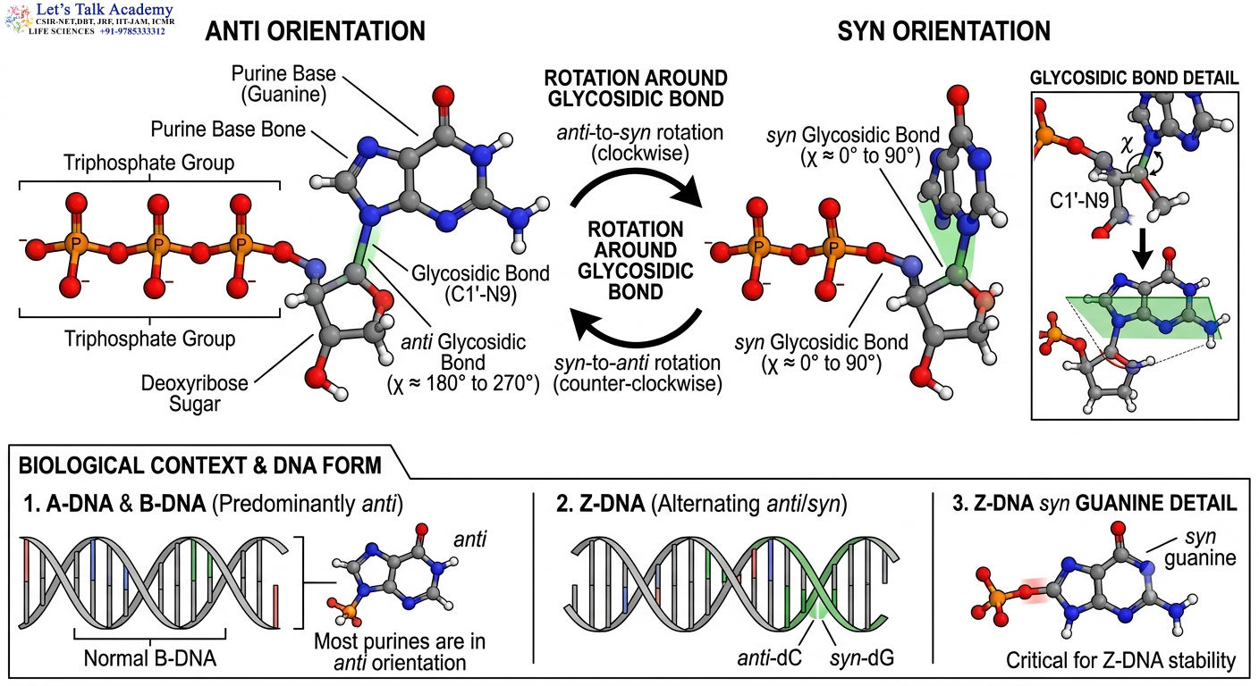
The nitrogenous base is attached to the sugar through a β-N-glycosidic bond.
Rotation around this bond determines how the base is positioned relative to the sugar.
Two major conformational states are called anti and syn.
4.1 Anti Conformation
In the anti conformation, the nitrogenous base is positioned away from the sugar.
A-DNA and B-DNA predominantly contain bases in the anti conformation.
This orientation is compatible with the regular arrangement of Watson–Crick base pairs within these right-handed helices.
4.2 Syn Conformation
In the syn conformation, the nitrogenous base rotates toward the sugar.
The syn orientation is uncommon in standard B-DNA but becomes particularly important in Z-DNA.
In canonical Z-DNA, purine residues such as guanine can adopt the syn conformation. Pyrimidines such as cytosine generally remain anti.
The alternation between syn and anti conformations is one of the key molecular changes associated with the B-to-Z transition.
5. A-DNA
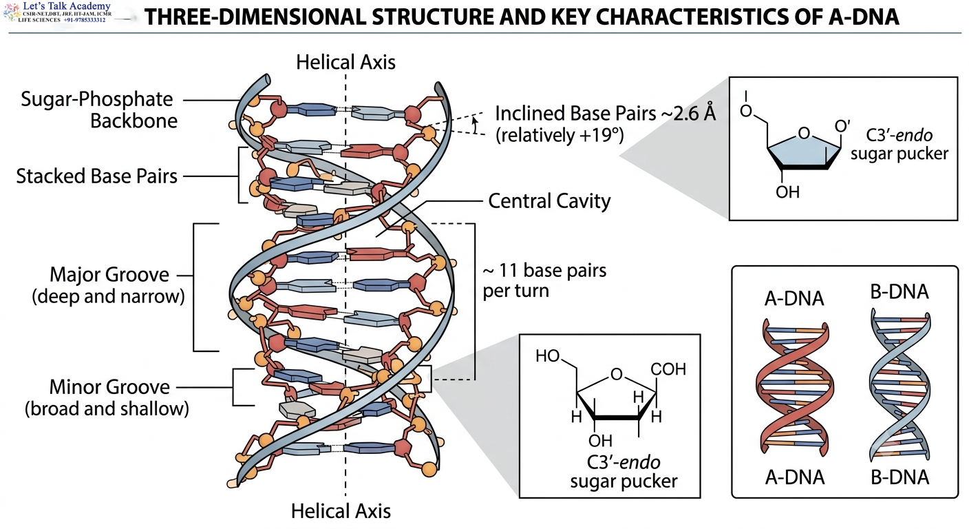
A-DNA is a right-handed double helix that differs from B-DNA in its overall dimensions and local molecular geometry.
It is shorter and wider than B-DNA and contains approximately 11 base pairs per turn.
A-DNA is generally associated with reduced hydration and is particularly important for understanding the structural properties of RNA-containing duplexes.
5.1 Overall Geometry of A-DNA
A-DNA contains two antiparallel strands wound around a common helical axis.
The sugar-phosphate backbone lies on the exterior, while the nitrogenous bases occupy the interior of the helix.
However, the base pairs in A-DNA are significantly inclined relative to the helical axis. This gives the molecule a broad and compact appearance.
The bases are also displaced away from the central axis compared with B-DNA, producing a central cavity along the axis of the helix.
This central cavity is one of the recognizable structural features of the A-form.
5.2 Helical Handedness of A-DNA
A-DNA is right-handed.
This means that A-DNA and B-DNA share the same general helical direction.
However, their structural dimensions are different because the sugar pucker, base-pair inclination and backbone geometry are different.
Thus, the terms “right-handed” and “left-handed” describe only one aspect of DNA structure and cannot by themselves completely identify a DNA conformation.
5.3 Number of Base Pairs per Turn
A-DNA contains approximately 11 base pairs per turn.
This is slightly higher than the approximately 10–10.5 base pairs per turn found in canonical B-DNA.
The higher number of base pairs per turn reflects the more compact geometry of A-DNA.
5.4 Rise per Base Pair
The rise per base pair in A-DNA is approximately 2.6 Å.
This is considerably smaller than the approximately 3.4 Å rise observed in canonical B-DNA.
The reduced rise means that successive base pairs are positioned closer together along the helical axis.
This contributes to the shorter pitch and compact longitudinal structure of A-DNA.
5.5 Helical Pitch of A-DNA
The helical pitch is the distance along the longitudinal axis corresponding to one complete turn of the helix.
A-DNA has a pitch of approximately 2.8–3.2 nm, depending on the structural model and sequence.
Because the number of base pairs per turn and the rise per base pair are both different from B-DNA, the overall pitch of A-DNA is also different.
5.6 Diameter of A-DNA
A-DNA is approximately 23–25 Å in diameter, making it wider than B-DNA.
The increased diameter results largely from the spatial arrangement of the base pairs and backbone.
The wider shape is an important visual and structural distinction between A-DNA and B-DNA.
5.7 Sugar Pucker of A-DNA
The predominant sugar-pucker conformation in A-DNA is C3′-endo.
This is one of the most useful molecular characteristics for distinguishing A-DNA from B-DNA.
The C3′-endo pucker changes the position of the phosphate backbone and brings successive base pairs closer together.
Therefore:
C3′-endo sugar pucker → A-form geometry
C2′-endo sugar pucker → B-form geometry
This relationship is not an isolated correlation. Sugar pucker affects the backbone torsion angles, base orientation and overall helical geometry.
5.8 Glycosidic Bond Orientation in A-DNA
The bases of A-DNA are predominantly in the anti conformation.
The anti orientation allows the bases to occupy the interior of the helix while forming normal Watson–Crick base pairs.
Therefore, both A-DNA and B-DNA generally have anti-oriented bases, even though their sugar conformations and helical geometries differ.
5.9 Base-Pair Inclination in A-DNA
A characteristic feature of A-DNA is the inclination of the base pairs.
The base pairs are tilted by approximately 20° relative to the helical axis in classical structural descriptions.
This is considerably different from B-DNA, in which the base pairs are approximately perpendicular to the helical axis.
The inclination contributes to the short, broad shape of A-DNA and helps create its characteristic groove dimensions.
5.10 Base Displacement in A-DNA
In A-DNA, the base pairs are displaced away from the central helical axis.
This produces a relatively prominent cavity along the central axis of the helix.
The bases are therefore positioned differently from those in B-DNA, where the base pairs occupy the central region of the helix more symmetrically.
5.11 Major Groove of A-DNA
The major groove of A-DNA is deep and narrow.
Because of the inclination and displacement of the base pairs, the chemical groups exposed in the major groove have a geometry different from that of B-DNA.
The narrower major groove can influence how proteins and small molecules interact with A-form DNA.
5.12 Minor Groove of A-DNA
The minor groove of A-DNA is broad and relatively shallow.
This is the opposite of the general groove relationship observed in B-DNA, where the major groove is wide and deep while the minor groove is narrower.
The difference in groove architecture changes the chemical surface available for molecular recognition.
5.13 Conditions Favoring A-DNA
A-DNA is favored by reduced hydration.
When water availability decreases, the energetic balance among different DNA conformations changes, and A-form geometry can become more favorable.
A-DNA can also be observed under certain conditions involving high salt concentrations or organic solvents.
The exact conditions required depend on the DNA sequence and molecular environment.
5.14 A-DNA and RNA
A-form geometry has particular importance in RNA biology.
RNA contains ribose rather than deoxyribose. Ribose has a 2′-OH group, which is absent from deoxyribose.
The presence of this additional hydroxyl group affects steric interactions and sugar conformation.
As a result, double-stranded RNA generally adopts an A-form-like helix.
DNA-RNA hybrid duplexes can also display A-form-like structural features.
Thus, A-DNA provides an important structural link between DNA chemistry and RNA structure.
6. B-DNA
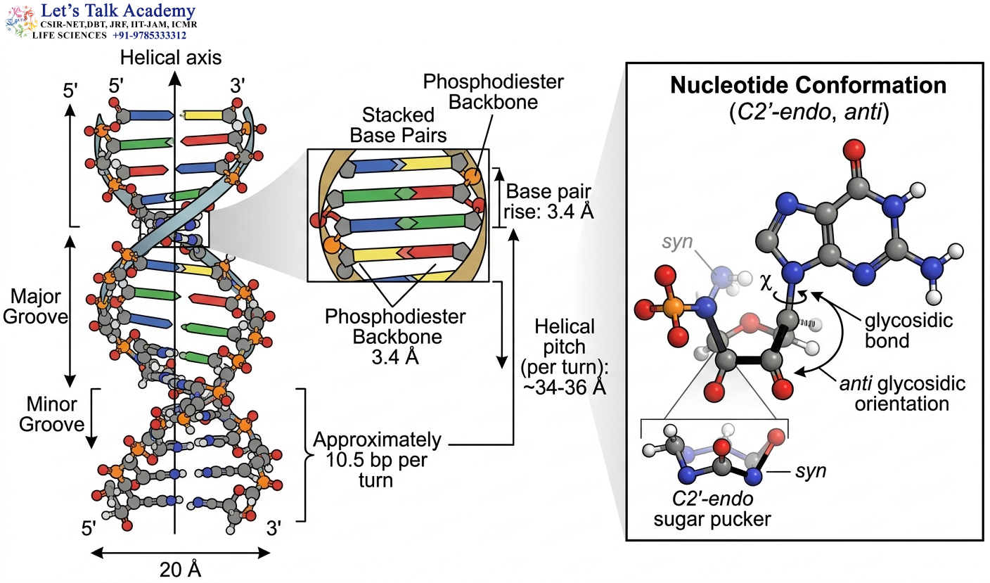
B-DNA is the classical form of the DNA double helix and is the structural form most commonly represented in biological discussions of genomic DNA.
It is a right-handed double helix consisting of two antiparallel strands connected through complementary Watson–Crick base pairs.
Under many aqueous physiological conditions, B-form geometry is favored.
6.1 General Architecture of B-DNA
The sugar-phosphate backbones are positioned on the exterior of the helix.
The nitrogenous bases are stacked inside the helix.
Adenine pairs with thymine, while guanine pairs with cytosine.
The bases are arranged approximately perpendicular to the helical axis in the idealized B-form.
This organization produces the familiar cylindrical double-helical appearance of DNA.
6.2 Helical Handedness
B-DNA is a right-handed helix.
The two strands wind around the central axis in a right-handed direction.
The right-handed geometry is compatible with the C2′-endo sugar pucker and anti glycosidic bond orientation characteristic of the B-form.
6.3 Number of Base Pairs per Turn
Canonical B-DNA contains approximately 10–10.5 base pairs per turn.
The frequently used value is approximately 10.5 base pairs per turn.
This means that a complete 360° rotation of the helix contains roughly 10 to 11 base pairs.
The exact helical twist is not identical throughout every DNA molecule. Sequence composition can cause local variations in helical twist.
6.4 Rise per Base Pair
The average rise between successive base pairs is approximately 3.4 Å.
Therefore, approximately 10.5 base pairs produce a helical pitch of roughly:
10.5 × 3.4 Å ≈ 35.7 Å
The commonly quoted pitch of B-DNA is approximately 34 Å or 3.4 nm, with the exact value depending on the structural model and sequence.
6.5 Diameter of B-DNA
The diameter of canonical B-DNA is approximately 20 Å or 2 nm.
This value is smaller than the diameter of A-DNA and larger than the diameter of Z-DNA.
The diameter is determined by the spatial arrangement of the bases and sugar-phosphate backbones around the helical axis.
6.6 Sugar Pucker
B-DNA predominantly contains the C2′-endo sugar pucker.
This geometry is one of the defining structural characteristics of B-DNA.
The C2′-endo conformation positions the sugar and phosphate backbone appropriately for the classical B-form helix.
6.7 Glycosidic Bond Orientation
The nitrogenous bases in B-DNA predominantly adopt the anti conformation.
The anti orientation positions the bases appropriately for Watson–Crick base pairing and stacking inside the helix.
6.8 Base-Pair Orientation
The base pairs of B-DNA are approximately perpendicular to the helical axis.
This differs from A-DNA, where the base pairs are significantly inclined.
The relatively perpendicular orientation of base pairs contributes to the familiar geometry of the B-form double helix.
7. Major and Minor Grooves of B-DNA
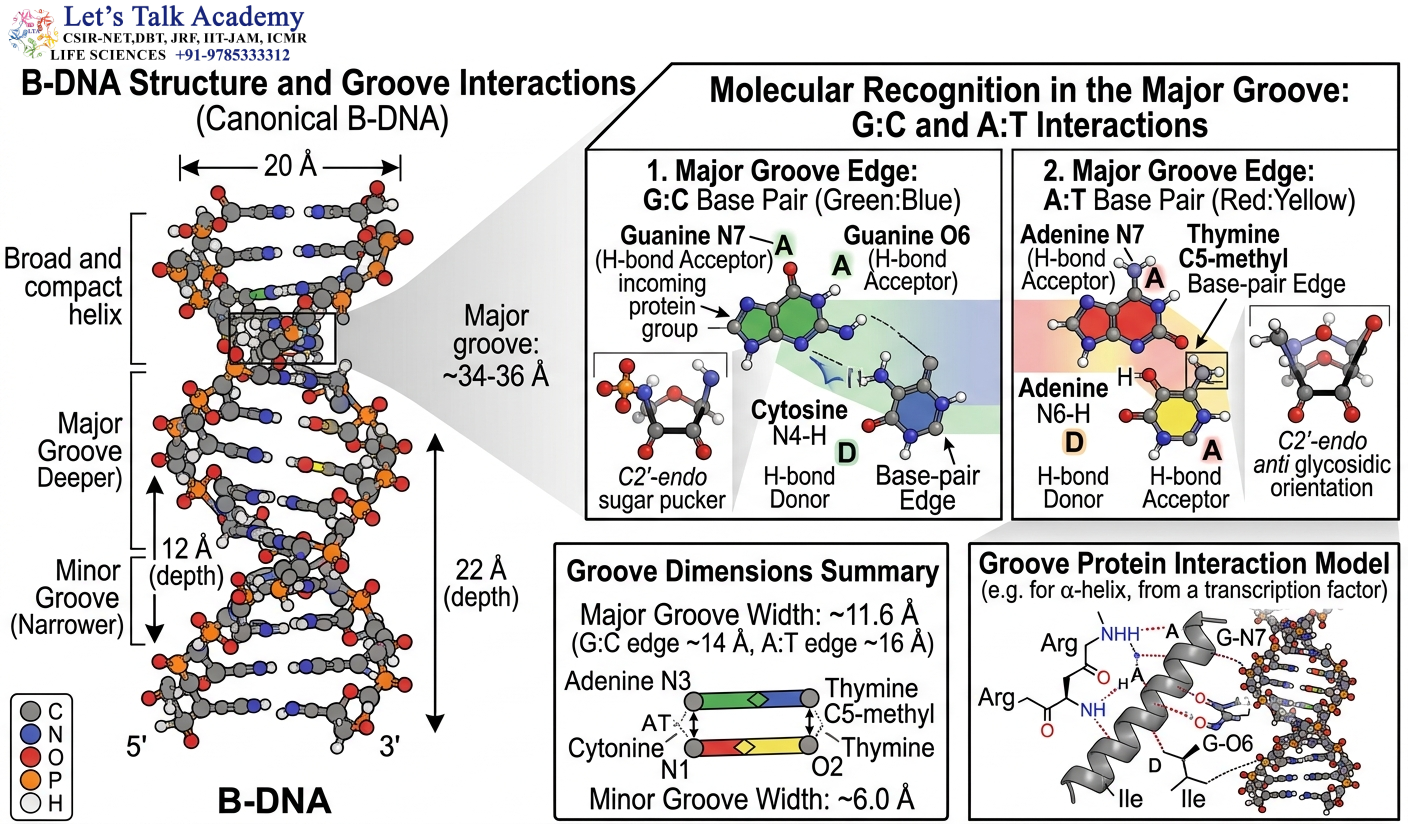
The two DNA strands do not divide the space around the helix equally.
Their attachment to the deoxyribose sugars creates two grooves:
Major groove
Minor groove
These grooves are among the most biologically important structural features of B-DNA because they provide surfaces through which proteins can recognize DNA.
7.1 Major Groove
The major groove is wide and relatively deep.
It exposes a large portion of the chemical information encoded by the edges of the base pairs.
Different base pairs present different arrangements of hydrogen-bond donors, hydrogen-bond acceptors and hydrophobic groups toward the major groove.
For this reason, many DNA-binding proteins can recognize specific DNA sequences without opening the double helix.
Sequence-specific transcription factors frequently use the major groove to identify their target DNA sequences.
7.2 Minor Groove
The minor groove is narrower than the major groove.
Although it exposes less sequence information than the major groove, it remains biologically important.
Proteins can recognize minor-groove width, curvature and electrostatic potential.
Some DNA-binding molecules and antibiotics bind preferentially in the minor groove.
The minor groove can therefore function as a structural recognition surface rather than merely being an empty space between the DNA strands.
7.3 Groove Width and DNA Recognition
Groove width can vary according to nucleotide sequence.
Certain sequences produce a narrower minor groove and stronger electrostatic potential.
DNA-binding proteins can recognize these structural differences through shape readout.
Therefore, DNA recognition can involve two complementary mechanisms:
Base readout: recognition of specific chemical groups associated with particular base pairs.
Shape readout: recognition of DNA curvature, groove width, electrostatic potential and flexibility.
8. Base Stacking in B-DNA
The nitrogenous bases are arranged in stacked layers inside the DNA helix.
Base stacking is a major contributor to DNA stability.
Adjacent aromatic bases interact through favorable van der Waals and hydrophobic interactions, along with electrostatic and dispersion-related effects.
The stability of a DNA sequence therefore depends not only on the number of hydrogen bonds but also on the identity and arrangement of neighboring bases.
This is why simply stating that G–C-rich DNA is always more stable because G–C contains three hydrogen bonds is an oversimplification.
Base stacking contributes substantially to the thermodynamics of DNA duplex formation.
9. B-DNA as a Dynamic Structure
Although B-DNA is commonly represented as a regular helix, actual DNA is not perfectly uniform.
The helical twist, roll, slide, rise and groove width can vary from one nucleotide step to another.
Sequence-dependent variations can produce:
- Local bending
- Curvature
- Groove narrowing
- Groove widening
- Altered flexibility
- Changes in base stacking
These variations are important for protein recognition and DNA packaging.
Thus, B-DNA should be viewed as a structural family rather than an absolutely rigid geometric object.
10. Z-DNA
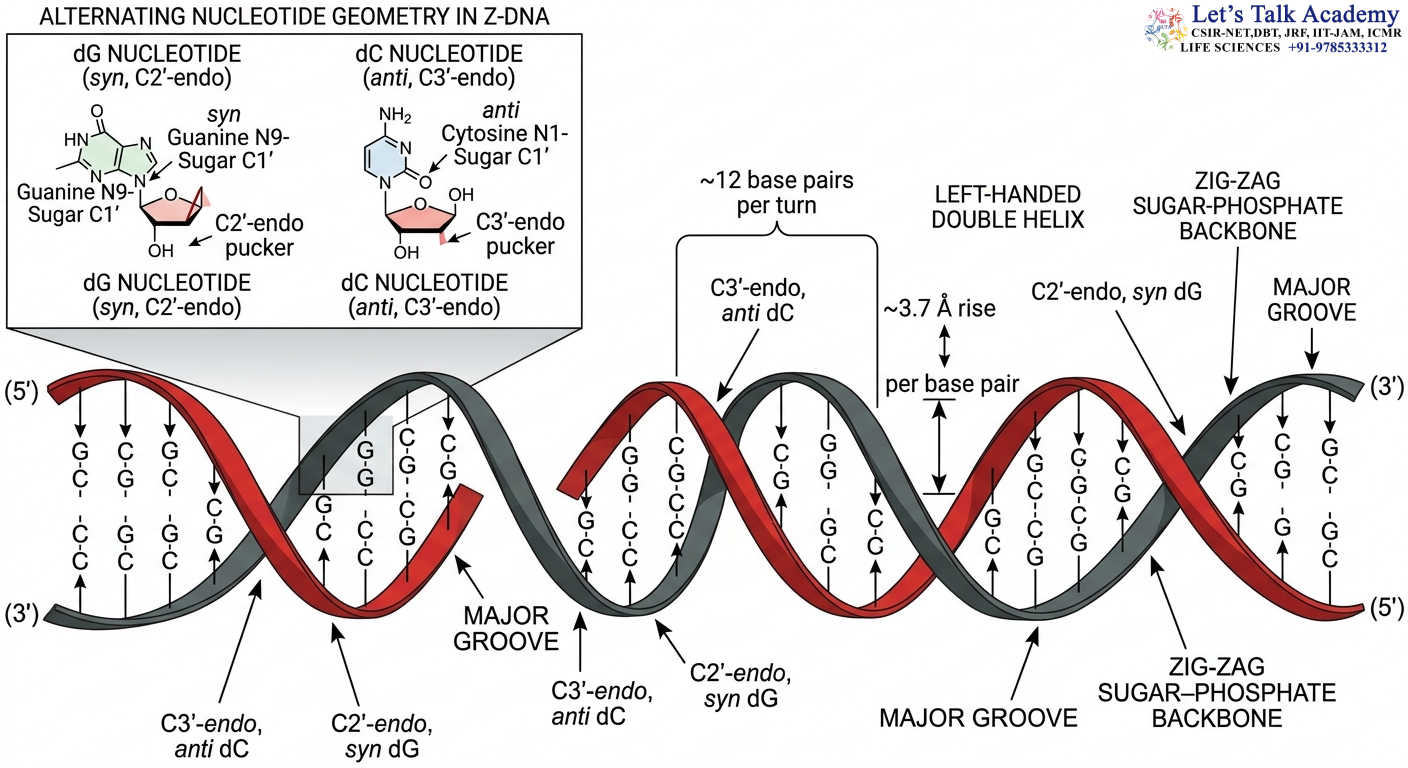
Z-DNA is a structurally distinctive form of DNA characterized by a left-handed double helix.
It is particularly associated with alternating purine-pyrimidine sequences and can be promoted by ionic conditions, negative supercoiling and other factors.
Its most visible structural characteristic is the zig-zag trajectory of the phosphate-sugar backbone.
10.1 Handedness
Z-DNA is left-handed.
This means that its overall helical direction is opposite to that of A-DNA and B-DNA.
The formation of a left-handed helix requires coordinated changes in nucleotide geometry rather than a simple rotation of the entire DNA molecule.
10.2 Number of Base Pairs per Turn
Z-DNA contains approximately 12 base pairs per turn.
Its structural repeat is approximately two base pairs.
This two-base-pair repeat arises because the two alternating types of nucleotide adopt different conformations.
10.3 Rise per Base Pair
The rise per base pair in Z-DNA is approximately 3.7 Å.
This is slightly greater than the rise of canonical B-DNA.
Combined with approximately 12 base pairs per turn, this gives a pitch of approximately 4.5 nm.
10.4 Diameter
Z-DNA has a diameter of approximately 18 Å.
It is therefore narrower than both A-DNA and B-DNA.
The narrow shape results from the distinctive arrangement of the backbone and bases.
11. Zig-Zag Backbone of Z-DNA
The name Z-DNA is derived from the zig-zag appearance of the phosphate-sugar backbone.
In B-DNA, the backbone follows a comparatively smooth helical trajectory.
In Z-DNA, alternating nucleotide conformations cause the backbone to move through a distinctive zig-zag path.
This geometry is produced by the combined effects of:
- Alternating sugar puckers
- Alternating glycosidic orientations
- Purine-pyrimidine sequence periodicity
- Changes in backbone torsion angles
Therefore, the zig-zag structure is a direct consequence of molecular geometry.
12. Anti and Syn Arrangement in Z-DNA
The anti/syn pattern is a central feature of Z-DNA.
In canonical alternating GC Z-DNA:
Cytosine → anti
Guanine → syn
This pattern alternates along the DNA chain.
The purine base can rotate around its glycosidic bond into the syn orientation, allowing the backbone to accommodate the left-handed structure.
This alternating orientation is fundamentally different from the predominantly anti arrangement found in A-DNA and B-DNA.
13. Sugar Pucker in Z-DNA
Z-DNA also displays alternating sugar geometry.
The purine and pyrimidine residues do not adopt identical sugar conformations.
This alternation is closely linked to the alternating anti and syn orientations of the bases.
The result is a repeated structural unit containing two different nucleotide geometries.
Therefore, Z-DNA can be understood as a structure in which sequence periodicity is translated directly into structural periodicity.
14. Groove Structure of Z-DNA
Z-DNA has an unusual groove architecture.
The conventional major groove is relatively flat or convex rather than resembling the wide, deep major groove of B-DNA.
The other groove is narrow and deep.
This unusual surface topology influences the interaction of proteins with Z-DNA.
A protein that recognizes B-DNA cannot necessarily recognize the same sequence in Z-form because the three-dimensional chemical landscape presented by the DNA has changed substantially.
15. Sequence Requirements for Z-DNA
Z-DNA formation is strongly dependent on nucleotide sequence.
Alternating purine-pyrimidine sequences are particularly favorable.
A classical example is:
5′-(GC)n-3′
where G and C alternate repeatedly.
Alternating sequences provide the structural periodicity required for the anti/syn pattern.
This sequence dependence is an important difference between Z-DNA and B-DNA.
B-DNA can occur across a broad range of sequences, whereas stable Z-DNA formation generally requires particular sequence and environmental conditions.
16. Ionic Environment and Z-DNA Formation
DNA contains negatively charged phosphate groups.
The phosphate groups repel one another because they carry similar negative charges.
Cations such as sodium, potassium, magnesium and other positively charged species can reduce this electrostatic repulsion through charge screening.
Under suitable conditions, increased ionic strength can therefore stabilize DNA conformations that would otherwise be energetically unfavorable.
Z-DNA formation can be promoted by ionic conditions, particularly in sequences that are intrinsically favorable for the Z-form.
The effect of ions is therefore both electrostatic and structural.
17. Negative Supercoiling and Z-DNA
DNA can become supercoiled when it experiences torsional stress.
Negative supercoiling introduces underwinding into DNA.
Certain alternating purine-pyrimidine sequences can respond to this torsional stress by undergoing a B-to-Z transition.
The transition from right-handed B-DNA to left-handed Z-DNA can absorb some of the torsional strain.
This provides a physical connection between DNA topology and alternative DNA structure.
18. B-to-Z DNA Transition
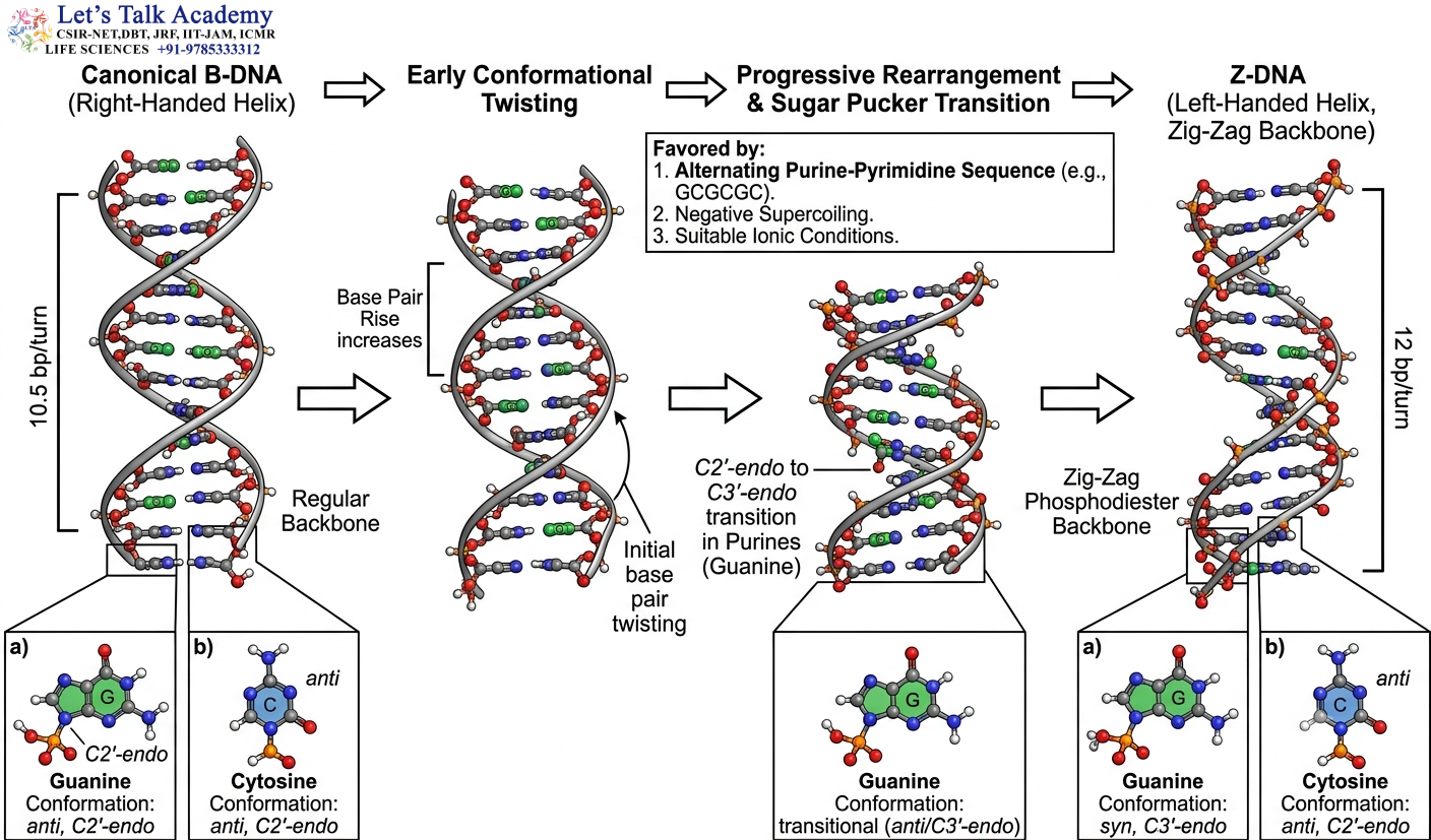
The B-to-Z transition is one of the most dramatic conformational changes known for DNA.
During this transition, the molecule changes from a right-handed helix to a left-handed helix.
The transition involves simultaneous changes in:
- Helical handedness
- Sugar pucker
- Glycosidic bond orientation
- Backbone torsion
- Groove geometry
- Base-pair arrangement
The change is therefore a coordinated molecular rearrangement.
18.1 Molecular Events During the Transition
In a typical alternating GC sequence, guanine changes from the anti orientation characteristic of B-DNA toward the syn orientation characteristic of Z-DNA.
The sugar conformation also changes.
These local changes alter the backbone trajectory.
As successive nucleotides undergo these changes, the entire segment develops the left-handed Z-DNA geometry.
18.2 Energetic Requirement
A conformational transition requires an energetic balance.
The B-form is usually favored under ordinary aqueous conditions.
However, the energetic cost of converting to Z-DNA can be reduced by:
- Alternating purine-pyrimidine sequences
- Negative supercoiling
- High ionic strength
- Cation binding
- Specific chemical modifications
- Binding of Z-DNA-recognizing proteins
When the factors favoring Z-DNA become sufficiently strong, the equilibrium can shift toward the Z-form.
19. B-Z Junction
A DNA molecule can contain B-DNA and Z-DNA segments simultaneously.
The boundary between them is called a B-Z junction.
The junction is structurally complex because the DNA must change from a right-handed geometry to a left-handed geometry.
Local base rearrangements can occur at the junction to accommodate the transition.
B-Z junctions demonstrate that different DNA conformations can coexist within the same DNA molecule.
This is important because DNA structure can vary locally rather than requiring the entire chromosome or DNA molecule to adopt one conformation.
20. Factors Controlling DNA Conformation
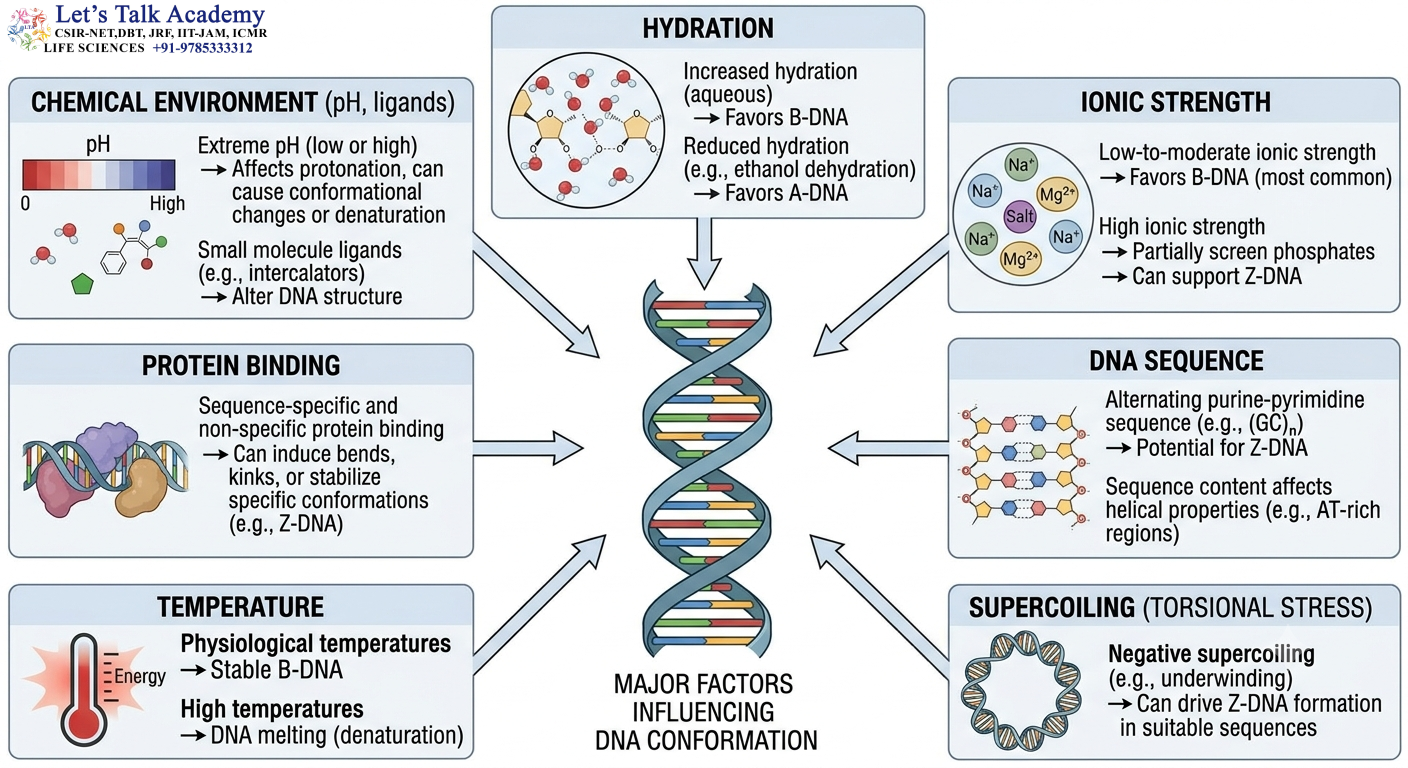
DNA conformation depends on multiple variables.
20.1 Hydration
Water molecules interact extensively with DNA.
High hydration generally favors B-form DNA under many conditions, whereas reduced hydration can promote A-form geometry.
Hydration affects the energetic balance of the backbone, bases and sugar residues.
20.2 Ionic Strength
Ions influence DNA by shielding the negative charges of phosphate groups.
Changes in ionic strength can therefore alter DNA stability and conformational preferences.
20.3 DNA Sequence
Different nucleotide sequences have different structural preferences.
Alternating GC sequences are particularly favorable for Z-DNA.
Other sequences may favor A-like or B-like local geometry.
20.4 Temperature
Temperature influences hydrogen bonding, base stacking and molecular motion.
Increasing temperature can destabilize the DNA duplex and eventually cause strand separation.
Before complete denaturation, temperature can also influence conformational equilibria.
20.5 Supercoiling
Supercoiling introduces torsional stress.
Positive and negative supercoiling can influence DNA twist and promote structural transitions.
20.6 Protein Binding
Proteins can bend, twist or otherwise deform DNA.
Some proteins specifically recognize alternative DNA conformations.
Protein binding can therefore shift the equilibrium between different structural states.
21. Detailed Comparison of A-DNA and B-DNA
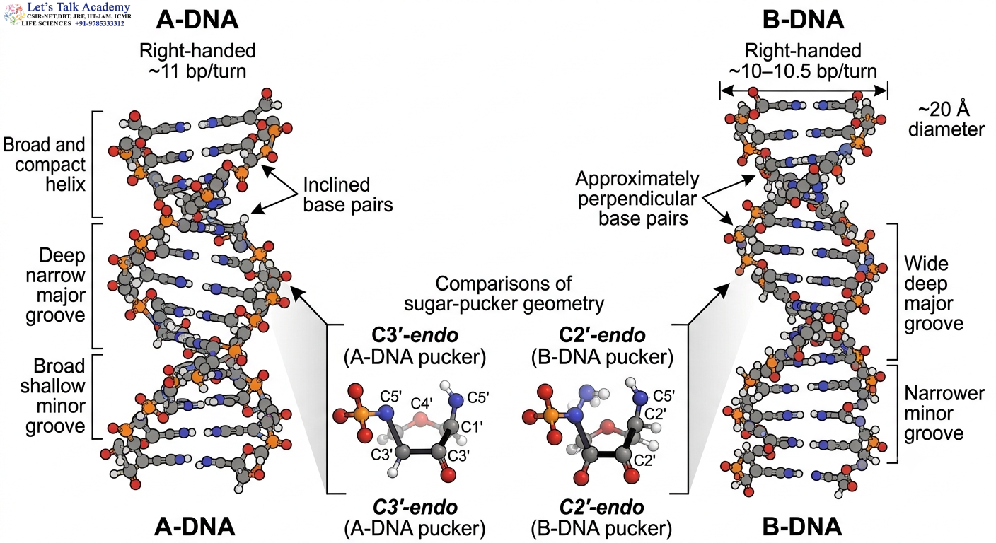
A-DNA and B-DNA are both right-handed helices, but their molecular architecture is significantly different.
A-DNA has a C3′-endo sugar pucker, while B-DNA predominantly has C2′-endo.
A-DNA contains approximately 11 base pairs per turn, while B-DNA contains approximately 10–10.5.
A-DNA has a smaller rise per base pair, making it more compact along the helical axis.
A-DNA is wider than B-DNA.
A-DNA has inclined base pairs, whereas B-DNA has approximately perpendicular base pairs.
A-DNA has a deep narrow major groove and broad shallow minor groove.
B-DNA has a wide deep major groove and a narrower minor groove.
These differences arise from changes in sugar geometry and backbone organization rather than from changes in the chemical identity of DNA.
22. Detailed Comparison of B-DNA and Z-DNA
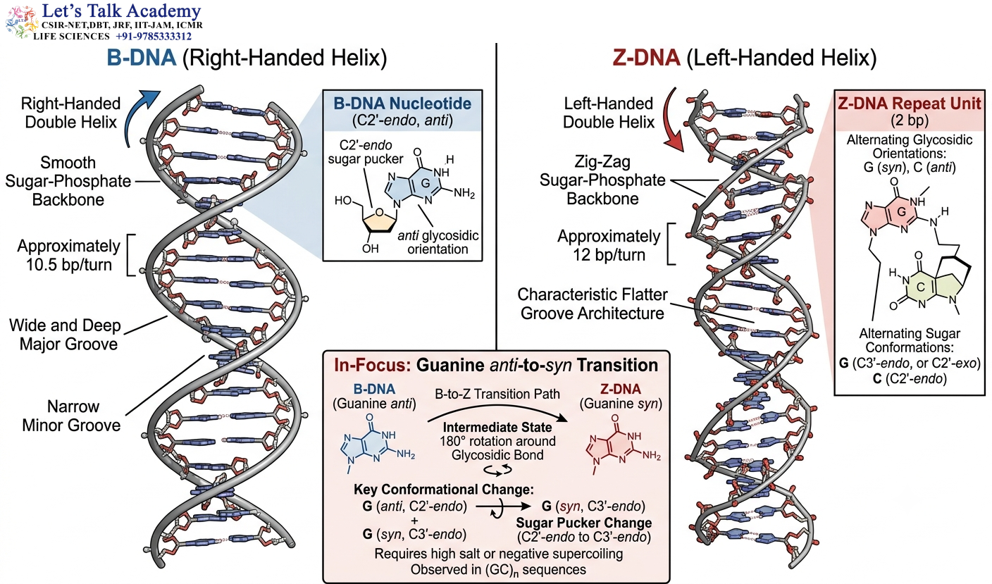
B-DNA is right-handed, whereas Z-DNA is left-handed.
B-DNA has a smooth helical backbone, whereas Z-DNA has a zig-zag backbone.
B-DNA predominantly contains anti-oriented bases, whereas canonical Z-DNA has alternating anti and syn orientations.
B-DNA has approximately 10–10.5 base pairs per turn, whereas Z-DNA has approximately 12.
B-DNA has a conventional wide major groove, whereas Z-DNA has a relatively flat or convex major-groove surface.
B-DNA is favored under many aqueous conditions, whereas Z-DNA requires particular sequence and environmental conditions for stable formation.
23. Detailed Comparison of A-DNA and Z-DNA
A-DNA and Z-DNA differ in both helical direction and molecular architecture.
A-DNA is right-handed, whereas Z-DNA is left-handed.
A-DNA predominantly uses C3′-endo sugar pucker and anti glycosidic orientation.
Z-DNA has alternating sugar geometry and alternating anti/syn glycosidic orientations.
A-DNA has approximately 11 base pairs per turn, whereas Z-DNA has approximately 12.
A-DNA has a broad structure with a central cavity, whereas Z-DNA is relatively narrow and has a characteristic zig-zag backbone.
24. Comprehensive Structural Comparison
Feature |
A-DNA |
B-DNA |
Z-DNA |
|---|---|---|---|
| Helical handedness | Right-handed | Right-handed | Left-handed |
| Approximate bp/turn | 11 | 10–10.5 | 12 |
| Diameter | ~23–25 Å | ~20 Å | ~18 Å |
| Rise/bp | ~2.6 Å | ~3.4 Å | ~3.7 Å |
| Pitch | ~2.8–3.2 nm | ~3.4 nm | ~4.5 nm |
| Sugar pucker | C3′-endo | C2′-endo | Alternating |
| Glycosidic orientation | Mainly anti | Mainly anti | Alternating anti/syn |
| Base-pair inclination | Significant | Low | Characteristic Z geometry |
| Backbone | Smooth right-handed | Smooth right-handed | Zig-zag |
| Major groove | Deep, narrow | Wide, deep | Flat/convex |
| Minor groove | Broad, shallow | Narrower | Narrow, deep |
| Structural repeat | 1 bp | 1 bp | 2 bp |
| Typical sequence preference | Variable | Broad | Alternating purine-pyrimidine |
| Hydration preference | Reduced hydration | Aqueous conditions | Environment-dependent |
| Major structural significance | RNA-like geometry | Classical DNA | Alternative left-handed DNA |
The values are approximate because DNA structural parameters vary with sequence, hydration, crystal environment and experimental conditions.
25. DNA Conformation and DNA Stability
DNA stability is determined by several forces acting simultaneously.
These include:
- Hydrogen bonding between complementary bases
- Base stacking
- Hydrophobic interactions
- Electrostatic interactions
- Solvent interactions
- Cation-mediated charge screening
- DNA topology
A conformational transition occurs when the energetic balance among these forces changes.
For example, reduced hydration can favor A-DNA, while negative supercoiling and appropriate sequence composition can favor Z-DNA.
Therefore, DNA conformation is closely connected with DNA thermodynamics.
26. DNA Conformation and Protein Recognition
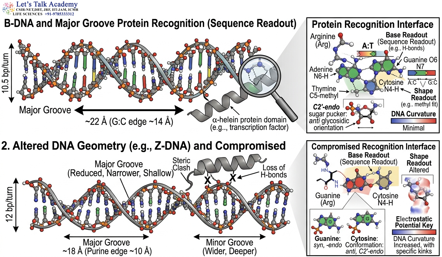
DNA-binding proteins interact with DNA through both sequence recognition and structural recognition.
A protein may recognize:
- Specific bases
- Major-groove chemical groups
- Minor-groove geometry
- DNA curvature
- Groove width
- Electrostatic potential
- Local flexibility
- Alternative DNA conformations
This explains why a change from B-DNA to Z-DNA can alter protein binding even though the nucleotide sequence itself has not changed.
The DNA molecule can therefore carry structural information in addition to its genetic sequence information.
27. DNA Conformation and Gene Regulation
DNA structure can influence accessibility to regulatory proteins.
Changes in local DNA shape can affect the ability of transcription factors to bind.
Z-DNA has been investigated in regions associated with transcription and torsional stress.
When RNA polymerase moves along DNA, it generates changes in DNA topology. Under suitable sequence conditions, these changes can promote alternative DNA conformations.
Therefore, DNA conformation can be connected to transcriptional regulation through the physical consequences of DNA torsional stress.
28. DNA Conformation and Replication
During DNA replication, the double helix must be locally unwound.
This process creates torsional stress in the surrounding DNA.
Topoisomerases help manage these changes in DNA topology.
The structural flexibility of DNA allows it to accommodate mechanical stress generated during replication.
Alternative conformations can therefore become relevant in regions experiencing unusual torsional conditions.
29. DNA Conformation and Transcription
Transcription involves movement of RNA polymerase along DNA.
The polymerase separates the DNA strands locally and changes the torsional state of DNA ahead of and behind the transcription complex.
This produces positive and negative supercoiling.
Under suitable sequence conditions, negative supercoiling can promote Z-DNA formation.
Therefore, transcription can influence DNA structure not only through protein binding but also through mechanical forces generated by the movement of the transcription machinery.
30. DNA Conformation and Genome Organization
DNA is packaged into higher-order structures in cells.
In eukaryotes, DNA interacts extensively with histones and other chromosomal proteins.
DNA bending and flexibility are essential for wrapping DNA around histone proteins.
Alternative conformations may also occur locally within chromosomes depending on sequence and molecular environment.
Thus, DNA conformation contributes to the three-dimensional organization of genetic material.
31. Relationship Between A-DNA and RNA
The comparison between A-DNA and RNA is structurally important.
RNA contains ribose, which has a 2′-OH group.
This hydroxyl group affects sugar pucker and steric interactions.
As a result, RNA duplexes generally adopt an A-form-like structure.
A-form geometry allows the 2′-OH group to be accommodated without the steric constraints that would arise in a conventional B-form duplex.
Therefore, the difference between ribose and deoxyribose has a direct effect on nucleic-acid structure.
32. Structural Polymorphism of DNA
A-DNA, B-DNA and Z-DNA demonstrate that DNA is conformationally polymorphic.
The same chemical polymer can change its three-dimensional architecture without changing its nucleotide sequence.
This property is important because biological molecules operate in constantly changing environments.
DNA is exposed to:
- Changes in hydration
- Changes in ion concentration
- Mechanical stress
- Protein binding
- Chemical modification
- Temperature changes
- Transcriptional activity
- Replication-associated stress
These conditions can alter the structural equilibrium of DNA.
33. Experimental Study of DNA Conformation
Different techniques are used to study DNA structure.
33.1 X-Ray Crystallography
X-ray crystallography can provide high-resolution structural information.
It can reveal:
- Base-pair geometry
- Sugar pucker
- Glycosidic orientation
- Helical handedness
- Groove architecture
- Backbone arrangement
- Ion binding
- Hydrogen-bonding patterns
Many fundamental structural parameters of DNA were established through crystallographic studies.
33.2 Nuclear Magnetic Resonance
NMR spectroscopy is useful for studying DNA in solution.
It can provide information about:
- Local conformation
- Sugar pucker
- Base orientation
- Hydrogen bonding
- Dynamics
- Structural transitions
NMR is particularly valuable because DNA can behave differently in solution compared with crystalline conditions.
33.3 Circular Dichroism
Circular dichroism spectroscopy is widely used to monitor DNA conformation.
Different DNA forms generate different CD spectral patterns because the electronic properties of stacked bases depend on their three-dimensional arrangement.
Changes in CD spectra can therefore indicate conformational transitions.
33.4 Molecular Modeling
Computational methods can be used to investigate DNA structure at atomic resolution.
Molecular dynamics simulations can examine:
- Base stacking
- Hydrogen bonding
- Groove width
- Backbone flexibility
- Ion interactions
- Conformational transitions
Computational studies complement experimental structural methods.
34. Structural Relationships That Explain the Three DNA Forms
The structural features of A-DNA, B-DNA and Z-DNA are interconnected.
A-DNA
C3′-endo sugar pucker
↓
Changes backbone geometry
↓
Base pairs become inclined
↓
Rise per base pair decreases
↓
Helix becomes shorter and wider
B-DNA
C2′-endo sugar pucker
↓
Classical backbone geometry
↓
Base pairs approximately perpendicular
↓
~3.4 Å rise per base pair
↓
Classical right-handed double helix
Z-DNA
Alternating sugar geometry
↓
Alternating anti/syn orientation
↓
Zig-zag backbone
↓
Left-handed helical geometry
↓
Characteristic two-base-pair repeating structure
These relationships demonstrate why structural parameters should be understood as a connected system rather than memorized as independent facts.


