
1. Introduction to Apoptosis
1.1 Definition of Apoptosis
Apoptosis is a genetically regulated and highly coordinated form of programmed cell death in which a cell actively dismantles itself and is subsequently removed without causing extensive damage to surrounding tissues.
The process involves a carefully controlled sequence of molecular events. These include activation of specific proteolytic enzymes called caspases, changes in mitochondrial function, degradation of nuclear and cytoplasmic proteins, fragmentation of DNA, membrane remodeling, formation of apoptotic bodies, and recognition of the dying cell by phagocytic cells.
Apoptosis is fundamentally different from accidental cellular destruction because it is controlled by an intracellular molecular program.
The simplified sequence is:
Death signal → Apoptotic signaling → Caspase activation → Cellular dismantling → Apoptotic-body formation → Phagocytic removal
1.2 Historical Background
The morphological features of apoptosis were systematically described in the early 1970s by John Kerr, Andrew Wyllie, and Alastair Currie.
They distinguished this form of cell death from necrosis based on characteristic structural changes such as cell shrinkage, chromatin condensation, nuclear fragmentation, and formation of membrane-bound apoptotic bodies.
The term apoptosis was derived from a Greek expression associated with the falling away of leaves from a tree, emphasizing the natural and controlled removal of cells.
1.3 Concept of Programmed Cell Death
Programmed cell death refers to genetically controlled cellular processes that lead to cell elimination.
Apoptosis is the classical example of programmed cell death, although modern cell biology recognizes several distinct regulated cell-death pathways.
The important principle is that cells are not simply passive victims of death signals. They contain molecular machinery capable of actively executing their own destruction.
1.4 Importance of Apoptosis
Apoptosis is required for:
- Embryonic development
- Tissue remodeling
- Maintenance of cell numbers
- Elimination of damaged cells
- Removal of potentially cancerous cells
- Immune-system regulation
- Elimination of unnecessary cells
- Maintenance of tissue homeostasis
Thus, apoptosis can be considered a major cellular quality-control mechanism.
2. Characteristics of Apoptosis
Apoptosis produces a characteristic series of morphological and biochemical changes.
2.1 Cell Shrinkage
One of the earliest morphological changes is reduction in cell volume.
The cytoplasm becomes more compact, and the cell separates from neighboring cells.
This contrasts with many forms of necrotic injury, where cells frequently undergo swelling.
2.2 Chromatin Condensation
Chromatin becomes highly condensed.
The condensed chromatin often accumulates near the nuclear membrane.
This process is called pyknosis.
2.3 Nuclear Fragmentation
The condensed nucleus subsequently breaks into several fragments.
This process is called karyorrhexis.
Nuclear fragmentation is one of the characteristic morphological features of apoptosis.
2.4 Membrane Blebbing
The plasma membrane develops outward protrusions known as membrane blebs.
Cytoskeletal rearrangement and changes in membrane-associated proteins contribute to this process.
2.5 Formation of Apoptotic Bodies
Eventually, portions of the cell become separated into small membrane-bound structures called apoptotic bodies.
These may contain:
- Cytoplasmic components
- Organelles
- Nuclear fragments
- Cellular proteins
Apoptotic bodies are subsequently recognized and engulfed.
2.6 Phosphatidylserine Exposure
During apoptosis, phosphatidylserine becomes exposed on the external surface of the plasma membrane.
Phosphatidylserine functions as an important “eat-me” signal.
Phagocytic cells recognize this signal and remove the apoptotic cell.
3. Apoptosis and Other Forms of Cell Death
3.1 Apoptosis vs Necrosis
| Feature | Apoptosis | Necrosis |
|---|---|---|
| Regulation | Highly regulated | Often associated with uncontrolled injury |
| Cell size | Usually decreases | Often increases |
| Membrane | Relatively preserved initially | Frequently disrupted |
| DNA | Fragmentation and condensation | Variable degradation |
| Cellular contents | Mostly contained | Often released |
| Inflammation | Usually limited | Often prominent |
| Physiological role | Very important | Usually pathological |
3.2 Apoptosis vs Necroptosis
Necroptosis is a regulated form of cell death that displays several morphological characteristics associated with necrosis.
Unlike classical apoptosis, necroptosis generally involves loss of plasma-membrane integrity.
Important regulatory proteins include:
- RIPK1
- RIPK3
- MLKL
Necroptosis can become particularly relevant when apoptotic signaling is blocked under certain cellular conditions.
3.3 Apoptosis vs Pyroptosis
Pyroptosis is a regulated inflammatory form of cell death.
It is strongly associated with:
- Caspase-1
- Gasdermin proteins
- Inflammasome signaling
- Inflammatory cytokines
Unlike classical apoptosis, pyroptosis generally results in formation of membrane pores and strong inflammatory signaling.
3.4 Apoptosis vs Autophagy-Associated Cell Death
Autophagy is primarily a cellular recycling and survival process.
Cells use autophagy to degrade damaged organelles and recycle cellular components.
Autophagy can interact with apoptosis, but autophagy itself should not automatically be considered a form of cell death.
Under particular circumstances, excessive or dysregulated autophagic processes may contribute to cell death.
4. Molecular Machinery of Apoptosis
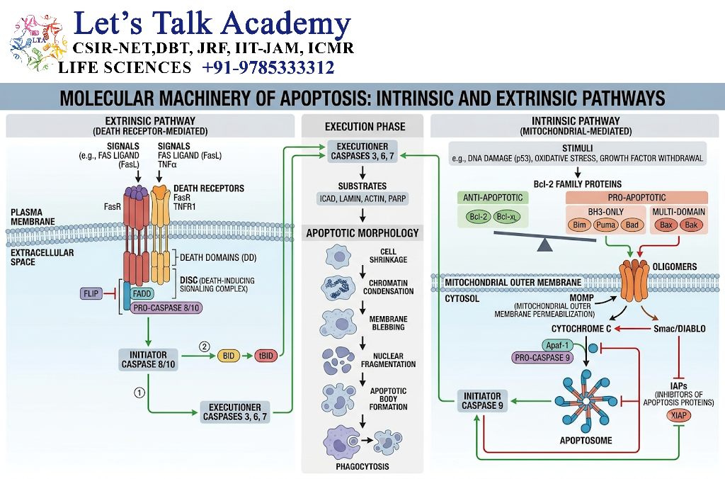
4.1 Caspases
Caspases are cysteine-dependent proteases that cleave specific protein substrates after aspartate residues.
They are central to the execution of apoptosis.
Caspases are initially synthesized as inactive precursors called procaspases.
4.2 Procaspases
Procaspases contain regulatory regions that allow them to be recruited to signaling complexes.
Activation involves molecular rearrangement and proteolytic processing that produces active caspase enzymes.
4.3 Initiator Caspases
Initiator caspases activate downstream apoptotic machinery.
Important examples include:
- Caspase-8
- Caspase-9
- Caspase-10
- Caspase-2
Caspase-8 and caspase-10 are mainly associated with extrinsic signaling, whereas caspase-9 is the major initiator of the mitochondrial pathway.
4.4 Executioner Caspases
The major executioner caspases include:
- Caspase-3
- Caspase-6
- Caspase-7
Once activated, these enzymes cleave numerous cellular substrates.
This produces:
Protein degradation → Cytoskeletal disruption → Nuclear dismantling → Apoptotic morphology
4.5 Caspase Activation Cascade
The caspase cascade can be represented as:
Apoptotic stimulus
↓
Initiator caspase activation
↓
Executioner caspase activation
↓
Cleavage of cellular substrates
↓
Cellular dismantling
↓
Apoptosis
5. Intrinsic Pathway of Apoptosis
5.1 Mitochondrial Pathway
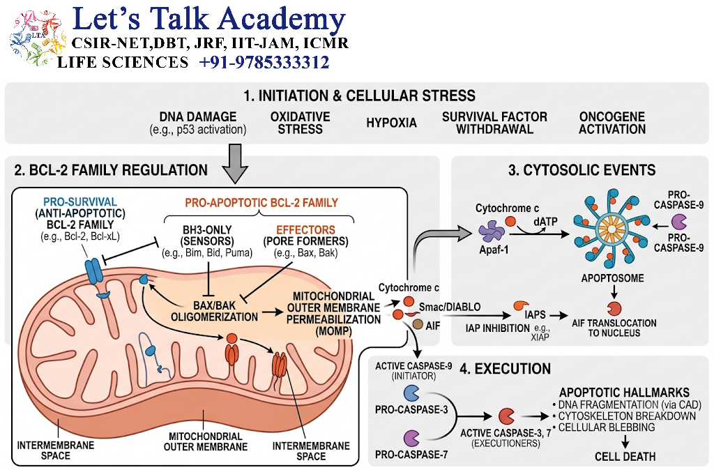
The intrinsic pathway is also called the mitochondrial pathway.
It is activated by intracellular disturbances rather than direct activation of an external death receptor.
Major triggers include:
- DNA damage
- Growth-factor withdrawal
- Oxidative stress
- Endoplasmic-reticulum stress
- Oncogenic stress
- Severe metabolic stress
5.2 Cellular Stress Signals
A cell continuously monitors its internal environment.
When stress becomes severe, pro-survival mechanisms may become insufficient.
The balance shifts toward pro-apoptotic signaling.
5.3 Mitochondrial Outer Membrane Permeabilization
The critical event of the mitochondrial pathway is mitochondrial outer membrane permeabilization (MOMP).
MOMP allows proteins from the mitochondrial intermembrane space to enter the cytosol.
The release of cytochrome c is particularly important.
5.4 Cytochrome c Release
Cytochrome c normally participates in mitochondrial electron transport.
After MOMP, it is released into the cytoplasm.
There it performs a completely different function by participating in apoptosome formation.
5.5 Apaf-1
Apaf-1, or apoptotic protease activating factor 1, is a central component of the intrinsic apoptotic pathway.
Cytochrome c binds Apaf-1 and promotes its conformational activation.
5.6 Apoptosome Formation
Cytochrome c and Apaf-1 participate in formation of the apoptosome.
The apoptosome recruits procaspase-9.
This provides a platform for caspase-9 activation.
5.7 Caspase-9 Activation
Activated caspase-9 acts as an initiator caspase.
It subsequently activates executioner caspases, particularly:
- Caspase-3
- Caspase-7
5.8 Activation of Executioner Caspases
Executioner caspases cleave hundreds of cellular proteins.
This leads to:
- Cytoskeletal collapse
- Nuclear changes
- DNA fragmentation
- Membrane remodeling
- Formation of apoptotic bodies
Intrinsic Pathway Flowchart
Cellular stress
↓
Bcl-2 family regulation
↓
Bax/Bak activation
↓
MOMP
↓
Cytochrome c release
↓
Apaf-1 activation
↓
Apoptosome formation
↓
Caspase-9
↓
Caspase-3/7
↓
Cellular dismantling
↓
Apoptosis
6. Bcl-2 Family of Proteins
6.1 Anti-Apoptotic Bcl-2 Proteins
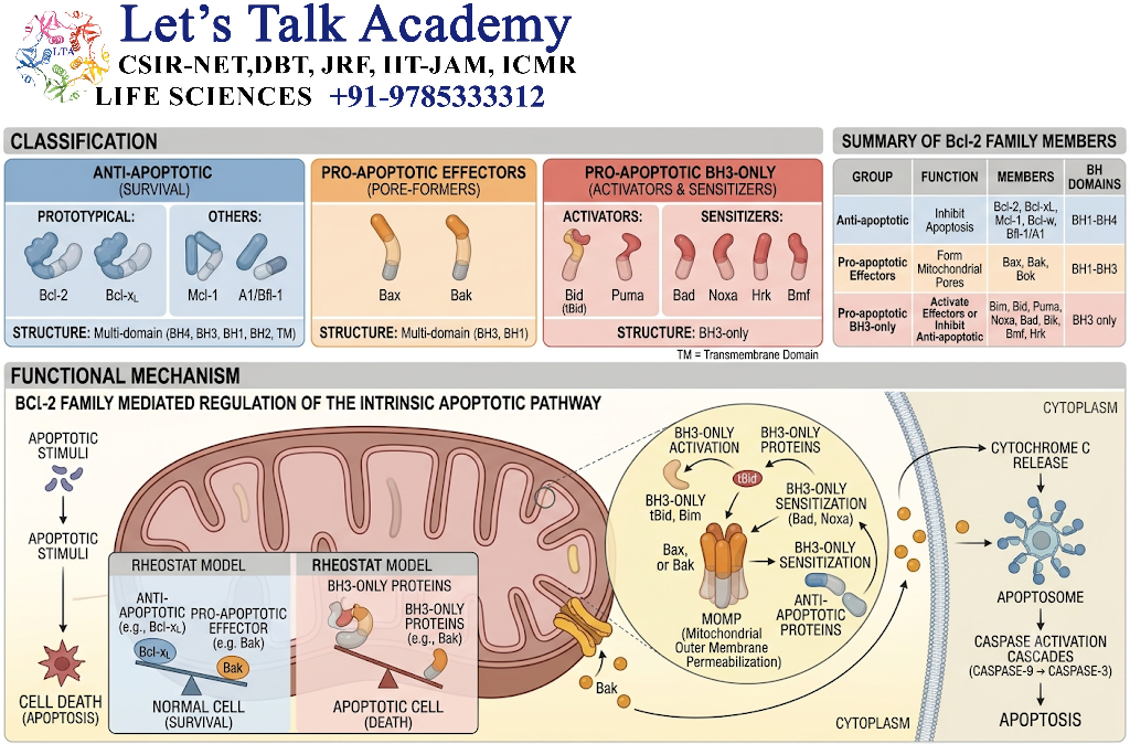
Anti-apoptotic proteins help preserve mitochondrial integrity.
Examples include:
- Bcl-2
- Bcl-xL
- Mcl-1
They suppress mitochondrial permeabilization by opposing pro-apoptotic proteins.
6.2 Pro-Apoptotic Multidomain Proteins
Important multidomain pro-apoptotic proteins include:
- Bax
- Bak
When activated, these proteins participate in formation of membrane structures that promote MOMP.
6.3 BH3-Only Proteins
BH3-only proteins are important sensors and regulators of cellular stress.
Examples include:
- Bim
- Bad
- Bid
- Puma
- Noxa
- Bmf
They can activate Bax/Bak or neutralize anti-apoptotic Bcl-2 proteins.
6.4 Bax and Bak
Bax and Bak are central effectors of mitochondrial outer membrane permeabilization.
Their activation promotes mitochondrial membrane permeabilization and release of apoptogenic factors.
6.5 Regulation of Mitochondrial Membrane Permeability
The mitochondrial apoptotic decision depends on the balance among:
Anti-apoptotic proteins ↔ Pro-apoptotic proteins
When pro-apoptotic activity dominates:
Bax/Bak activation → MOMP → Cytochrome c release
6.6 Balance Between Survival and Death Signals
The Bcl-2 family operates like a molecular decision-making network.
Strong survival signaling → Mitochondrial integrity maintained
Strong death signaling → MOMP → Apoptosis
7. Extrinsic Pathway of Apoptosis
7.1 Death Receptors
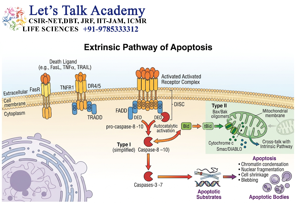
The extrinsic pathway begins with extracellular death signals.
Death receptors belong primarily to the tumor necrosis factor receptor superfamily.
Examples include:
- Fas/CD95
- TNFR family receptors
- TRAIL receptors
7.2 Fas/Fas Ligand Pathway
Fas ligand binds to Fas receptors on target cells.
This promotes receptor clustering and recruitment of intracellular signaling proteins.
The pathway is particularly important in immune-mediated elimination of unwanted cells.
7.3 TNF Receptor Pathway
Tumor necrosis factor can bind TNF receptors and activate signaling pathways that may promote survival, inflammation, or cell death depending on cellular context and signaling configuration.
Thus, TNF signaling is more complex than a simple apoptosis switch.
7.4 TRAIL Receptors
TRAIL, or TNF-related apoptosis-inducing ligand, can activate specific death receptors and initiate apoptotic signaling.
TRAIL receptors are studied extensively in relation to selective elimination of abnormal cells.
7.5 FADD
FADD, or Fas-associated protein with death domain, is an adaptor protein involved in death-receptor signaling.
FADD connects activated receptors to downstream caspase activation machinery.
7.6 DISC Formation
The death-inducing signaling complex (DISC) forms following activation of certain death receptors.
The DISC recruits procaspase-8 and/or procaspase-10.
7.7 Caspase-8 and Caspase-10 Activation
Once activated, caspase-8 or caspase-10 can:
- Activate executioner caspases directly
- Amplify mitochondrial apoptosis through Bid
Extrinsic Pathway Flowchart
Death ligand
↓
Death receptor
↓
Receptor clustering
↓
FADD recruitment
↓
DISC formation
↓
Caspase-8/10 activation
↓
Executioner caspases
↓
Apoptosis
8. Crosstalk Between Apoptotic Pathways
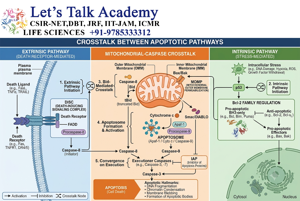
8.1 Caspase-8 and Bid
Activated caspase-8 can cleave Bid, a BH3-only Bcl-2 family protein.
8.2 tBid Formation
Caspase-8 converts Bid into truncated Bid (tBid).
tBid can promote mitochondrial apoptotic signaling.
8.3 Connection with Mitochondrial Pathway
The pathway becomes:
Death receptor → Caspase-8 → Bid → tBid → Bax/Bak → MOMP
Thus, an extracellular signal can activate the mitochondrial pathway.
8.4 Amplification of Apoptotic Signaling
This crosstalk can amplify the apoptotic response.
Therefore, intrinsic and extrinsic apoptosis should not be viewed as completely isolated pathways.
9. Execution Phase of Apoptosis
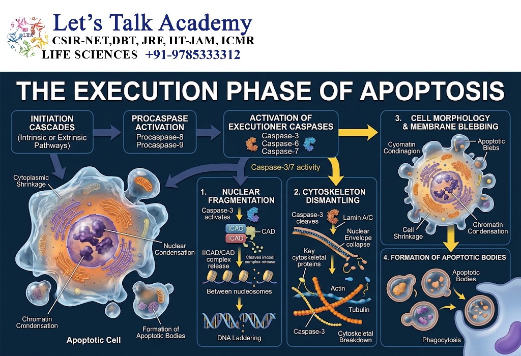
9.1 Caspase-3 Activation
Caspase-3 is one of the major executioner caspases.
Its activation produces extensive cleavage of cellular substrates.
9.2 Caspase-6 Activation
Caspase-6 participates in cleavage of specific nuclear and cytoskeletal substrates.
9.3 Caspase-7 Activation
Caspase-7 has overlapping substrate specificity with caspase-3 and contributes to cellular dismantling.
9.4 Cleavage of Cellular Proteins
Executioner caspases cleave proteins involved in:
- Cytoskeleton
- Nuclear structure
- DNA repair
- Cell adhesion
- Cellular signaling
9.5 Cytoskeletal Disruption
Caspase-mediated cleavage of cytoskeletal and associated proteins contributes to:
- Cell shrinkage
- Membrane blebbing
- Cellular fragmentation
9.6 Nuclear Dismantling
Nuclear proteins are cleaved and chromatin undergoes condensation.
The nucleus eventually fragments into smaller structures.
10. DNA Fragmentation During Apoptosis
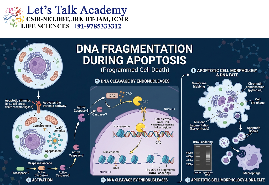
10.1 CAD
CAD, or caspase-activated DNase, is an endonuclease responsible for much of the characteristic DNA fragmentation observed during apoptosis.
10.2 ICAD
CAD is normally associated with an inhibitor called ICAD.
ICAD prevents CAD from acting under normal conditions.
10.3 Caspase-Mediated ICAD Cleavage
Executioner caspases cleave ICAD.
This removes the inhibition imposed on CAD.
10.4 CAD Activation
Once released from ICAD inhibition, CAD can access chromosomal DNA.
10.5 Chromosomal DNA Fragmentation
CAD cleaves DNA at regions between nucleosomes, producing characteristic DNA fragments.
The simplified mechanism is:
Caspase-3 → ICAD cleavage → CAD activation → DNA cleavage
11. Morphological Changes During Apoptosis
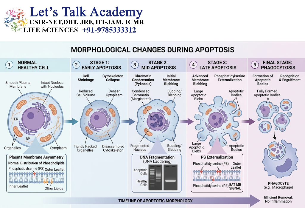
11.1 Pyknosis
Pyknosis refers to condensation of nuclear chromatin.
11.2 Karyorrhexis
Karyorrhexis refers to fragmentation of the nucleus.
11.3 Membrane Blebbing
The plasma membrane forms blebs due to cytoskeletal and membrane-associated changes.
11.4 Apoptotic-Body Formation
The cell divides into membrane-bound apoptotic bodies.
11.5 Cellular Fragmentation
Cellular components become packaged into fragments that can be efficiently removed by phagocytes.
12. Recognition and Removal of Apoptotic Cells
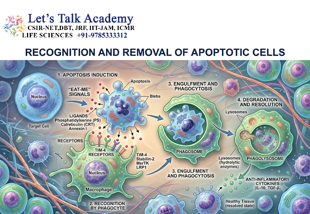
12.1 “Eat-Me” Signals
Apoptotic cells expose molecular signals that tell surrounding cells and phagocytes that they should be removed.
12.2 Phosphatidylserine
Phosphatidylserine is one of the best-characterized apoptotic recognition signals.
It becomes exposed on the external surface of the plasma membrane.
12.3 Recognition by Phagocytes
Macrophages and other phagocytes express receptors and use bridging molecules to recognize apoptotic cells.
12.4 Engulfment
After recognition, the phagocyte surrounds and internalizes the apoptotic cell or apoptotic bodies.
12.5 Degradation of Apoptotic Bodies
The engulfed material is delivered to intracellular degradative compartments where cellular components are broken down.
12.6 Efferocytosis
The process of recognizing and clearing apoptotic cells is called efferocytosis.
It is important for maintaining tissue homeostasis and preventing prolonged exposure of dying-cell material.
13. Regulation of Apoptosis
13.1 Regulation by Bcl-2 Family
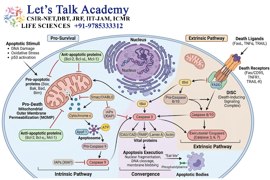
Bcl-2 family proteins regulate mitochondrial integrity and therefore strongly influence intrinsic apoptosis.
13.2 Regulation by IAP Proteins
Inhibitor of apoptosis proteins (IAPs) can suppress caspase activity.
Important members include:
- XIAP
- c-IAP1
- c-IAP2
13.3 SMAC/DIABLO
Mitochondria can release SMAC/DIABLO, which counteracts certain IAP-mediated inhibition of caspases.
This helps promote efficient apoptotic execution.
13.4 Survival Signaling
Cells receive survival signals from:
- Growth factors
- Hormones
- Extracellular matrix
- Neighboring cells
Loss of important survival signals can promote apoptosis.
13.5 PI3K-AKT Pathway
The PI3K-AKT pathway is an important survival signaling pathway.
AKT can promote cell survival by regulating several proteins involved in apoptosis and metabolism.
13.6 Growth-Factor Signaling
Growth factors activate intracellular pathways that generally favor cell survival and proliferation.
Withdrawal of essential growth signals can shift the balance toward apoptosis.
14. p53 and Apoptosis
14.1 DNA Damage and p53
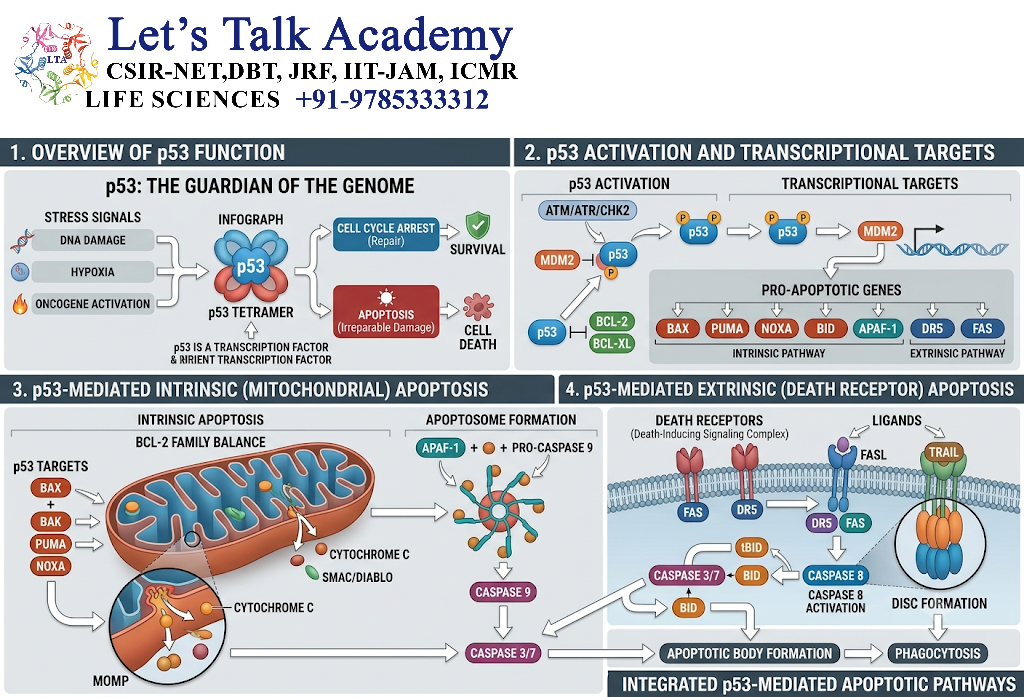
DNA damage activates signaling pathways that can stabilize and activate p53.
p53 then determines whether the cell should:
- Pause the cell cycle
- Repair DNA
- Enter senescence
- Undergo apoptosis
14.2 p53 Stabilization
Under normal conditions, p53 is maintained at relatively low levels through regulated degradation.
DNA damage can interfere with this degradation and promote p53 accumulation.
14.3 p53-Dependent Cell-Cycle Arrest
p53 can induce expression of p21, a CDK inhibitor.
The pathway is:
DNA damage → p53 → p21 → CDK inhibition → Cell-cycle arrest
14.4 PUMA and NOXA
When damage is severe, p53 can increase expression of pro-apoptotic proteins such as:
- PUMA
- NOXA
These proteins influence the Bcl-2 family network and promote mitochondrial apoptosis.
14.5 p53-Mediated Mitochondrial Apoptosis
The pathway can be summarized as:
DNA damage → p53 activation → Pro-apoptotic gene expression → Bax/Bak activation → MOMP → Caspases → Apoptosis
15. Apoptosis and Cell-Cycle Regulation
15.1 Cell-Cycle Arrest
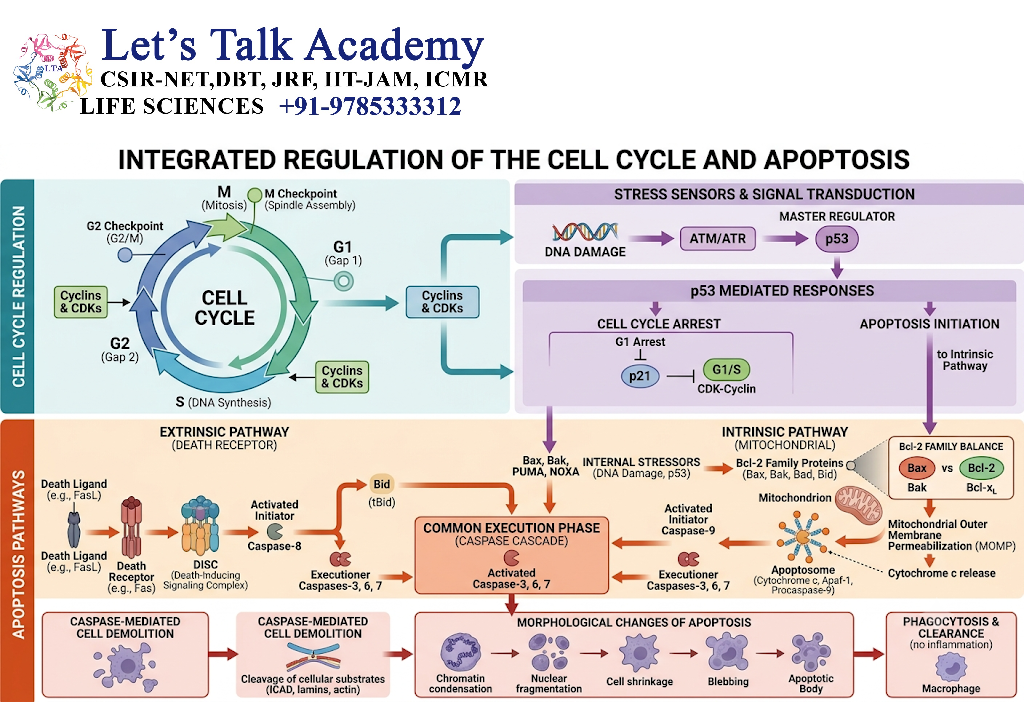
Apoptosis is closely linked to cell-cycle checkpoints.
If cellular damage is detected, the cell may stop progressing through the cell cycle.
15.2 DNA-Damage Checkpoints
Important checkpoint pathways include:
- ATM
- ATR
- Chk1
- Chk2
- p53
These pathways can prevent damaged DNA from being transmitted to daughter cells.
15.3 Relationship Between p53 and Apoptosis
p53 can function as a molecular decision maker.
If damage is repairable:
p53 → Cell-cycle arrest → Repair
If damage is severe:
p53 → Pro-apoptotic signaling → Apoptosis
15.4 Cell-Cycle Failure and Apoptosis
Cells that cannot properly control replication or chromosome integrity may activate apoptotic pathways.
This prevents propagation of genetically unstable cells.
16. Apoptosis in Development
16.1 Embryonic Development
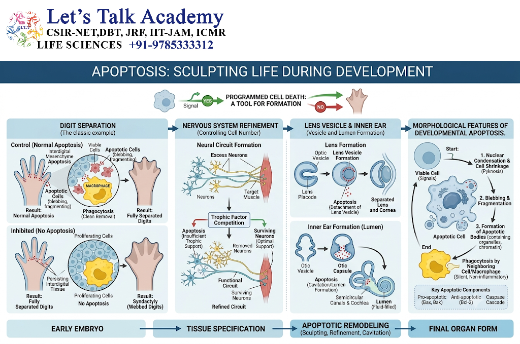
Apoptosis is essential during embryonic development.
It removes cells that are:
- Unnecessary
- Incorrectly positioned
- No longer required
- Produced in excess
16.2 Tissue Remodeling
Developing tissues undergo continuous remodeling.
Apoptosis removes temporary structures and helps establish the final architecture of organs.
16.3 Limb Development
During limb development, apoptosis contributes to separation of developing digits.
Without appropriate programmed cell removal, tissue structures may remain abnormally connected.
16.4 Nervous-System Development
During nervous-system development, neurons are produced in large numbers.
Some neurons fail to establish appropriate survival relationships and are eliminated through apoptosis.
16.5 Removal of Unnecessary Cells
Apoptosis therefore helps sculpt tissues and organs into their final functional forms.
17. Apoptosis in the Immune System
17.1 Elimination of Self-Reactive Lymphocytes
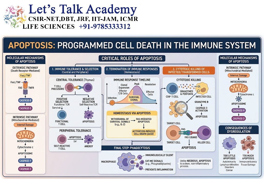
Developing immune cells that strongly recognize self-components can be eliminated through programmed cell death.
This contributes to immune tolerance.
17.2 Termination of Immune Responses
After an immune response has eliminated a threat, many activated lymphocytes are no longer required.
Apoptosis helps reduce their population.
17.3 Cytotoxic T-Cell-Mediated Apoptosis
Cytotoxic T lymphocytes can induce apoptosis in target cells.
Targets may include:
- Virus-infected cells
- Abnormal cells
- Certain transformed cells
17.4 Natural Killer Cell-Mediated Apoptosis
Natural killer cells can also induce apoptosis in susceptible target cells.
17.5 Perforin-Granzyme Pathway
Cytotoxic lymphocytes release:
- Perforin
- Granzymes
Perforin facilitates delivery of granzymes into target cells.
Granzymes can activate apoptotic machinery, including caspase-dependent and other death pathways.
18. Apoptosis and Cancer
18.1 Loss of Apoptotic Control
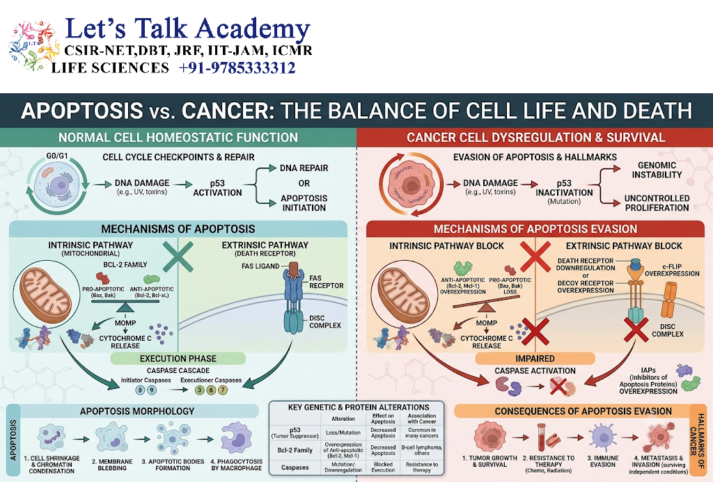
One of the important features of cancer is the ability of abnormal cells to survive when they should normally be eliminated.
18.2 p53 Abnormalities
Loss or alteration of p53 function can allow cells containing extensive DNA damage to survive.
18.3 Bcl-2 Dysregulation
Increased anti-apoptotic Bcl-2 family activity can protect abnormal cells from mitochondrial apoptosis.
18.4 Caspase Inhibition
Cancer cells may develop mechanisms that reduce caspase activation or increase anti-apoptotic signaling.
18.5 Apoptosis Resistance
Apoptosis resistance can result from multiple changes:
- Defective death receptors
- Altered Bcl-2 family balance
- Abnormal p53
- Increased survival signaling
- Increased IAP activity
- Reduced caspase activation
18.6 Cancer-Cell Survival
The consequence can be represented as:
Genetic damage
↓
Failure of apoptosis
↓
Abnormal cell survives
↓
Further mutations
↓
Uncontrolled proliferation
↓
Tumor development
19. Apoptosis and Disease
19.1 Excessive Apoptosis
Excessive or inappropriate apoptosis can contribute to loss of important cells.
This has been investigated in several degenerative and neurological conditions.
19.2 Insufficient Apoptosis
Failure to eliminate abnormal cells can contribute to:
- Cancer
- Persistent immune-cell survival
- Certain autoimmune disorders
19.3 Neurodegenerative Disorders
Abnormal regulation of cell death pathways has been implicated in several neurodegenerative diseases.
However, the exact contribution of apoptosis varies among diseases and disease stages.
19.4 Autoimmune Conditions
Defective removal of self-reactive immune cells or abnormal regulation of immune-cell survival can contribute to autoimmunity.
19.5 Cancer
Defective apoptotic control is a major mechanism allowing damaged cells to survive and proliferate.
19.6 Tissue Degeneration
When cell loss exceeds regenerative capacity, excessive cell death can contribute to tissue degeneration.
20. Experimental Detection of Apoptosis
Apoptosis can be detected using morphological, biochemical, and molecular methods.
20.1 Morphological Detection
Microscopy can reveal:
- Cell shrinkage
- Chromatin condensation
- Nuclear fragmentation
- Membrane blebbing
- Apoptotic bodies
20.2 DNA Fragmentation Assays
DNA fragmentation can be detected using biochemical methods.
A characteristic nucleosomal DNA fragmentation pattern may be observed under suitable experimental conditions.
20.3 TUNEL Assay
The TUNEL assay detects DNA strand breaks by enzymatically labeling exposed DNA ends.
It is widely used to identify cells undergoing apoptosis or other processes involving DNA fragmentation.
TUNEL positivity should be interpreted in the appropriate biological context because DNA breaks are not exclusively specific to apoptosis.
20.4 Annexin V Assay
Annexin V binds phosphatidylserine exposed on the outer surface of apoptotic cells.
It is commonly used to detect early apoptotic changes.
20.5 Caspase Activity Assays
Caspase activation can be measured using:
- Fluorogenic substrates
- Colorimetric assays
- Immunological techniques
- Activity-based probes
20.6 Flow Cytometry
Flow cytometry can combine several markers to distinguish:
- Viable cells
- Early apoptotic cells
- Late apoptotic cells
- Membrane-compromised cells
A commonly used approach combines Annexin V with a membrane-impermeable viability dye.
20.7 Western Blotting
Western blotting can detect:
- Caspase cleavage
- PARP cleavage
- Bcl-2 family proteins
- Other apoptosis-related proteins
21. Apoptosis in Biotechnology and Medicine
21.1 Apoptosis as a Therapeutic Target
Because apoptosis controls cell survival, its regulatory pathways are important targets in biomedical research.
21.2 Pro-Apoptotic Cancer Therapies
Some therapeutic strategies attempt to selectively increase death signaling in cancer cells.
The objective is to eliminate abnormal cells while minimizing toxicity to normal cells.
21.3 Death-Receptor-Based Approaches
Death-receptor pathways have been investigated as potential therapeutic targets.
21.4 Bcl-2-Targeting Strategies
Because Bcl-2 family proteins control mitochondrial apoptosis, drugs that alter their activity can influence apoptotic sensitivity.
One important therapeutic concept is the use of BH3-mimetic compounds, which can inhibit selected anti-apoptotic Bcl-2 family proteins.
21.5 Caspase-Based Approaches
Caspases are attractive mechanistic targets, although therapeutic manipulation must be carefully controlled because apoptosis is essential for normal tissue maintenance.
22. Integrated Apoptosis Pathway
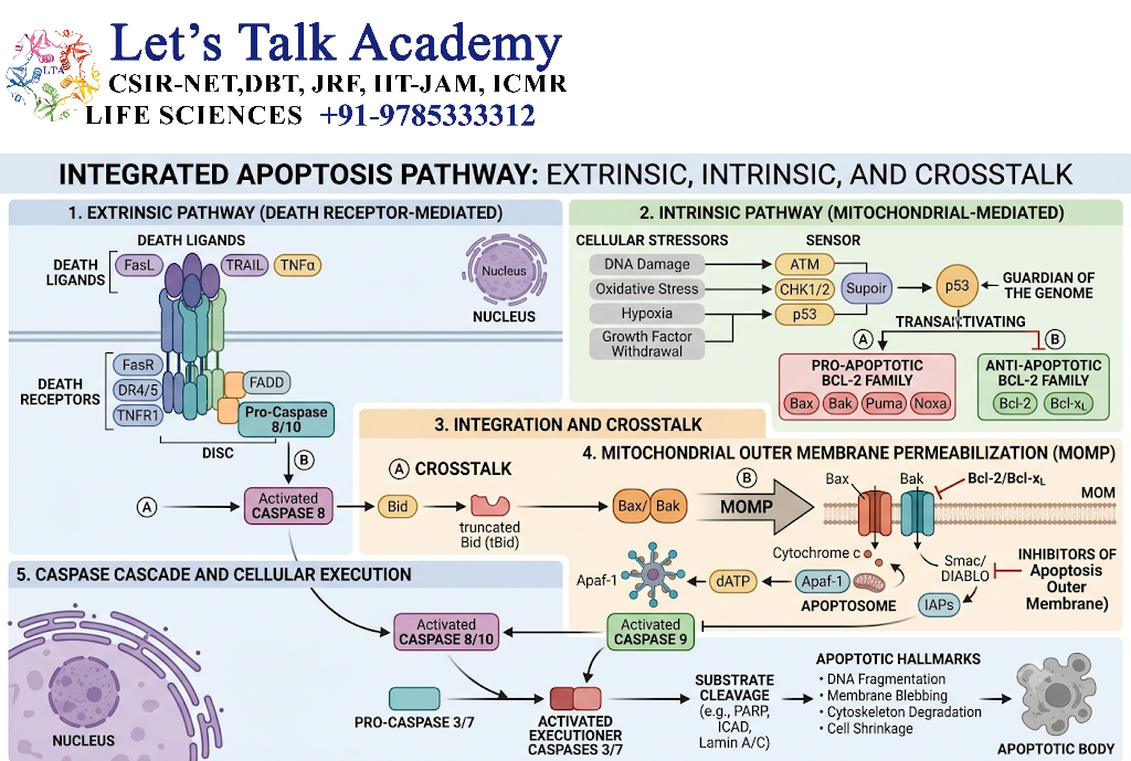
22.1 Intrinsic Pathway Flowchart
Cellular stress
↓
Bcl-2 family regulation
↓
Bax/Bak activation
↓
MOMP
↓
Cytochrome c release
↓
Apaf-1
↓
Apoptosome
↓
Caspase-9
↓
Caspase-3/7
↓
Apoptosis
22.2 Extrinsic Pathway Flowchart
Death ligand
↓
Death receptor
↓
FADD
↓
DISC
↓
Caspase-8/10
↓
Executioner caspases
↓
Apoptosis
22.3 Caspase Cascade
The caspase cascade is:
Procaspase activation → Initiator caspase → Executioner caspase → Substrate cleavage → Cellular dismantling
This cascade provides amplification because a relatively small upstream signal can activate many downstream proteolytic events.
22.4 Pathway Crosstalk
The major connection is:
Caspase-8 → Bid → tBid → Mitochondrial pathway
Therefore, extrinsic signaling can amplify intrinsic apoptosis.
22.5 Cell Survival vs Cell Death Decision
The final cellular outcome depends on the balance between competing signals.
Survival signals > Death signals → Cell survival
Death signals > Survival signals → Apoptosis
23. Apoptosis and Biological Significance
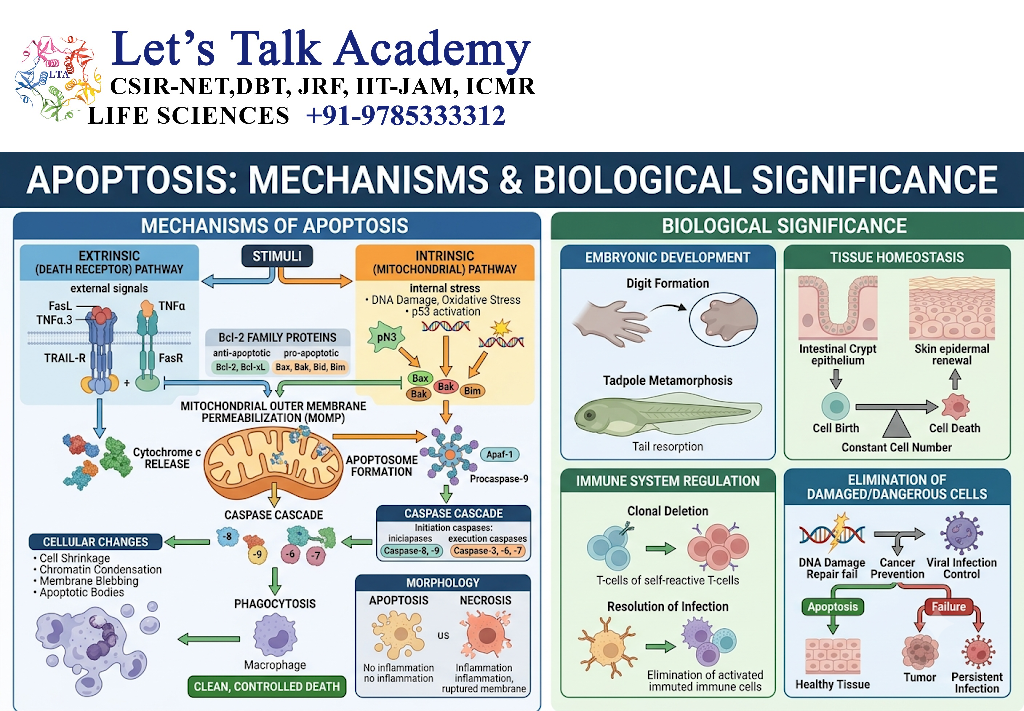
23.1 Tissue Homeostasis
Apoptosis maintains appropriate cell numbers by balancing cellular proliferation and cell elimination.
23.2 Development
It shapes tissues and organs during embryogenesis and development.
23.3 Genome Protection
Apoptosis eliminates cells carrying severe or irreparable genetic damage.
23.4 Immune Regulation
It removes unnecessary or potentially harmful immune cells.
23.5 Removal of Damaged Cells
Apoptosis prevents severely damaged cells from continuing to function or proliferate.
24. Important Regulatory Proteins
| Protein/Complex | Major Function |
|---|---|
| Bcl-2 | Anti-apoptotic mitochondrial regulator |
| Bcl-xL | Anti-apoptotic protein |
| Bax | Pro-apoptotic mitochondrial effector |
| Bak | Pro-apoptotic mitochondrial effector |
| Bid | Connects extrinsic and intrinsic pathways |
| PUMA | Pro-apoptotic p53 target |
| NOXA | Pro-apoptotic BH3-only protein |
| Cytochrome c | Promotes apoptosome formation |
| Apaf-1 | Core component of apoptosome |
| Caspase-8 | Extrinsic initiator caspase |
| Caspase-9 | Intrinsic initiator caspase |
| Caspase-3 | Major executioner caspase |
| Caspase-7 | Executioner caspase |
| FADD | Death-receptor adaptor |
| p53 | DNA-damage and stress regulator |
| p21 | CDK inhibitor |
| XIAP | Inhibits caspases |
| SMAC/DIABLO | Antagonizes IAP-mediated inhibition |
| CAD | DNA-fragmenting endonuclease |
| ICAD | Inhibitor of CAD |
25. Comparison of Intrinsic and Extrinsic Apoptosis
| Feature | Intrinsic Pathway | Extrinsic Pathway |
|---|---|---|
| Other name | Mitochondrial pathway | Death-receptor pathway |
| Main trigger | Intracellular stress | Extracellular death signal |
| Main organelle | Mitochondria | Plasma membrane |
| Major initiator caspase | Caspase-9 | Caspase-8/10 |
| Major platform | Apoptosome | DISC |
| Key adaptor | Apaf-1 | FADD |
| Important signal | Cytochrome c | Death-receptor activation |
| Bcl-2 family | Central | Can amplify through Bid |
| Executioners | Caspase-3/7 | Caspase-3/7 |
26. Key Terms
Apoptosis: Regulated programmed cell death.
Caspase: Cysteine-dependent protease involved in programmed cell death.
Procaspase: Inactive precursor form of a caspase.
Initiator caspase: Caspase that initiates the downstream apoptotic cascade.
Executioner caspase: Caspase that cleaves cellular substrates and executes apoptosis.
MOMP: Mitochondrial outer membrane permeabilization.
Apoptosome: Multiprotein complex involved in caspase-9 activation.
DISC: Death-inducing signaling complex.
Bcl-2 family: Family of proteins regulating mitochondrial apoptosis.
Cytochrome c: Mitochondrial protein released during MOMP that participates in apoptosome formation.
Phosphatidylserine: Membrane phospholipid exposed during apoptosis and recognized as an “eat-me” signal.
Efferocytosis: Recognition and clearance of apoptotic cells.
Pyknosis: Chromatin condensation.
Karyorrhexis: Nuclear fragmentation.
CAD: Caspase-activated DNase.
ICAD: Inhibitor of CAD.
27. Important Points
- Apoptosis is a highly regulated form of cell death.
- It is essential for development and tissue homeostasis.
- Caspases are central enzymes of apoptosis.
- Initiator caspases activate executioner caspases.
- The intrinsic pathway is controlled primarily through mitochondria.
- MOMP is a critical event in mitochondrial apoptosis.
- Cytochrome c participates in apoptosome formation.
- Caspase-9 is the major initiator caspase of the intrinsic pathway.
- Death receptors initiate the extrinsic pathway.
- Caspase-8 is a major initiator caspase of the extrinsic pathway.
- Bid connects extrinsic and intrinsic pathways.
- Bcl-2 family proteins regulate mitochondrial apoptosis.
- p53 can induce apoptosis following severe DNA damage.
- Phosphatidylserine acts as an important apoptotic recognition signal.
- Apoptotic cells are generally removed through efferocytosis.
- Defective apoptosis can contribute to cancer.
- Excessive apoptosis can contribute to loss of functional cells.
- Apoptosis is important in embryonic development and immune regulation.
28. Conceptual Flowcharts
28.1 Complete Intrinsic Pathway
DNA damage / cellular stress
↓
p53 and stress signaling
↓
BH3-only proteins
↓
Bax/Bak activation
↓
Mitochondrial outer membrane permeabilization
↓
Cytochrome c release
↓
Apaf-1
↓
Apoptosome
↓
Caspase-9
↓
Caspase-3/7
↓
CAD activation + protein cleavage
↓
Nuclear fragmentation + apoptotic bodies
↓
Phagocytic removal
28.2 Complete Extrinsic Pathway
Death ligand
↓
Death receptor
↓
FADD
↓
DISC
↓
Caspase-8/10
↙ ↘
Executioner caspases Bid → tBid
↓ ↓
Apoptosis Mitochondrial pathway
28.3 Cellular Decision-Making Model
Cellular condition
↓
Survival signals vs stress signals
↓
Is damage manageable?
↙ ↘
Yes No
↓ ↓
Repair / survival Apoptotic signaling
↓
Caspase activation
↓
Cellular dismantling
↓
Apoptosis
29. Summary
Apoptosis is a highly regulated form of programmed cell death that plays a fundamental role in development, tissue homeostasis, immune regulation, and protection against genetically damaged cells.
The molecular machinery of apoptosis is centered around caspases. Initiator caspases activate executioner caspases, which cleave numerous cellular substrates and produce the characteristic structural changes of apoptosis.
Two major apoptotic pathways are recognized. The intrinsic pathway is primarily controlled by mitochondria and is activated by intracellular stress such as DNA damage, growth-factor withdrawal, and oxidative stress. Bcl-2 family proteins regulate mitochondrial outer membrane permeabilization. Following MOMP, cytochrome c is released and participates with Apaf-1 in formation of the apoptosome, leading to activation of caspase-9 and subsequently executioner caspases.
The extrinsic pathway begins with activation of death receptors such as Fas. Receptor activation promotes formation of the DISC and activation of caspase-8 or caspase-10. These enzymes can directly activate executioner caspases and can also connect with the mitochondrial pathway through Bid.
The final stage of apoptosis involves extensive cellular dismantling. Chromatin condenses, DNA becomes fragmented, the nucleus breaks apart, the plasma membrane develops blebs, and apoptotic bodies form. Phosphatidylserine exposure facilitates recognition and removal of apoptotic cells through efferocytosis.
Apoptosis is tightly regulated by Bcl-2 family proteins, IAPs, survival signaling pathways, p53, and other regulatory systems. Proper regulation is essential because both excessive and insufficient apoptosis can cause disease.
30. Concluding Perspective
Apoptosis represents one of the most sophisticated mechanisms by which multicellular organisms control the life and death of individual cells.
Rather than being a simple process of cellular destruction, apoptosis is an organized molecular program involving signal detection, pathway activation, mitochondrial regulation, caspase cascades, cellular dismantling, and phagocytic clearance.
The central concept can be summarized as:
Cellular stress or death signal → Decision between survival and death → Initiator pathway → Caspase cascade → Controlled cellular dismantling → Clearance
The intrinsic and extrinsic pathways provide different mechanisms for initiating apoptosis, while their interaction allows cells to integrate multiple signals. Bcl-2 family proteins regulate the mitochondrial decision, death receptors transmit extracellular signals, caspases execute the process, and phagocytic mechanisms complete the removal of dying cells.
The biological importance of apoptosis extends from the earliest stages of embryonic development to maintenance of adult tissues. It removes unnecessary cells, shapes organs, regulates immune responses, eliminates severely damaged cells, and protects organisms from uncontrolled cellular proliferation.
At the same time, failure of apoptotic regulation can have serious consequences. Excessive cell death may contribute to tissue degeneration, whereas failure to eliminate abnormal cells can promote cancer and other pathological conditions.
Therefore, apoptosis can be viewed as a fundamental cellular quality-control and homeostatic mechanism, maintaining the balance between cell survival and cell elimination throughout the life of an organism.


