
1. Necrosis and Autophagy
Cell survival depends on a delicate balance between growth, adaptation, repair, and cell death. Cells continuously respond to changes in their environment, including nutrient availability, oxygen levels, toxic substances, infections, mechanical stress, and signals from neighboring cells. When cellular damage becomes severe or when specific intracellular pathways are activated, cells may undergo different forms of death or degradation.
Necrosis and autophagy are two important cellular processes that are often discussed together because both can occur during cellular stress and injury. However, they are fundamentally different processes. Necrosis is traditionally described as an uncontrolled form of cell death associated with severe cellular injury, whereas autophagy is primarily a cellular recycling and quality-control mechanism that usually promotes survival. Under certain circumstances, excessive or dysregulated autophagy can also contribute to cell death.
Understanding these processes requires attention to cellular morphology as well as molecular mechanisms. Changes in the plasma membrane, mitochondria, lysosomes, cytoplasm, and intracellular signaling pathways provide important clues about what is happening inside a stressed cell.
1.1 Overview of Cellular Stress and Cell Fate
Cells are exposed to many types of stress during their lifetime. These include:
- Oxidative stress
- Nutrient deprivation
- Hypoxia
- DNA damage
- Endoplasmic reticulum stress
- Mitochondrial dysfunction
- Infection
- Exposure to toxins
- Physical injury
- Excessive metabolic activity
A mild or temporary stress can activate protective mechanisms. Autophagy is one of the major mechanisms that helps cells adapt to such conditions. If the damage is too severe, however, cellular homeostasis may fail, leading to cell death.
The outcome of cellular stress therefore depends on several factors, including the intensity and duration of the stress, the cell type, metabolic condition, availability of nutrients, mitochondrial status, and activation of intracellular signaling pathways.
1.2 Necrosis and Autophagy Are Not the Same Process
Necrosis and autophagy differ significantly in their biological functions.
Necrosis is generally characterized by cellular swelling, loss of plasma membrane integrity, leakage of intracellular components, and inflammation.
Autophagy, in contrast, involves the sequestration and lysosomal degradation of intracellular components. It allows the cell to remove damaged proteins and organelles and recycle their constituent molecules.
An important point is that autophagy itself should not automatically be considered a form of cell death. In most physiological situations, autophagy is a pro-survival mechanism. Its primary function is to maintain cellular homeostasis.
2. Necrosis
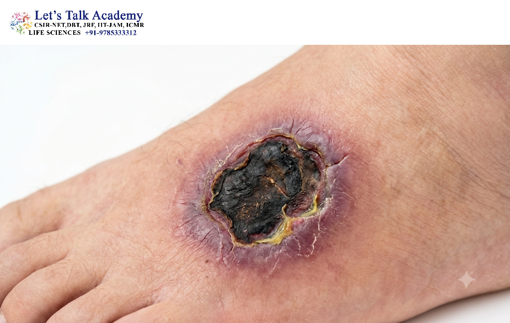
Necrosis is a form of cell death traditionally associated with acute cellular injury. It occurs when cellular damage disrupts essential structures and biochemical processes to such an extent that the cell can no longer maintain homeostasis.
Unlike programmed forms of cell death, classical necrosis was historically considered an accidental and uncontrolled process. Modern cell biology, however, has demonstrated that some forms of necrotic cell death can be regulated at the molecular level. These include pathways such as necroptosis, pyroptosis, and ferroptosis, although they have distinct molecular mechanisms and should not simply be treated as classical necrosis.
2.1 Definition of Necrosis
Necrosis can be defined as a form of cellular death characterized by severe cellular injury, loss of membrane integrity, cellular swelling, organelle damage, and release of intracellular contents into the surrounding tissue.
The release of intracellular molecules can activate inflammatory responses in neighboring cells and immune cells.
2.2 Causes of Necrosis
Necrosis can result from a wide range of damaging conditions.
2.2.1 Physical Injury
Extreme temperature, radiation, mechanical trauma, and other physical stresses can damage cellular membranes and organelles.
2.2.2 Chemical Injury
Toxic chemicals, excessive concentrations of certain drugs, pollutants, and metabolic toxins can interfere with cellular metabolism and membrane integrity.
2.2.3 Ischemia
Ischemia occurs when blood flow to a tissue is reduced or interrupted. This decreases the delivery of oxygen and nutrients and prevents the efficient removal of metabolic waste.
Severe ischemia can cause ATP depletion, ion imbalance, mitochondrial dysfunction, and eventually cell death.
2.2.4 Hypoxia
Oxygen deficiency can impair mitochondrial oxidative phosphorylation and reduce ATP production. Prolonged or severe hypoxia may therefore contribute to necrotic cell injury.
2.2.5 Infection
Certain infectious agents can directly damage cells or trigger strong inflammatory responses that result in cellular injury and death.
2.2.6 Oxidative Stress
Excessive production of reactive oxygen species (ROS) can damage proteins, lipids, DNA, and cellular membranes. If antioxidant defenses are overwhelmed, severe oxidative damage can contribute to cell death.
3. Morphological Features of Necrosis
Necrotic cells show characteristic morphological changes that can be observed using microscopy.
3.1 Cellular Swelling
One of the earliest features of necrotic injury is cellular swelling.
Damage to ATP-dependent ion pumps, particularly the sodium-potassium pump, disrupts ionic homeostasis. Sodium and water accumulate inside the cell, causing the cell and its organelles to swell.
3.2 Cytoplasmic Changes
The cytoplasm may become more intensely stained because of changes in protein composition and cellular organization. Eventually, extensive cellular damage causes breakdown of the cytoplasmic structure.
3.3 Mitochondrial Damage
Mitochondria can swell and lose their normal structural organization. Severe mitochondrial dysfunction reduces ATP production and can further accelerate cellular injury.
3.4 Nuclear Changes
Three classical nuclear changes are associated with necrosis:
Pyknosis: The nucleus becomes smaller and highly condensed.
Karyorrhexis: The condensed nucleus fragments into multiple pieces.
Karyolysis: The nuclear material gradually dissolves because of enzymatic degradation.
These changes are important morphological indicators of irreversible cellular injury.
3.5 Plasma Membrane Rupture
In classical necrosis, the plasma membrane eventually loses its integrity. Intracellular proteins, enzymes, nucleic acids, and other molecules are released into the extracellular environment.
These molecules can act as damage-associated molecular patterns (DAMPs) and stimulate inflammatory responses.
4. Molecular Events During Necrosis
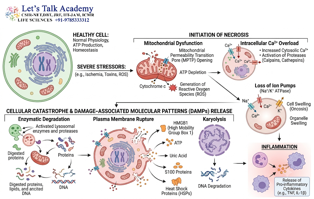
Although classical necrosis is not considered a highly organized programmed process, several biochemical events contribute to its progression.
4.1 ATP Depletion
ATP is essential for maintaining cellular homeostasis. It powers ion pumps, protein synthesis, cytoskeletal organization, and numerous metabolic reactions.
When ATP levels fall severely, membrane ion gradients cannot be maintained. This promotes cellular swelling and contributes to membrane damage.
4.2 Calcium Overload
Loss of calcium homeostasis causes intracellular calcium concentration to rise.
Excess calcium activates several enzymes, including:
- Proteases
- Phospholipases
- Endonucleases
These enzymes can damage proteins, membrane lipids, and DNA.
4.3 Reactive Oxygen Species
Mitochondrial dysfunction and impaired antioxidant systems can increase ROS production. ROS further damage cellular components and create a positive feedback loop of oxidative injury.
4.4 Lysosomal Damage
Damage to lysosomal membranes can release hydrolytic enzymes into the cytoplasm. These enzymes contribute to the degradation of cellular structures.
4.5 Membrane Breakdown
Progressive damage to phospholipids, membrane proteins, and cytoskeletal components eventually compromises plasma membrane integrity.
5. Inflammation Associated with Necrosis
One of the major biological consequences of necrosis is inflammation.
When the plasma membrane ruptures, intracellular molecules are released into the extracellular environment. Some of these molecules are recognized by receptors of the innate immune system.
Examples of important DAMPs include:
- ATP
- HMGB1
- Nuclear DNA
- Mitochondrial components
- Certain intracellular proteins
Recognition of these molecules can stimulate inflammatory signaling and recruitment of immune cells.
Therefore, necrosis can influence not only the dying cell but also the surrounding tissue.
6. Regulated Forms of Necrotic Cell Death
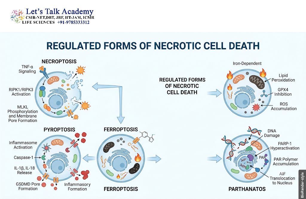
Modern cell biology has revealed that some forms of cell death that resemble necrosis are regulated by specific signaling pathways.
6.1 Necroptosis
Necroptosis is a regulated form of necrotic cell death. It is morphologically associated with cellular swelling and plasma membrane disruption but is controlled by defined signaling proteins.
Important components include:
- RIPK1
- RIPK3
- MLKL
Activation of RIPK3 promotes phosphorylation and activation of MLKL, which contributes to plasma membrane disruption.
6.2 Pyroptosis
Pyroptosis is an inflammatory form of programmed cell death associated particularly with infection and immune responses.
It commonly involves inflammatory caspases and gasdermin proteins. Activated gasdermins form pores in the plasma membrane, resulting in cellular swelling, membrane disruption, and release of inflammatory molecules.
6.3 Ferroptosis
Ferroptosis is an iron-dependent form of regulated cell death characterized by excessive lipid peroxidation.
It is strongly associated with:
- Iron metabolism
- Reactive oxygen species
- Polyunsaturated fatty acid-containing membrane lipids
- Glutathione metabolism
- GPX4 activity
Ferroptosis is mechanistically distinct from classical necrosis and apoptosis.
7. Autophagy
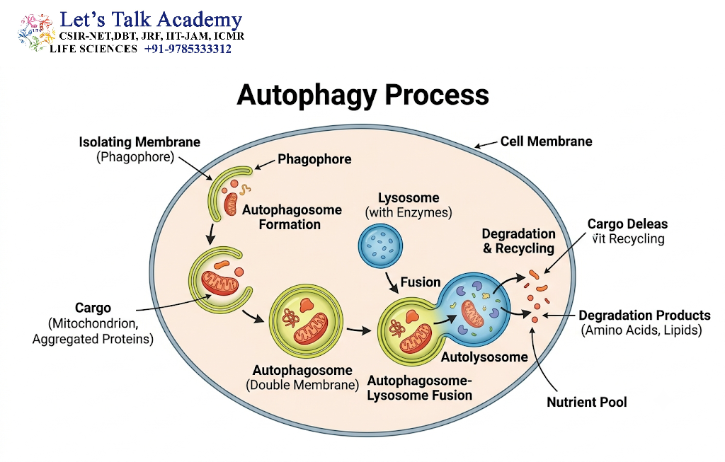
Autophagy is an evolutionarily conserved cellular process responsible for the degradation and recycling of intracellular components.
The word autophagy comes from Greek terms meaning “self-eating.” Despite this name, autophagy is not simply destruction of the cell. Instead, it is an organized recycling system that helps cells maintain quality control and survive under stressful conditions.
Autophagy is particularly important during nutrient deprivation because it allows cells to recycle macromolecules and generate building blocks for essential cellular processes.
7.1 Definition of Autophagy
Autophagy is a lysosome-dependent degradation pathway through which cytoplasmic proteins, damaged organelles, protein aggregates, and other cellular components are delivered to lysosomes for degradation and recycling.
Autophagy occurs at a basal level in healthy cells but can be strongly induced under stress.
8. Major Types of Autophagy
Three major forms of autophagy are commonly described:
- Macroautophagy
- Microautophagy
- Chaperone-mediated autophagy
8.1 Macroautophagy
Macroautophagy is the best-characterized form of autophagy.
During macroautophagy, a portion of the cytoplasm or a cellular organelle is enclosed within a double-membrane structure called an autophagosome.
The autophagosome subsequently fuses with a lysosome, producing an autolysosome, where the enclosed material is degraded.
The degradation products can then be released and reused by the cell.
8.2 Microautophagy
In microautophagy, the lysosomal membrane directly invaginates and engulfs cytoplasmic material.
The material is subsequently degraded within the lysosome.
8.3 Chaperone-Mediated Autophagy
Chaperone-mediated autophagy differs from macroautophagy because the target proteins are transported individually across the lysosomal membrane.
A cytosolic chaperone, particularly Hsc70, recognizes proteins containing specific targeting motifs. These proteins interact with the lysosomal membrane protein LAMP2A and are transported into the lysosomal lumen for degradation.
9. Mechanism of Macroautophagy
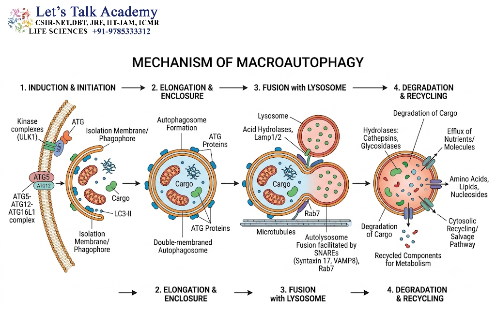
Macroautophagy proceeds through several coordinated stages.
9.1 Initiation
Autophagy begins when the cell senses conditions such as nutrient deprivation, energy stress, or damaged cellular components.
The regulation of autophagy involves major signaling pathways, especially mTOR and AMPK.
Under nutrient-rich conditions, mTORC1 generally suppresses autophagy.
When nutrients are limited or cellular energy is low, mTORC1 activity decreases and AMPK signaling can promote autophagy.
9.2 Phagophore Formation
The first visible membrane structure of macroautophagy is the phagophore, also called the isolation membrane.
It begins as a small membrane structure that expands around the cellular material targeted for degradation.
9.3 Autophagosome Formation
The phagophore expands and eventually encloses its cargo.
Once the edges of the membrane fuse, a double-membrane vesicle called the autophagosome is formed.
9.4 Fusion with the Lysosome
The autophagosome moves toward lysosomal compartments and fuses with a lysosome.
The resulting structure is known as an autolysosome.
9.5 Degradation and Recycling
Lysosomal hydrolases degrade the enclosed material.
Proteins are broken down into amino acids, lipids into fatty acids and other components, and nucleic acids into nucleotides and related molecules.
These products can be recycled for cellular metabolism and biosynthesis.
10. Molecular Regulation of Autophagy
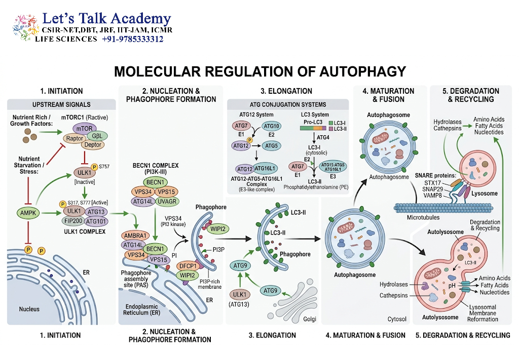
Autophagy is controlled by a complex network of signaling proteins.
10.1 mTOR Pathway
The mechanistic target of rapamycin complex 1 (mTORC1) is a major negative regulator of autophagy.
When nutrients and growth signals are abundant, mTORC1 activity promotes anabolic processes and suppresses autophagy.
When nutrient availability decreases, mTORC1 activity is inhibited, allowing autophagy to be activated.
10.2 AMPK Pathway
AMP-activated protein kinase (AMPK) acts as an important sensor of cellular energy status.
When the cellular AMP/ATP or ADP/ATP ratio increases, AMPK becomes activated.
AMPK promotes catabolic pathways and can stimulate autophagy.
Thus, AMPK and mTOR often have opposing effects on autophagy.
10.3 ULK1 Complex
The ULK1 complex is an important component of autophagy initiation.
It helps connect nutrient-sensing pathways with the molecular machinery responsible for autophagosome formation.
10.4 Beclin-1 and Class III PI3K
The Beclin-1-containing class III phosphatidylinositol 3-kinase complex contributes to nucleation of the autophagic membrane.
VPS34, a class III PI3K, generates phosphatidylinositol 3-phosphate (PI3P), which helps recruit proteins required for phagophore development.
10.5 ATG Proteins
Autophagy-related proteins, commonly abbreviated as ATG proteins, coordinate different stages of autophagy.
Important ATG proteins participate in:
- Autophagy initiation
- Membrane nucleation
- Membrane expansion
- Cargo selection
- Autophagosome formation
- Autophagosome maturation
11. LC3 and Autophagy
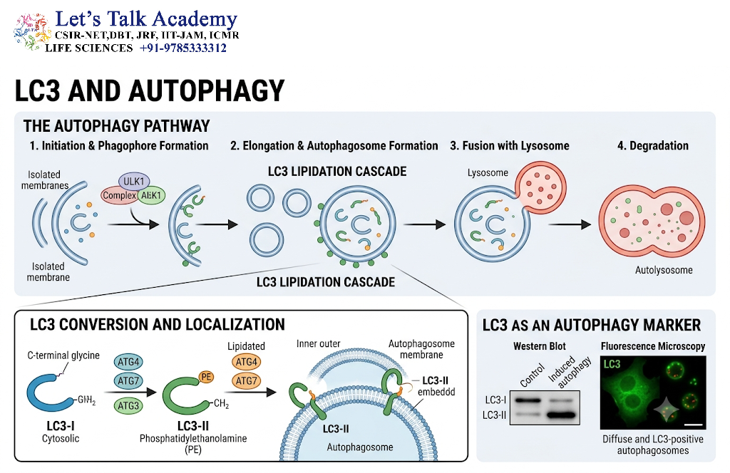
LC3 is one of the most widely used molecular markers associated with autophagy.
LC3 is initially synthesized as a precursor and processed to produce LC3-I. During autophagy, LC3-I is conjugated with phosphatidylethanolamine to generate LC3-II.
LC3-II becomes associated with autophagosomal membranes.
Therefore, LC3-II levels and LC3-positive structures are commonly used to study autophagy. However, LC3 accumulation alone does not always mean that autophagic degradation is increased because accumulation may also occur when lysosomal degradation is blocked.
For this reason, autophagic flux is an important concept.
12. Autophagic Flux
Autophagic flux refers to the complete process of autophagy from formation of autophagic structures to their degradation.
A simplified sequence is:
Cargo selection → phagophore formation → autophagosome formation → lysosomal fusion → degradation → recycling
Measuring autophagic flux provides more information than simply measuring the number of autophagosomes.
For example, a large accumulation of autophagosomes may indicate either increased autophagosome production or impaired degradation.
This distinction is essential when interpreting experimental data.
13. Functions of Autophagy
Autophagy performs numerous physiological functions.
13.1 Nutrient Recycling
During nutrient deprivation, autophagy provides amino acids, fatty acids, and other metabolites that can be reused by the cell.
13.2 Removal of Damaged Organelles
Autophagy helps eliminate damaged or dysfunctional organelles.
Selective forms include:
- Mitophagy – degradation of mitochondria
- Pexophagy – degradation of peroxisomes
- ER-phagy – degradation of portions of the endoplasmic reticulum
- Ribophagy – degradation of ribosomes
13.3 Protein Quality Control
Autophagy helps remove protein aggregates and damaged proteins that cannot be efficiently degraded by other systems.
13.4 Cellular Adaptation to Stress
During starvation, hypoxia, and other stress conditions, autophagy helps cells adapt by recycling intracellular materials.
13.5 Maintenance of Cellular Homeostasis
Basal autophagy continuously removes damaged components and contributes to long-term cellular quality control.
14. Autophagy and Cell Survival
Autophagy is generally considered a survival mechanism.
When nutrients are scarce, cells can degrade non-essential components and recycle their molecular constituents.
For example, during starvation, autophagy can provide amino acids and other metabolites needed for energy production and biosynthesis.
However, autophagy can have different effects depending on the intensity and duration of stress.
Moderate autophagy may promote survival, whereas persistent or dysregulated autophagy may be associated with cellular dysfunction and, in some contexts, cell death.
Therefore, the relationship between autophagy and cell death should not be oversimplified.
15. Autophagy-Dependent Cell Death
The term autophagic cell death has historically been used when extensive autophagy is observed in dying cells.
However, it is important to distinguish between:
Autophagy occurring in a dying cell and cell death that is mechanistically dependent on the autophagy machinery.
These are not necessarily the same.
In many situations, autophagy increases as a protective response to stress, even when the cell ultimately dies through another mechanism.
Therefore, demonstrating that autophagy is present is not sufficient to conclude that autophagy caused the death of the cell.
16. Relationship Between Necrosis and Autophagy
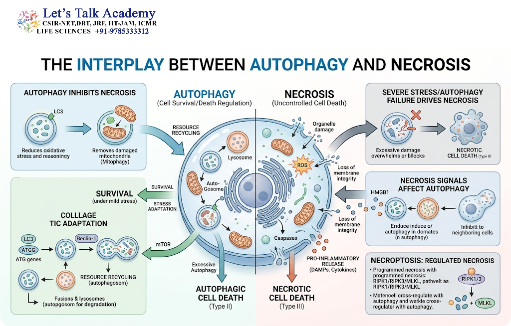
Necrosis and autophagy can occur in the same tissue or under the same stressful conditions, but their cellular functions are fundamentally different.
Autophagy generally attempts to maintain cellular homeostasis by removing damaged components and recycling nutrients.
Necrosis represents severe cellular injury accompanied by loss of membrane integrity.
The two processes may interact because autophagy can influence mitochondrial function, ROS production, energy availability, and inflammatory signaling, all of which can affect whether a stressed cell survives or progresses toward cell death.
17. Necrosis Versus Autophagy
| Feature | Necrosis | Autophagy |
|---|---|---|
| Basic nature | Cell death/injury-associated process | Cellular degradation and recycling pathway |
| Major function | Results in loss of cellular integrity | Maintains cellular quality control |
| Plasma membrane | Often becomes disrupted | Generally remains intact during autophagy |
| Cell size | Commonly increases | Usually does not show classical necrotic swelling |
| Organelles | Become damaged and swollen | Selected organelles may be selectively degraded |
| Lysosomes | May contribute to cellular degradation after damage | Central to degradation process |
| Inflammation | Commonly associated with inflammation | Usually not inherently inflammatory |
| ATP | Severe depletion is common in classical necrosis | Helps maintain energy and nutrient availability |
| Molecular regulation | Classical necrosis may be unregulated; regulated necrotic pathways also exist | Highly regulated by ATG proteins and nutrient-sensing pathways |
| Major outcome | Loss of cellular integrity and death | Recycling, adaptation, and quality control |
18. Key Differences in Cellular Morphology
The morphological differences between necrosis and autophagy are particularly important.
During necrosis, the cell typically becomes swollen, organelles become damaged, and the plasma membrane eventually ruptures.
During autophagy, the cytoplasm contains autophagic structures, particularly double-membrane autophagosomes. These structures deliver material to lysosomes for degradation.
Thus, membrane integrity is an important distinction between classical necrosis and autophagy.
19. Autophagy and Mitochondrial Quality Control
Mitochondria are major targets of selective autophagy.
The selective removal of damaged mitochondria is called mitophagy.
Damaged mitochondria can generate excessive ROS and may become inefficient in ATP production. Their removal prevents accumulation of dysfunctional organelles.
One well-studied mitophagy pathway involves PINK1 and Parkin.
When mitochondria lose their membrane potential, PINK1 can accumulate on the outer mitochondrial membrane and promote Parkin-dependent ubiquitination of mitochondrial proteins. These signals facilitate recognition of damaged mitochondria by the autophagic machinery.
20. Autophagy and Oxidative Stress
Autophagy and oxidative stress are closely interconnected.
Excessive ROS can damage proteins and organelles, which may stimulate autophagy.
At the same time, autophagy can remove damaged mitochondria and other ROS-producing structures, thereby reducing oxidative stress.
This creates a protective feedback system:
Cellular stress → ROS generation → damage to organelles → autophagy activation → removal of damaged organelles → reduced cellular stress
When this protective mechanism becomes insufficient, persistent oxidative damage can contribute to cell death.
21. Autophagy and Disease
Dysregulation of autophagy has been associated with many diseases.
21.1 Cancer
Autophagy has a complex relationship with cancer.
In some circumstances, autophagy can suppress tumor development by maintaining cellular quality and limiting the accumulation of damaged organelles.
In established tumors, however, autophagy may help cancer cells survive nutrient limitation, hypoxia, and metabolic stress.
Therefore, autophagy can have both tumor-suppressive and tumor-supportive effects depending on the cellular and disease context.
21.2 Neurodegenerative Disorders
Neurons are particularly dependent on efficient protein and organelle quality-control systems.
Defective autophagy can contribute to accumulation of damaged proteins and organelles, which has been implicated in several neurodegenerative disorders.
21.3 Metabolic Disorders
Autophagy contributes to metabolic adaptation and the regulation of lipid and glucose metabolism. Altered autophagy can therefore influence metabolic homeostasis.
21.4 Infectious Diseases
Autophagy can help cells eliminate intracellular pathogens and process pathogen-derived material.
Some microorganisms, however, have evolved mechanisms to evade or manipulate autophagic pathways.
22. Experimental Detection of Autophagy
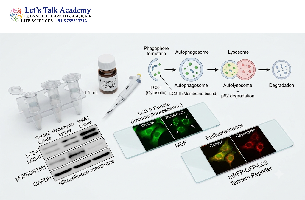
Several experimental approaches can be used to investigate autophagy.
22.1 LC3 Analysis
Conversion of LC3-I to LC3-II and changes in LC3-associated structures are commonly studied.
22.2 p62/SQSTM1
p62 is a selective autophagy receptor and is itself degraded through autophagy.
Changes in p62 levels can therefore provide information about autophagic activity, although interpretation should be combined with other measurements.
22.3 Fluorescence Microscopy
Fluorescently tagged LC3 or other autophagy-related proteins can be used to visualize autophagic structures.
22.4 Electron Microscopy
Transmission electron microscopy can reveal the characteristic double-membrane structure of autophagosomes.
22.5 Autophagic Flux Assays
Flux assays are particularly important because they distinguish increased autophagosome formation from impaired degradation.
23. Experimental Detection of Necrosis
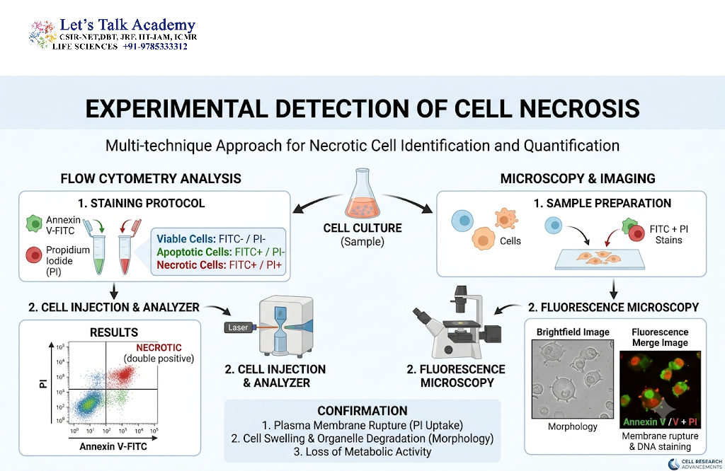
Necrotic cell death can be investigated using several approaches.
23.1 Plasma Membrane Integrity
Loss of membrane integrity can be detected using membrane-impermeable dyes.
23.2 LDH Release
Lactate dehydrogenase (LDH) is normally present inside cells. When the plasma membrane is damaged, LDH can be released into the surrounding medium.
Therefore, extracellular LDH activity can serve as an indicator of membrane damage.
23.3 Morphological Analysis
Microscopy can reveal:
- Cellular swelling
- Organelle swelling
- Nuclear changes
- Membrane rupture
- Loss of cellular architecture
23.4 Detection of Inflammatory Signals
In necrotic and regulated inflammatory cell death, release of intracellular molecules can activate inflammatory pathways.
24. Necrosis, Autophagy, and Cellular Decision-Making
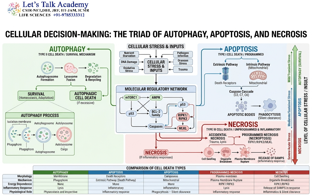
Cells do not respond to stress using a single pathway. Instead, multiple signaling networks interact continuously.
For example:
Nutrient availability → mTOR/AMPK signaling → autophagy regulation
Mitochondrial damage → ROS generation → stress responses → autophagy or cell death
Severe ATP depletion → ion imbalance → cellular swelling → membrane damage
The final cellular outcome depends on the balance between protective and damaging signals.
25. Important Molecular Concepts
Several molecular concepts are especially important for understanding necrosis and autophagy.
25.1 mTOR
A major regulator of cellular growth and nutrient availability that generally suppresses autophagy when active.
25.2 AMPK
An energy sensor that becomes activated during cellular energy stress and can stimulate autophagy.
25.3 ULK1
An important component of the autophagy initiation machinery.
25.4 Beclin-1
A key regulator of autophagosome nucleation through its association with class III PI3K complexes.
25.5 LC3
An autophagy-associated protein widely used as a marker of autophagosomal membranes.
25.6 p62/SQSTM1
A selective autophagy receptor that links ubiquitinated cargo with the autophagic machinery and is itself degraded during autophagy.
25.7 RIPK1, RIPK3, and MLKL
Important proteins involved in necroptosis, a regulated form of necrotic cell death.
26. Integrated Comparison of Cell Death and Autophagy
Cellular stress can be understood as a progressive process.
Under mild stress:
Stress → adaptation → autophagy → cellular recovery
Under persistent stress:
Stress → organelle damage → increased ROS → impaired homeostasis
Under severe injury:
Severe damage → ATP depletion → ion imbalance → membrane damage → necrotic cell death
This simplified framework demonstrates why autophagy and necrosis should not be viewed as interchangeable processes.
27. Biological Significance
Necrosis and autophagy have major implications for tissue physiology and disease.
Autophagy protects cells by maintaining intracellular quality control, recycling nutrients, and removing damaged organelles.
Necrotic cell death, on the other hand, can contribute to tissue injury and inflammation because cellular contents are released into the extracellular environment.
The balance between these processes is therefore important for maintaining tissue homeostasis.


