
1. Introduction
The cytoskeleton is a dynamic and highly organized network of protein filaments present throughout the cytoplasm of eukaryotic cells. The term “cytoskeleton” may suggest a rigid internal framework, but in reality, it is a flexible and continuously changing system. It provides mechanical strength, maintains cell shape, organizes cellular components, facilitates intracellular transport, participates in cell division, and enables cells to move.
Unlike a fixed skeletal framework, the cytoskeleton is continuously assembled, disassembled, rearranged, and remodeled according to the needs of the cell. These changes allow cells to respond rapidly to their surroundings and perform specialized functions.
The cytoskeleton consists primarily of three major types of filamentous structures:
- Microtubules
- Microfilaments or actin filaments
- Intermediate filaments
Each filament system has a characteristic structure, composition, mechanical property, and cellular function. Although these systems can be studied independently, they frequently work together to coordinate complex cellular activities.
Cellular motility is one of the most important functions associated with the cytoskeleton. Movement may occur at the level of the entire cell, as in crawling cells, or at the level of specific cellular components, such as chromosomes, vesicles, organelles, and cilia. Cytoskeletal proteins and their associated motor proteins generate the forces required for these movements.
2. General Organization of the Cytoskeleton
The cytoskeleton extends through the cytoplasm and forms an interconnected network. Different filament systems occupy different regions of the cell and perform specialized functions.
Microtubules are generally organized from microtubule-organizing centers and extend toward the cell periphery. They are particularly important for maintaining cell polarity and serving as tracks for long-distance intracellular transport.
Actin filaments are especially abundant near the plasma membrane, where they participate in cell shape changes, cell adhesion, membrane movement, and migration.
Intermediate filaments form mechanically strong networks throughout the cytoplasm and often connect the cell surface with the nucleus and other cellular structures.
The three systems are interconnected through numerous accessory proteins. Therefore, cytoskeletal organization should be considered an integrated system rather than three completely independent networks.
3. Major Components of the Cytoskeleton
3.1 Microtubules
Microtubules are hollow cylindrical structures approximately 25 nm in diameter. They are composed primarily of the protein tubulin.
A microtubule is constructed from repeating units of α-tubulin and β-tubulin. These two proteins form a heterodimer, and the heterodimers assemble longitudinally to form protofilaments. Typically, 13 protofilaments associate laterally to produce the cylindrical microtubule.
Microtubules possess structural polarity. One end is called the plus end, while the other is called the minus end.
The plus end generally undergoes faster growth and shrinkage, whereas the minus end is often anchored at a microtubule-organizing center.
3.1.1 Dynamic Instability of Microtubules
Microtubules are highly dynamic structures. They alternate between phases of growth and shrinkage, a behavior known as dynamic instability.
The GTP bound to β-tubulin plays an important role in this process. Newly incorporated tubulin carries GTP, but GTP is hydrolyzed after incorporation into the microtubule.
When a stable GTP-tubulin cap is present at the growing end, the microtubule tends to remain extended. Loss of this stabilizing cap can result in rapid depolymerization, known as catastrophe.
A transition from shrinking to growing is called rescue.
Dynamic instability allows microtubules to rapidly reorganize in response to cellular requirements.
3.2 Actin Filaments
Actin filaments, also called microfilaments, are thin protein polymers approximately 7 nm in diameter.
They are formed by polymerization of globular actin, commonly called G-actin, into filamentous actin or F-actin.
Like microtubules, actin filaments are polarized and have structurally distinct plus and minus ends. Actin polymerization is particularly important near the plasma membrane.
Actin filaments form networks and bundles that contribute to:
- Cell shape
- Cell migration
- Muscle contraction
- Cytokinesis
- Cell adhesion
- Membrane protrusion
- Endocytosis
- Intracellular organization
Actin filaments interact extensively with motor proteins, particularly myosin, to generate mechanical force.
3.3 Intermediate Filaments
Intermediate filaments are approximately 10 nm in diameter, making them intermediate in size between microfilaments and microtubules.
Unlike microtubules and actin filaments, intermediate filaments are not composed of one universal protein. Instead, they are formed from a large family of related proteins.
Examples include:
- Keratins
- Vimentin
- Desmin
- Neurofilaments
- Nuclear lamins
Intermediate filaments are especially important for mechanical stability. They can withstand tensile stress and help prevent cells from becoming damaged when subjected to physical forces.
Unlike actin filaments and microtubules, intermediate filaments generally do not have the same type of intrinsic structural polarity.
4. Comparison of the Three Cytoskeletal Systems
| Feature | Microtubules | Actin Filaments | Intermediate Filaments |
|---|---|---|---|
| Approximate diameter | 25 nm | 7 nm | 10 nm |
| Major subunit | α/β-tubulin | Actin | Various IF proteins |
| Polarity | Present | Present | Generally absent |
| Main role | Transport, organization, chromosome movement | Motility, contraction, cell shape | Mechanical strength |
| Major motor proteins | Kinesin, dynein | Myosin | No major associated motor system |
| Dynamic behavior | Highly dynamic | Dynamic | Relatively stable |
| Important role in cell division | Spindle formation | Cytokinesis | Structural support |
5. Functions of the Cytoskeleton
The cytoskeleton performs numerous functions that are essential for cellular organization and survival.
5.1 Maintenance of Cell Shape
The cytoskeleton provides an internal framework that determines and maintains cellular morphology.
Actin networks support the cell cortex and help maintain the shape of the plasma membrane. Microtubules provide internal organization and resist certain types of compressive forces. Intermediate filaments provide tensile strength.
Cell shape can therefore be understood as a result of the coordinated activity of all three cytoskeletal systems.
5.2 Intracellular Transport
Cells must transport proteins, vesicles, organelles, and other materials between different regions of the cytoplasm.
Microtubules act as long-distance tracks for molecular motors.
Two major microtubule-associated motor proteins are:
- Kinesins
- Dyneins
Most kinesins move cargo toward the microtubule plus end, whereas cytoplasmic dynein generally transports cargo toward the minus end.
Actin filaments also function as tracks for myosin motors, particularly for short-range transport near the cell cortex.
5.3 Cell Division
The cytoskeleton plays a central role in both mitosis and cytokinesis.
Microtubules form the mitotic spindle, which captures and separates chromosomes during cell division.
Actin and myosin form the contractile ring during cytokinesis. Contraction of this ring produces a cleavage furrow that eventually divides the cell into two daughter cells.
5.4 Cell Polarity
The cytoskeleton contributes to the establishment and maintenance of cellular polarity.
Polarized cells, such as epithelial cells and neurons, have distinct regions with different structures and functions. Cytoskeletal organization allows proteins and organelles to be selectively distributed to these regions.
5.5 Mechanical Strength
Intermediate filaments provide strong mechanical support. They are particularly important in tissues exposed to mechanical stress.
For example, keratin networks help epithelial cells resist stretching and mechanical injury.
5.6 Cell Adhesion
Cytoskeletal components interact with cell-adhesion molecules and extracellular matrix receptors.
Actin filaments are associated with structures such as:
- Focal adhesions
- Adherens junctions
Intermediate filaments are associated with:
- Desmosomes
- Hemidesmosome-related structures
Through these connections, forces can be transmitted between the extracellular environment and the interior of the cell.
6. Cytoskeleton and Cellular Motility
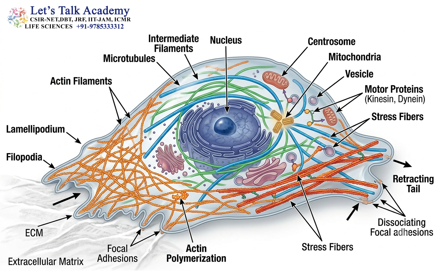
Cellular motility refers to the ability of a cell or cellular component to undergo directed movement.
Motility can occur through several mechanisms, including:
- Actin-based crawling
- Myosin-driven contraction
- Microtubule-based transport
- Ciliary movement
- Flagellar movement
- Chromosome movement
- Vesicular and organelle transport
The cytoskeleton provides both the structural tracks and the force-generating machinery required for these processes.
7. Actin-Based Cell Motility

Actin is the principal cytoskeletal component involved in many forms of cell crawling.
A migrating cell continuously changes its shape. It extends its front edge, forms attachments to the surrounding surface, pulls its cell body forward, and releases attachments at the rear.
This process involves highly coordinated actin remodeling.
7.1 Formation of Cell Protrusions
Migrating cells frequently produce specialized membrane protrusions.
Two major examples are:
- Lamellipodia
- Filopodia
7.1.1 Lamellipodia
Lamellipodia are broad, sheet-like protrusions at the leading edge of migrating cells.
They contain branched actin networks that grow toward the plasma membrane and push the membrane forward.
7.1.2 Filopodia
Filopodia are thin, finger-like protrusions containing parallel bundles of actin filaments.
They can function as sensory structures and help cells explore their environment.
8. Molecular Mechanism of Cell Crawling
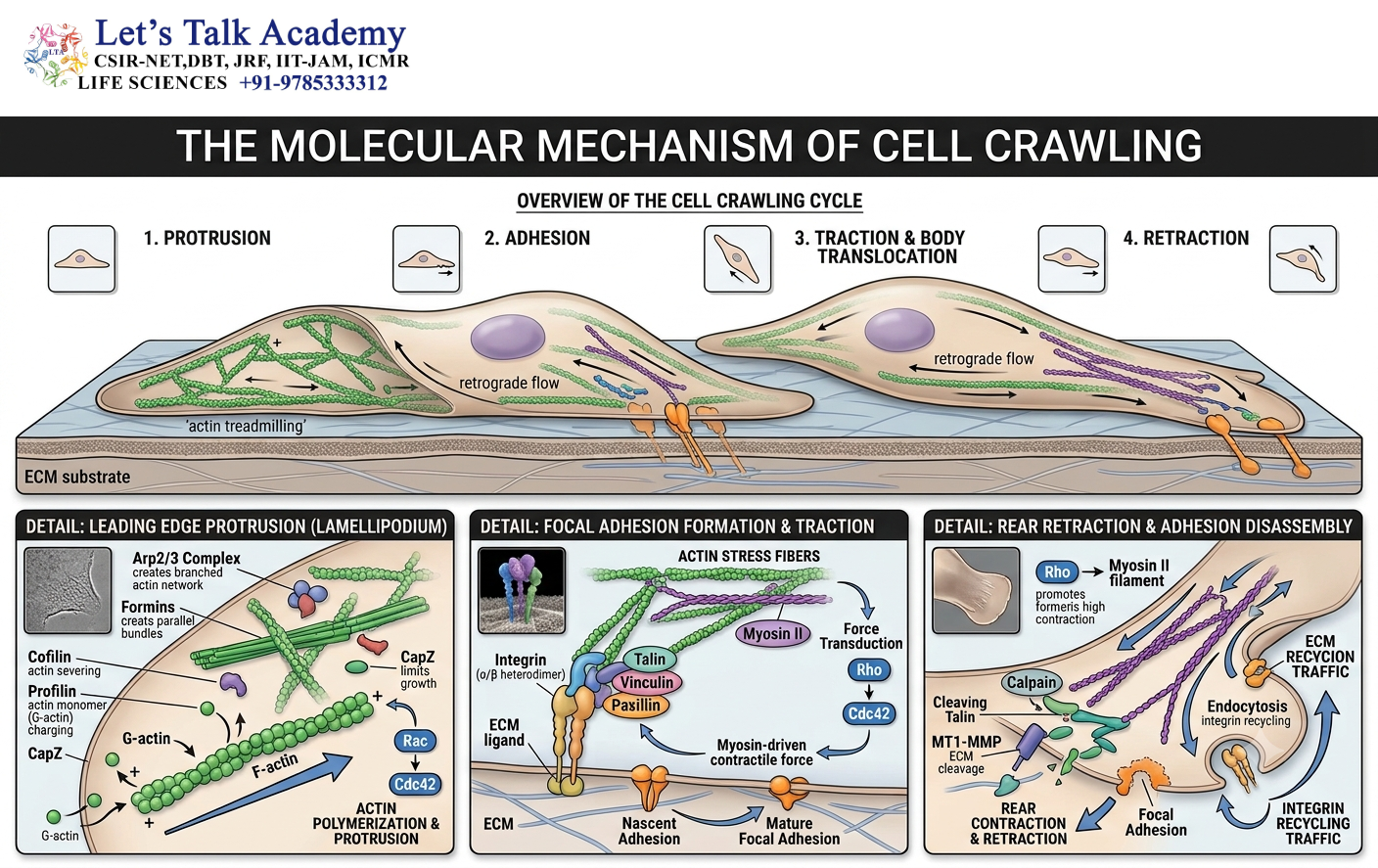
Cell migration is a multistep process.
8.1 Polarization
The cell establishes a front and a rear. Cytoskeletal components become asymmetrically organized.
8.2 Protrusion
Actin polymerization at the leading edge pushes the plasma membrane forward.
8.3 Adhesion
The protruding region forms attachments with the extracellular matrix through adhesion receptors such as integrins.
8.4 Contraction
Myosin motors interact with actin filaments and generate contractile forces.
8.5 Rear Retraction
The rear portion of the cell releases its attachments and moves forward.
These events occur repeatedly and in a coordinated manner, resulting in directional cell movement.
9. Role of Myosin in Motility
Myosins are actin-associated motor proteins that convert chemical energy into mechanical work.
They generally contain:
- A motor domain
- A neck region
- A tail domain
The motor domain interacts with actin and binds or hydrolyzes ATP.
The ATPase cycle produces conformational changes in the myosin molecule, resulting in movement along actin filaments or generation of tension.
Different myosin families perform different functions.
9.1 Myosin II
Myosin II is particularly important in:
- Muscle contraction
- Cytokinesis
- Cell migration
- Cell contractility
Myosin II molecules can assemble into bipolar filaments. These structures can pull actin filaments relative to one another and generate contraction.
10. Muscle Contraction and the Cytoskeleton
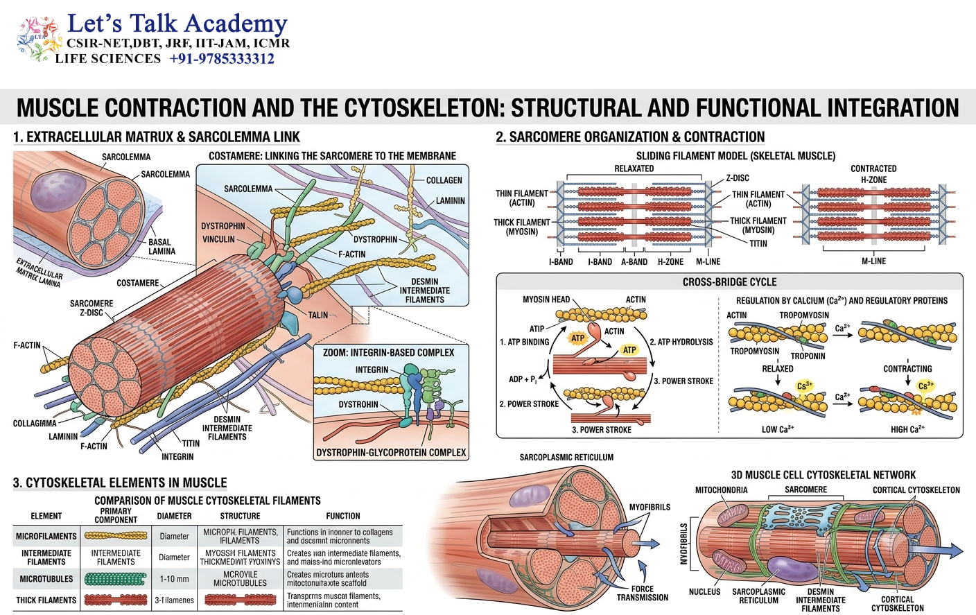
Muscle contraction is one of the best-characterized examples of actin–myosin interaction.
In skeletal muscle, thin filaments are primarily composed of actin, while thick filaments contain myosin.
The sliding-filament mechanism involves movement of actin relative to myosin.
Calcium ions play a critical regulatory role. An increase in cytosolic Ca²⁺ allows regulatory proteins associated with actin to expose myosin-binding sites, enabling contraction.
The process can be summarized as:
Ca²⁺ increase → regulatory protein activation → actin–myosin interaction → ATP-dependent cross-bridge cycling → force generation
11. Microtubules and Intracellular Motility
Microtubules are particularly important for intracellular transport.
They provide polarized tracks along which motor proteins transport cargo.
11.1 Kinesin
Kinesins are generally plus-end-directed microtubule motors.
They participate in the transport of:
- Vesicles
- Protein complexes
- Organelles
- Other intracellular cargo
11.2 Dynein
Dyneins are generally minus-end-directed motors.
Cytoplasmic dynein transports cargo toward microtubule-organizing centers and is involved in several important cellular processes.
Dynein also plays a major role in the movement of cilia and flagella.
12. Cilia and Flagella
Cilia and eukaryotic flagella are specialized structures based on microtubules.
The internal axoneme of many motile cilia and flagella has a characteristic 9 + 2 arrangement:
- Nine outer microtubule doublets
- Two central singlet microtubules
Dynein motor proteins associated with the microtubule doublets generate sliding forces.
Because the microtubules are structurally constrained, sliding is converted into bending.
This produces the characteristic beating movement of cilia and flagella.
13. Mechanism of Ciliary and Flagellar Movement

The movement of cilia and flagella depends on ATP-driven activity of axonemal dynein.
The basic sequence is:
ATP hydrolysis → dynein conformational change → microtubule sliding → bending of axoneme → coordinated movement
Ciliary beating can move fluid across the surface of cells.
For example, epithelial cilia in the respiratory tract help move mucus and trapped particles toward the pharynx.
Flagella can generate propulsion by producing waves along their length.
14. Cytoskeleton in Intracellular Transport
The cytoskeleton acts as an intracellular highway system.
Long microtubules provide routes for movement across large distances within the cell, while actin filaments often support transport over shorter distances.
Cargo can include:
- Secretory vesicles
- Endocytic vesicles
- Mitochondria
- Lysosomes
- Peroxisomes
- Protein complexes
- mRNA-containing complexes
Motor proteins recognize specific cargo through adaptor proteins and transport them along cytoskeletal tracks.
15. Cytoskeleton in Neuronal Transport
Neurons can have extremely long axons. Transporting materials through such long distances requires an efficient cytoskeletal system.
Microtubules extend along the axon and provide tracks for motor proteins.
Kinesin generally mediates anterograde transport, moving cargo toward the axon terminal.
Dynein generally mediates retrograde transport, moving cargo toward the neuronal cell body.
This transport system is essential for maintaining neuronal structure and function.
16. Cytoskeleton and Cytokinesis
At the end of nuclear division, the cytoplasm must also divide.
In animal cells, an actin–myosin contractile ring forms beneath the plasma membrane.
The ring contracts progressively and produces a cleavage furrow.
Eventually, the furrow deepens and separates the daughter cells.
Thus, actin and myosin convert molecular energy into mechanical force to complete cytokinesis.
17. Cytoskeleton and Cell Junctions

Cytoskeletal elements are strongly associated with cell junctions.
17.1 Adherens Junctions
Adherens junctions connect neighboring cells through adhesion proteins and link these proteins to actin filaments.
They help tissues resist mechanical stress and maintain coordinated cellular organization.
17.2 Desmosomes
Desmosomes connect intermediate filament networks between neighboring cells.
They are particularly important in tissues exposed to mechanical stress, such as epithelial tissues.
17.3 Focal Adhesions
Focal adhesions connect actin filaments inside the cell with extracellular matrix components through integrins and associated proteins.
They are especially important during cell migration because they provide temporary traction points.
18. Regulation of Cytoskeletal Dynamics
Cytoskeletal organization is tightly regulated.
Important regulatory mechanisms include:
- GTP and ATP hydrolysis
- Actin-binding proteins
- Microtubule-associated proteins
- Motor proteins
- Signaling pathways
- Small GTPases
- Calcium signaling
- Phosphorylation and dephosphorylation
Small GTPases of the Rho family, including Rho, Rac, and Cdc42, are particularly important in regulating actin organization.
Their coordinated activity helps determine whether a cell forms protrusions, develops contractile structures, or changes its polarity.
19. Cytoskeleton-Associated Proteins
The cytoskeleton does not function alone. Numerous accessory proteins regulate filament assembly, organization, stability, and interaction with other cellular components.
Important classes include:
19.1 Actin-Binding Proteins
These regulate actin filament:
- Nucleation
- Elongation
- Branching
- Bundling
- Severing
- Capping
Examples include profilin, cofilin, gelsolin, fimbrin, and α-actinin.
19.2 Microtubule-Associated Proteins
These proteins influence microtubule stability and organization.
Examples include:
- Tau
- MAP2
- Kinesins
- Dyneins
19.3 Intermediate Filament-Associated Proteins
These proteins help organize intermediate filament networks and connect them to other cellular structures.
20. Cytoskeleton and Cell Migration
Cell migration is essential for many biological processes, including:
- Embryonic development
- Wound healing
- Immune responses
- Tissue remodeling
- Nervous system development
During migration, the cytoskeleton continuously changes.
Actin polymerization drives protrusion, adhesion structures provide traction, myosin generates contractile force, and microtubules help coordinate polarity and intracellular transport.
Therefore, cell migration is not controlled by a single cytoskeletal component. It is an integrated process involving multiple cytoskeletal systems.
21. Cytoskeletal Remodeling
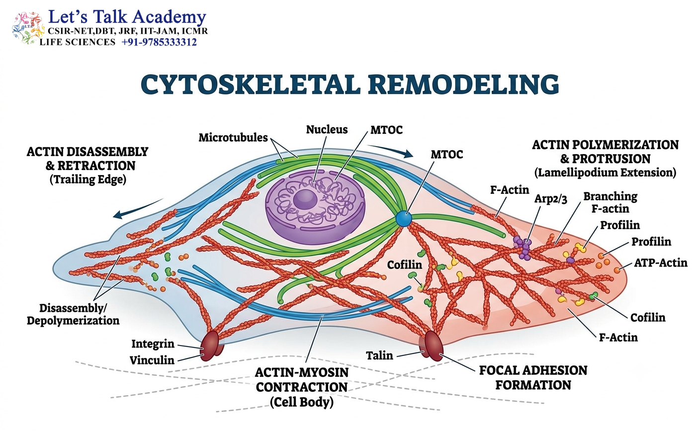
Cytoskeletal remodeling refers to the controlled alteration of filament organization.
Remodeling can involve:
- Polymerization
- Depolymerization
- Branching
- Cross-linking
- Severing
- Bundling
- Reorientation
This remodeling allows cells to rapidly respond to external stimuli.
For example, when a cell receives a migration signal, actin filaments can be rapidly reorganized at the leading edge.
22. Cytoskeleton and Signal Transduction
The cytoskeleton is not simply an endpoint of cellular signaling. It can also participate in signal transduction.
Cell-surface receptors can communicate with cytoskeletal proteins through adaptor and signaling molecules.
Mechanical forces transmitted through adhesion complexes can influence intracellular signaling pathways.
Thus, the cytoskeleton contributes to both chemical signaling and mechanotransduction.
23. Cytoskeleton and Mechanotransduction
Mechanotransduction is the conversion of mechanical stimuli into biochemical signals.
Cells continuously experience mechanical forces from:
- Neighboring cells
- Extracellular matrix
- Fluid flow
- Tissue deformation
The cytoskeleton helps transmit these forces from the plasma membrane toward intracellular structures.
Actin filaments, intermediate filaments, adhesion complexes, and nuclear structures can work together to determine how a cell responds to mechanical stress.
24. Cytoskeleton and the Nucleus

The cytoskeleton is physically and functionally connected to the nucleus.
Connections between cytoskeletal elements and the nuclear envelope allow mechanical forces to be transmitted between the cell surface, cytoplasm, and nucleus.
This connection can influence:
- Nuclear shape
- Chromatin organization
- Gene regulation
- Cellular mechanical responses
The nucleus is therefore not mechanically isolated from the rest of the cell.
25. Cytoskeleton During Mitosis
Microtubules undergo extensive reorganization during mitosis.
The interphase microtubule network is dismantled and reorganized into the mitotic spindle.
The spindle contains different populations of microtubules, including:
- Kinetochore microtubules
- Interpolar microtubules
- Astral microtubules
These microtubules cooperate to position and segregate chromosomes accurately.
25.1 Kinetochore Microtubules
These microtubules interact with kinetochores assembled at chromosome centromeres.
25.2 Interpolar Microtubules
These extend toward the opposite spindle pole and interact with microtubules from the other pole.
25.3 Astral Microtubules
These extend toward the cell cortex and help position and orient the mitotic spindle.
26. Cytoskeleton in Plants
Plant cells also possess cytoskeletal systems consisting of microtubules and actin filaments.
Because plant cells have rigid cell walls, their cytoskeleton has specialized roles in:
- Cell division
- Cell expansion
- Vesicle trafficking
- Organelle movement
- Cell polarity
Plant cortical microtubules are particularly important in determining the orientation of cellulose deposition and therefore influence cell growth.
Actin networks facilitate cytoplasmic streaming and intracellular transport.
27. Cytoplasmic Streaming

Cytoplasmic streaming is the directed movement of cytoplasm within certain cells.
It is strongly associated with actin filaments and myosin motors.
Cytoplasmic streaming facilitates the redistribution of:
- Organelles
- Vesicles
- Nutrients
- Other cellular materials
It is particularly prominent in large plant cells.
28. Cytoskeleton and Organelle Positioning
The cytoskeleton helps maintain the spatial arrangement of organelles.
Microtubules and actin filaments provide physical tracks for motor-driven positioning.
For example, mitochondria can be transported to regions with high energy requirements, while vesicles can be directed toward specific cellular compartments.
Thus, intracellular organization depends heavily on cytoskeletal architecture.
29. Functional Integration of Cytoskeletal Systems
The three cytoskeletal systems do not function independently.
Microtubules, actin filaments, and intermediate filaments interact directly or indirectly through cross-linking proteins and cellular structures.
For example:
Microtubules → long-distance transport and organization
Actin → cortical organization, contraction, and migration
Intermediate filaments → mechanical stability
Together, these systems allow cells to maintain both flexibility and structural integrity.
30. Important Molecular Concepts
Several concepts are fundamental for understanding cytoskeletal function.
30.1 Polymerization and Depolymerization
Filaments are formed by addition of protein subunits and can be disassembled when subunits are removed.
30.2 Structural Polarity
Microtubules and actin filaments possess plus and minus ends, which influence their growth and interactions with motor proteins.
30.3 Motor Activity
Motor proteins convert ATP hydrolysis into mechanical movement.
30.4 Dynamic Instability
Microtubules can switch rapidly between growth and shrinkage.
30.5 Treadmilling
Actin filaments can undergo a process in which subunits are preferentially added at one end and removed from the other, producing apparent movement of the filament while maintaining approximately constant length.
31. Cytoskeleton and Cellular Motility: Integrated Model

Cellular motility can be understood as the coordinated interaction of three major processes:
Force generation + cytoskeletal remodeling + adhesion/traction
Actin polymerization can push the membrane outward.
Myosin can generate contractile forces.
Microtubules can regulate polarity and transport materials.
Adhesion complexes connect the cytoskeleton to the extracellular environment.
The combined action of these mechanisms allows a cell to convert molecular activity into organized movement.
32. Biological Importance of the Cytoskeleton
The cytoskeleton is essential for virtually every aspect of cellular organization.
Its major functions include:
- Maintaining cell shape
- Organizing intracellular components
- Facilitating intracellular transport
- Supporting cell migration
- Enabling muscle contraction
- Controlling cytokinesis
- Organizing the mitotic spindle
- Supporting ciliary and flagellar movement
- Maintaining tissue integrity
- Participating in mechanotransduction
- Establishing cell polarity
- Supporting neuronal transport
- Positioning organelles
- Connecting cells with their extracellular environment
33. Key Differences Between Cytoskeletal Components
A useful way to understand the cytoskeleton is to associate each filament system with its dominant physical and biological role.
Microtubules are suited for long-range organization, intracellular transport, and chromosome movement because of their large diameter, rigidity, and polarity.
Actin filaments are particularly suited for cell-surface remodeling, contraction, and migration because they can rapidly reorganize and interact with myosin.
Intermediate filaments are specialized for mechanical strength because of their high tensile resistance and relatively stable organization.
These properties are complementary rather than mutually exclusive.


