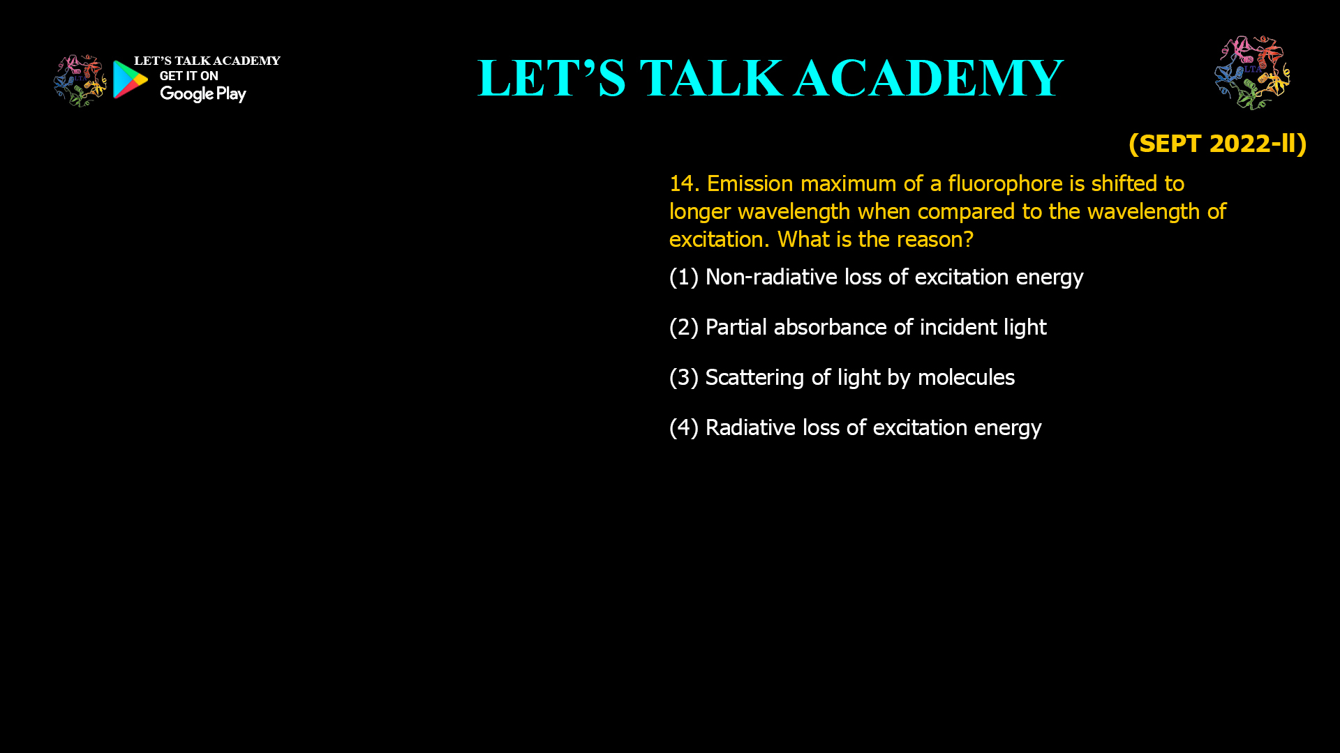
14. Emission maximum of a fluorophore is shifted to longer wavelength when compared to the wavelength of excitation. What is the reason?
(1) Non-radiative loss of excitation energy
(2) Partial absorbance of incident light
(3) Scattering of light by molecules
(4) Radiative loss of excitation energy
Why is the Emission Maximum of a Fluorophore Shifted to a Longer Wavelength? Understanding the Stokes Shift
Fluorescence spectroscopy is one of the most powerful analytical techniques used in biochemistry, molecular biology, biotechnology, medicine, environmental science, and materials research. A fundamental property of fluorescence is that the emitted light usually possesses a longer wavelength and lower energy than the light used to excite the fluorophore. This phenomenon is known as the Stokes shift and forms the basis of fluorescence imaging, flow cytometry, fluorescence microscopy, FRET analysis, and numerous biochemical assays.
Correct Answer
Option (1): Non-radiative Loss of Excitation Energy
The correct answer is Option (1). After a fluorophore absorbs an excitation photon, the molecule is promoted from the ground electronic state to a higher excited electronic state. However, fluorescence emission does not occur immediately. Before emitting light, the excited molecule rapidly loses a portion of its energy through non-radiative processes such as vibrational relaxation and internal conversion. These processes release energy as heat to the surrounding environment without producing photons.
Because part of the absorbed energy has already been lost before fluorescence occurs, the emitted photon contains less energy than the absorbed photon. Since photon energy is inversely proportional to wavelength, lower-energy photons possess longer wavelengths. Consequently, the fluorescence emission maximum appears at a longer wavelength than the excitation wavelength. This shift toward longer wavelengths is known as the Stokes shift.
Understanding the Stokes Shift
The Stokes shift represents the difference between the wavelength of maximum excitation and the wavelength of maximum fluorescence emission. In almost all conventional fluorophores, the emission spectrum is displaced toward the red region of the spectrum because the excited molecule undergoes rapid energy relaxation before emitting light.
Immediately after excitation, the molecule occupies one of the higher vibrational levels of the excited electronic state. Within picoseconds, vibrational relaxation transfers this excess vibrational energy to surrounding solvent molecules through molecular collisions. Internal conversion may also occur, allowing transitions between electronic states without photon emission. Only after these non-radiative relaxation processes does fluorescence occur from the lowest vibrational level of the excited state to one of the vibrational levels of the ground state.
Since some excitation energy has already been dissipated as heat, the emitted fluorescence possesses lower energy and therefore a longer wavelength than the absorbed light. This energy difference between absorption and emission forms the physical basis of the Stokes shift.
Role of the Jablonski Diagram
The Jablonski diagram provides a graphical representation of fluorescence processes. Vertical upward arrows represent photon absorption, while downward straight arrows indicate fluorescence emission. Curved arrows within the excited state illustrate non-radiative processes such as vibrational relaxation and internal conversion. These non-radiative transitions reduce the internal energy of the excited molecule before fluorescence occurs, explaining why the emitted photon is less energetic than the absorbed photon.
Understanding the Jablonski diagram is essential for interpreting fluorescence spectra, fluorescence lifetime measurements, energy transfer experiments, and photophysical behavior of fluorophores in biological systems.
Why Option (1) is Correct – Non-radiative Loss of Excitation Energy
Non-radiative relaxation allows the excited molecule to lose excess energy without emitting photons. This energy is transferred to molecular vibrations and eventually dissipated as heat into the surrounding solvent. As a result, fluorescence originates from a lower-energy excited state than the one initially populated during excitation. Consequently, the emitted photon carries less energy, producing fluorescence at a longer wavelength. This mechanism is responsible for the Stokes shift observed in almost every fluorescent molecule.
Why Option (2) is Incorrect – Partial Absorbance of Incident Light
Partial absorption simply means that not every incident photon is absorbed by the sample. It affects the intensity of excitation reaching the fluorophore and may influence fluorescence intensity, but it does not alter the energy of the emitted photons. Therefore, partial absorbance cannot explain why fluorescence emission shifts toward longer wavelengths.
Why Option (3) is Incorrect – Scattering of Light by Molecules
Scattering changes the direction of light propagation rather than its intrinsic energy. Elastic scattering, such as Rayleigh scattering, preserves wavelength, while Raman scattering involves different physical mechanisms unrelated to fluorescence emission maxima. Consequently, molecular scattering cannot account for the characteristic red shift observed in fluorescence spectra.
Why Option (4) is Incorrect – Radiative Loss of Excitation Energy
Radiative processes involve the emission of photons. Fluorescence itself is a radiative process, but the wavelength shift occurs before fluorescence emission, during the non-radiative relaxation stage. Since the energy reduction takes place prior to photon emission, radiative loss cannot explain the Stokes shift.
Importance of the Stokes Shift in Fluorescence Spectroscopy
A large Stokes shift is highly desirable in fluorescence experiments because it allows the excitation light and emitted fluorescence to be separated efficiently using optical filters. This minimizes background interference, improves signal-to-noise ratio, and enhances sensitivity in fluorescence microscopy, confocal imaging, flow cytometry, fluorescence resonance energy transfer (FRET), biosensors, and clinical diagnostics. Fluorophores with larger Stokes shifts generally provide clearer imaging and more accurate quantitative measurements.
Applications in Biological Research
The principle of the Stokes shift is extensively applied in protein fluorescence studies, nucleic acid detection, immunofluorescence assays, live-cell imaging, enzyme activity measurements, fluorescence polarization, molecular interaction studies, and super-resolution microscopy. Researchers use the predictable separation between excitation and emission wavelengths to detect specific biomolecules with high sensitivity while reducing interference from excitation light.
Final Answer
Correct Option: (1) Non-radiative loss of excitation energy.
When a fluorophore absorbs light, it first reaches an excited electronic state and rapidly undergoes vibrational relaxation and internal conversion, losing part of its excitation energy as heat through non-radiative processes. Fluorescence emission then occurs from this lower-energy excited state, producing photons with lower energy and therefore longer wavelengths than the excitation light. This phenomenon is known as the Stokes shift, making Option (1) the correct answer.


