
1. Introduction to Protein Degradation
Proteins are dynamic molecules that continuously undergo synthesis, modification, folding, functioning, and degradation. Although much emphasis is placed on protein synthesis, the removal of proteins is equally important for maintaining normal cellular function. Protein degradation is the controlled process by which proteins are broken down into smaller peptides and amino acids. These products can subsequently be recycled for the synthesis of new proteins or used in other metabolic processes.
Protein degradation is not simply a mechanism for destroying unwanted proteins. It is an essential regulatory process that controls the abundance, activity, localization, and lifetime of many proteins. Cells selectively remove proteins that are damaged, misfolded, incorrectly assembled, no longer required, or whose continued activity could interfere with cellular regulation.
The cellular protein pool is therefore maintained through a balance between protein synthesis and protein degradation. This balance is commonly referred to as protein homeostasis or proteostasis.
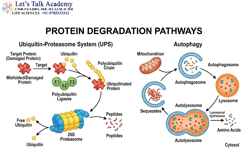
In eukaryotic cells, two major systems account for much of intracellular protein degradation:
- Ubiquitin–proteasome system (UPS)
- Autophagy–lysosome system
These pathways are not completely independent. They can cooperate and compensate for one another, particularly during conditions such as starvation, oxidative stress, protein misfolding, and other forms of cellular stress.
2. Importance of Protein Degradation
Protein degradation is essential because proteins have different lifetimes. Some proteins remain functional for many days, whereas others are synthesized and degraded within minutes or hours.
The controlled removal of proteins performs several important functions.
2.1 Maintenance of Protein Homeostasis
Protein homeostasis ensures that the correct amount of each protein is maintained inside a cell. Excess accumulation of proteins can disturb cellular processes, whereas insufficient levels of essential proteins can impair cellular function.
Protein degradation works together with protein synthesis and folding mechanisms to maintain a balanced proteome.
2.2 Removal of Damaged Proteins
Proteins can become damaged because of:
- Oxidative stress
- Heat stress
- Mutations
- Chemical modification
- Errors during protein synthesis
- Incorrect folding
- Environmental stress
Damaged proteins may lose their normal function or form toxic aggregates. Cells therefore recognize and eliminate many such proteins through regulated degradation pathways.
2.3 Regulation of Protein Activity
Protein degradation can function as a molecular switch.
When a regulatory protein is degraded, its cellular activity may rapidly decrease. This is particularly important in processes such as:
- Cell-cycle progression
- Signal transduction
- Transcriptional regulation
- Apoptosis
- Immune responses
- Development
- Metabolic regulation
Thus, degradation can regulate biological processes just as effectively as protein synthesis.
2.4 Removal of Short-Lived Regulatory Proteins
Many regulatory proteins are deliberately short-lived. Their rapid degradation allows cells to respond quickly to changes in their environment.
For example, degradation of specific cell-cycle regulators allows the cell to progress from one stage of the cell cycle to another.
2.5 Recycling of Cellular Components
Degradation converts proteins and other macromolecules into smaller molecules. Amino acids generated from protein breakdown can be reused for new protein synthesis or diverted into metabolic pathways.
This recycling becomes particularly important during nutrient limitation and starvation.
3. Major Pathways of Protein Degradation
Protein degradation in cells occurs through several mechanisms. The most important eukaryotic pathways are:
3.1 Ubiquitin–Proteasome System
The ubiquitin–proteasome system is a highly selective pathway that generally targets individual proteins for degradation. It is particularly important for the degradation of many short-lived, damaged, misfolded, and regulatory proteins.
3.2 Autophagy–Lysosome System
Autophagy delivers cellular material to lysosomes, where hydrolytic enzymes degrade the material. It is particularly important for long-lived proteins, protein aggregates, and larger cellular structures such as organelles.
3.3 Other Specialized Proteolytic Systems
Cells also possess specialized degradation mechanisms associated with particular cellular compartments, including the endoplasmic reticulum and mitochondria. These pathways contribute to protein quality control and organelle maintenance.
4. Ubiquitin–Proteasome System
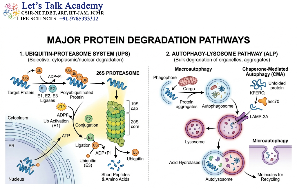
The ubiquitin–proteasome system is one of the best-characterized mechanisms of selective intracellular protein degradation.
The basic principle is relatively simple:
Target protein → ubiquitination → recognition by proteasome → unfolding → proteolysis → peptide products
The system depends on two major components:
- Ubiquitin
- 26S proteasome
Ubiquitin acts as a molecular tag, while the proteasome functions as the degradation machinery.
5. Ubiquitin: The Molecular Tag
Ubiquitin is a small, highly conserved protein consisting of 76 amino acids. It can be covalently attached to other proteins and thereby influence their fate.
Ubiquitination does not always mean that a protein will be degraded. Different ubiquitin modifications can produce different cellular outcomes.
Depending on the type and arrangement of ubiquitin modification, ubiquitination can influence:
- Protein degradation
- Protein trafficking
- DNA repair
- Cellular signaling
- Endocytosis
- Protein localization
- Regulation of protein activity
Therefore, ubiquitin should be considered not merely a degradation label but an important regulatory signal.
6. Mechanism of Ubiquitination
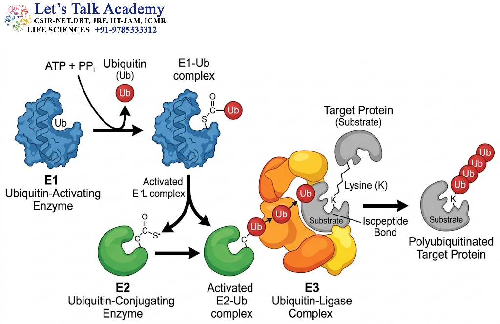
Ubiquitination generally involves three major enzyme classes:
- E1 – Ubiquitin-activating enzyme
- E2 – Ubiquitin-conjugating enzyme
- E3 – Ubiquitin ligase
These enzymes work sequentially to attach ubiquitin to a target protein.
6.1 Activation of Ubiquitin by E1
The first step is the activation of ubiquitin by an E1 enzyme.
This process requires ATP. The C-terminal region of ubiquitin is activated and linked to E1 through a high-energy intermediate.
This activation prepares ubiquitin for subsequent transfer.
6.2 Transfer to E2
Activated ubiquitin is transferred from E1 to an E2 ubiquitin-conjugating enzyme.
The E2 enzyme therefore acts as a carrier of activated ubiquitin.
6.3 Recognition by E3
The E3 ubiquitin ligase plays a major role in substrate specificity.
The E3 enzyme recognizes a particular target protein or degradation signal, commonly called a degron, and facilitates the transfer of ubiquitin to the substrate.
Because cells contain many different E3 ligases, different proteins can be selectively recognized and regulated.
6.4 Formation of Polyubiquitin Chains
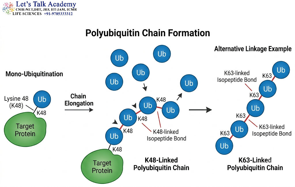
Additional ubiquitin molecules can be attached to the ubiquitin already present on the substrate.
This produces a polyubiquitin chain.
The exact linkage between ubiquitin molecules is biologically important because different ubiquitin-chain architectures can generate different cellular signals.
For example, K48-linked polyubiquitin chains are classically associated with proteasomal degradation, whereas K63-linked ubiquitin chains frequently participate in signaling, DNA repair, and trafficking rather than serving as the conventional proteasomal degradation signal.
7. Deubiquitinating Enzymes
Ubiquitination is a reversible process.
Deubiquitinating enzymes (DUBs) remove ubiquitin molecules from proteins or ubiquitin chains.
DUBs perform several functions, including:
- Editing ubiquitin chains
- Regulating ubiquitin-chain length
- Recycling ubiquitin
- Preventing inappropriate degradation
- Regulating protein stability
- Controlling ubiquitin-dependent signaling
Therefore, cellular protein degradation depends not only on ubiquitin attachment but also on precise regulation of ubiquitin removal.
8. The 26S Proteasome
The 26S proteasome is a large ATP-dependent proteolytic complex responsible for degrading many ubiquitinated proteins.
It can be broadly divided into:
- 20S core particle
- 19S regulatory particles
The 20S core contains the proteolytic chamber, while the 19S regulatory particles recognize ubiquitinated substrates, remove or process ubiquitin chains, and facilitate substrate unfolding and entry into the proteolytic chamber.
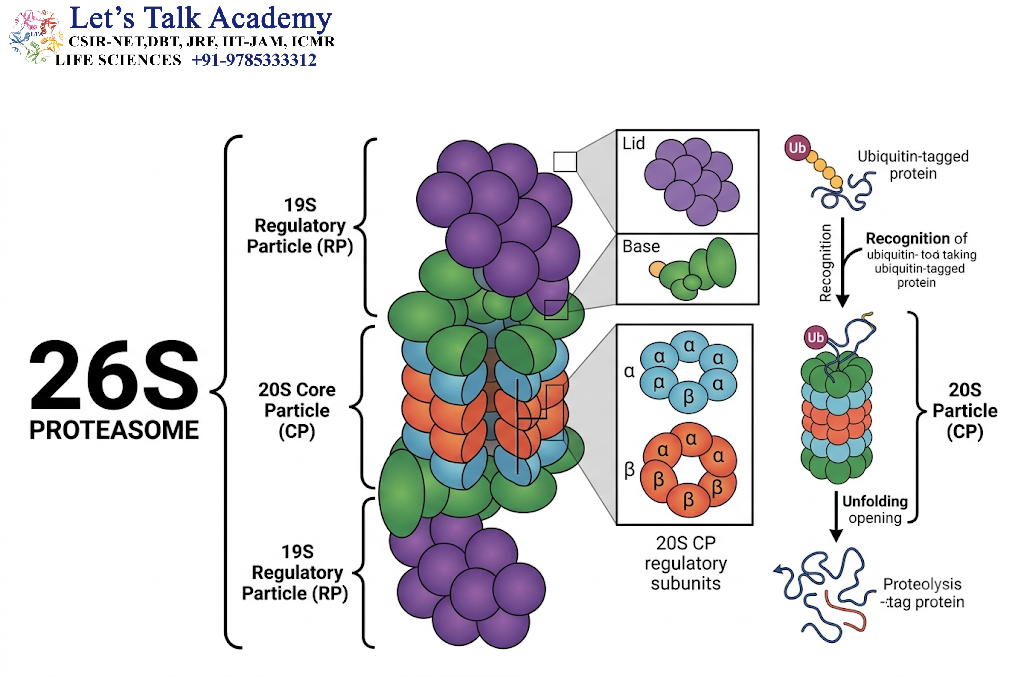
8.1 20S Core Particle
The 20S core has a cylindrical structure composed of multiple protein rings.
Its internal chamber contains proteolytic active sites.
The structure provides a protected environment in which proteins can be broken down into smaller peptide fragments.
8.2 19S Regulatory Particle
The 19S regulatory particle controls access to the proteolytic chamber.
Its major functions include:
- Recognition of ubiquitinated proteins
- Processing of ubiquitin chains
- ATP-dependent unfolding of substrates
- Translocation of unfolded proteins into the core
Thus, the proteasome functions as a highly organized molecular machine rather than simply as a nonspecific protease.
9. Steps of Proteasomal Protein Degradation
The overall process can be summarized as follows:
Step 1: A target protein is identified through a degradation signal.
Step 2: The protein is ubiquitinated by the E1–E2–E3 enzymatic system.
Step 3: A suitable ubiquitin chain is assembled.
Step 4: The ubiquitinated substrate is recognized by the proteasome.
Step 5: Ubiquitin is removed and recycled.
Step 6: The substrate protein is unfolded using ATP-dependent mechanisms.
Step 7: The unfolded protein enters the 20S proteolytic chamber.
Step 8: Proteolytic active sites cleave the protein into peptide fragments.
Step 9: Peptides are released and can subsequently be processed into amino acids.
10. Degrons and Protein Stability
A degron is a sequence, structural feature, or molecular characteristic that can promote recognition of a protein by a degradation system.
The presence or exposure of a degron can determine whether a protein is stable or rapidly degraded.
Degrons may be:
- Sequence-based
- Structure-based
- Created by post-translational modification
- Exposed following protein damage
- Located at the N-terminal or C-terminal region
- Associated with internal regions of proteins
The concept of degrons helps explain how cells distinguish proteins that should remain stable from proteins that should be eliminated.
11. N-End Rule Pathway
The N-end rule pathway is a regulatory mechanism in which the identity of the amino acid at the N-terminus of a protein can influence its stability.
Some N-terminal residues are associated with relatively stable proteins, whereas others can contribute to recognition by degradation machinery.
The N-terminal residue can therefore function as part of a degradation signal.
This pathway illustrates an important principle of protein degradation: the lifetime of a protein can be encoded within its molecular structure.
12. Autophagy and Lysosomal Protein Degradation
Autophagy is another major cellular degradation pathway.
The word autophagy means “self-eating.” In biological terms, it refers to pathways through which cellular material is delivered to lysosomes for degradation.
Unlike the proteasome, which is highly suited to the degradation of individual proteins, autophagy can eliminate larger structures, including:
- Protein aggregates
- Long-lived proteins
- Damaged organelles
- Portions of cytoplasm
- Certain intracellular pathogens
Autophagy therefore serves both as a degradation pathway and as a cellular quality-control mechanism.
13. Major Forms of Autophagy
Autophagy is not a single pathway. Several forms have been described.
13.1 Macroautophagy
Macroautophagy involves the formation of a double-membrane structure called an autophagosome.
The general sequence is:
Cargo selection → phagophore formation → membrane expansion → autophagosome formation → fusion with lysosome → degradation
The lysosome contains hydrolytic enzymes that break down the material delivered by the autophagosome.
13.2 Microautophagy
During microautophagy, the lysosomal or vacuolar membrane directly invaginates and captures cytoplasmic material.
The captured material is subsequently degraded within the lysosomal compartment.
13.3 Chaperone-Mediated Autophagy
Chaperone-mediated autophagy differs from macroautophagy because selected proteins are delivered directly across the lysosomal membrane with the assistance of molecular chaperones.
This provides a selective mechanism for the degradation of particular soluble proteins.
14. Molecular Mechanism of Macroautophagy
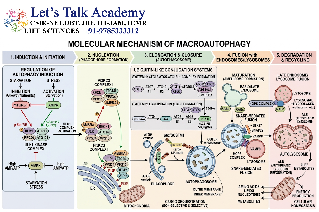
Macroautophagy is a tightly regulated multistep process.
14.1 Initiation
Autophagy is often stimulated when cells experience nutrient deprivation or other forms of stress.
A key regulatory component is the ULK complex, which initiates the autophagy program.
14.2 Phagophore Formation
A membrane structure known as the phagophore begins to form.
This structure expands around cellular material selected for degradation.
14.3 Autophagosome Formation
The growing phagophore eventually closes to produce a double-membrane vesicle known as the autophagosome.
14.4 Lysosomal Fusion
The autophagosome fuses with a lysosome.
This produces an environment in which lysosomal hydrolases can degrade the enclosed material.
14.5 Breakdown and Recycling
Proteins and other macromolecules are broken down into smaller components.
The resulting molecules can then be released and reused by the cell.
The molecular machinery involved in autophagy includes proteins such as ULK1, Beclin-1, VPS34, ATG5, ATG12, ATG16L1, and LC3. LC3 processing and membrane association are important features of autophagosome formation.
15.Selective Autophagy
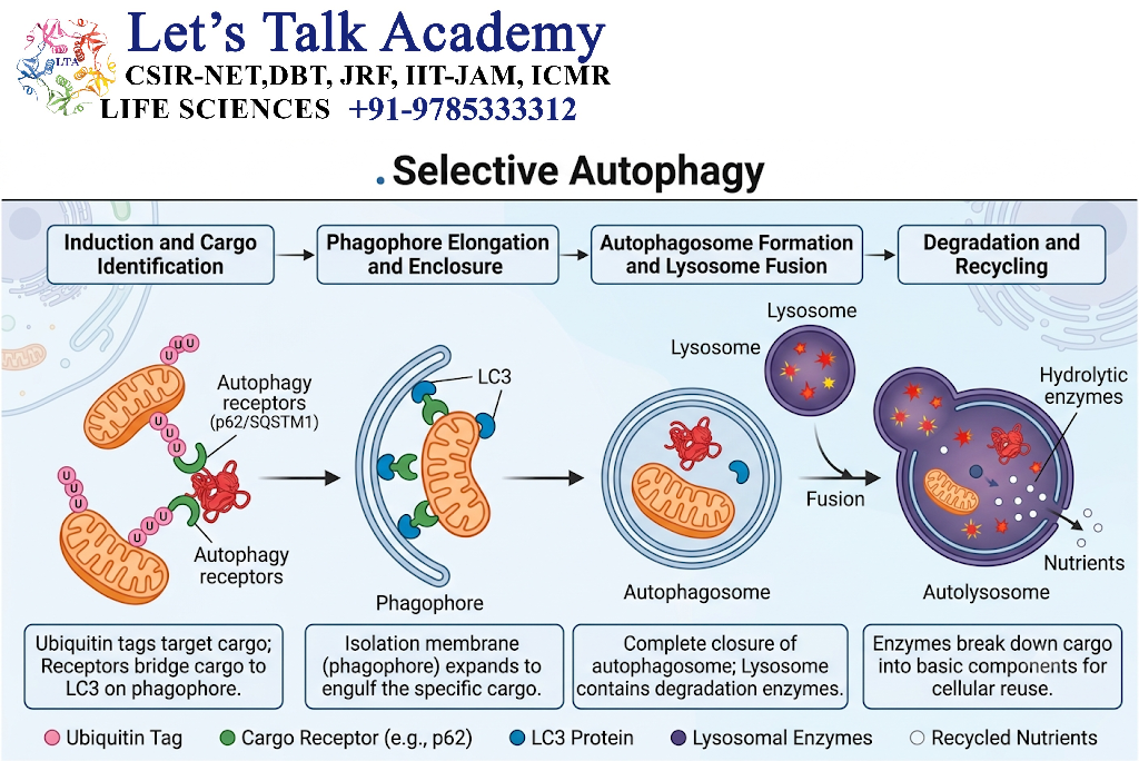
Autophagy was traditionally viewed as a relatively nonspecific bulk degradation pathway. However, many forms of autophagy are highly selective.
Selective autophagy can target:
- Protein aggregates
- Damaged mitochondria
- Endoplasmic reticulum
- Peroxisomes
- Ribosomes
- Intracellular pathogens
Cargo receptors can recognize specific substrates and connect them to the autophagic machinery.
For example, proteins such as p62/SQSTM1 can participate in the recognition and delivery of ubiquitinated cargo to autophagosomes.
16. Protein Quality Control
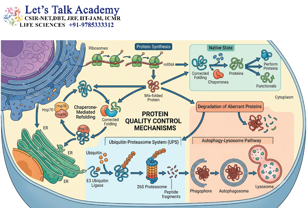
Protein quality control is a fundamental function of degradation systems.
Newly synthesized proteins must fold correctly to acquire their functional structures. Molecular chaperones assist in folding and can sometimes help misfolded proteins return to their native state.
However, if a protein cannot be repaired, it may be targeted for degradation.
The basic quality-control sequence can therefore be represented as:
Protein synthesis → folding → functional protein
or, when folding fails:
Protein synthesis → misfolding → chaperone recognition → refolding
If refolding is unsuccessful:
Misfolded protein → degradation → recycling
This coordinated system prevents the accumulation of potentially toxic proteins.
17. Protein Degradation During Cellular Stress
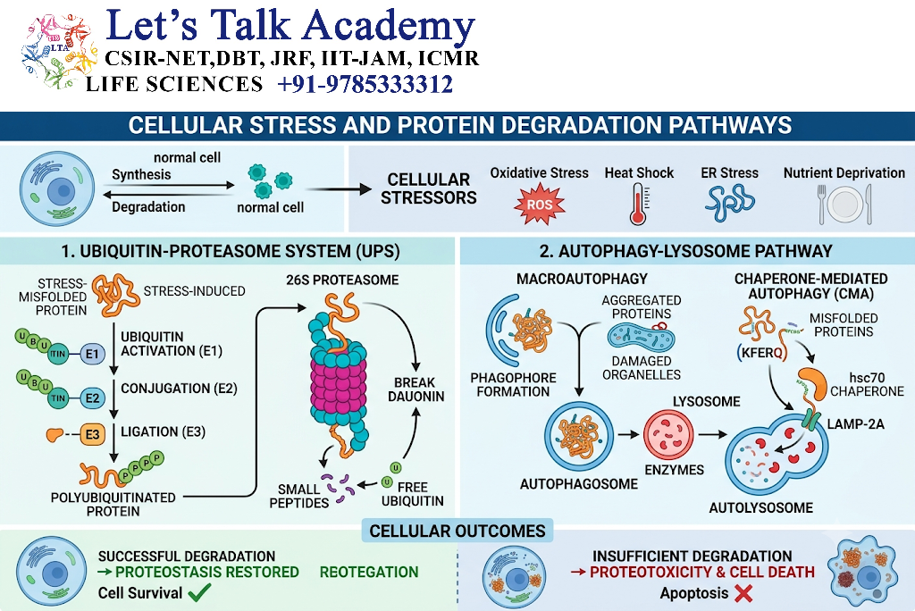
Cells experience various forms of stress, including:
- Heat stress
- Oxidative stress
- Nutrient deprivation
- Endoplasmic reticulum stress
- Hypoxia
- Proteotoxic stress
Under these conditions, protein degradation becomes especially important.
Misfolded and aggregated proteins can accumulate when protein synthesis and folding systems become overloaded. The ubiquitin–proteasome system and autophagy can cooperate to remove these abnormal proteins.
18. Endoplasmic Reticulum-Associated Degradation
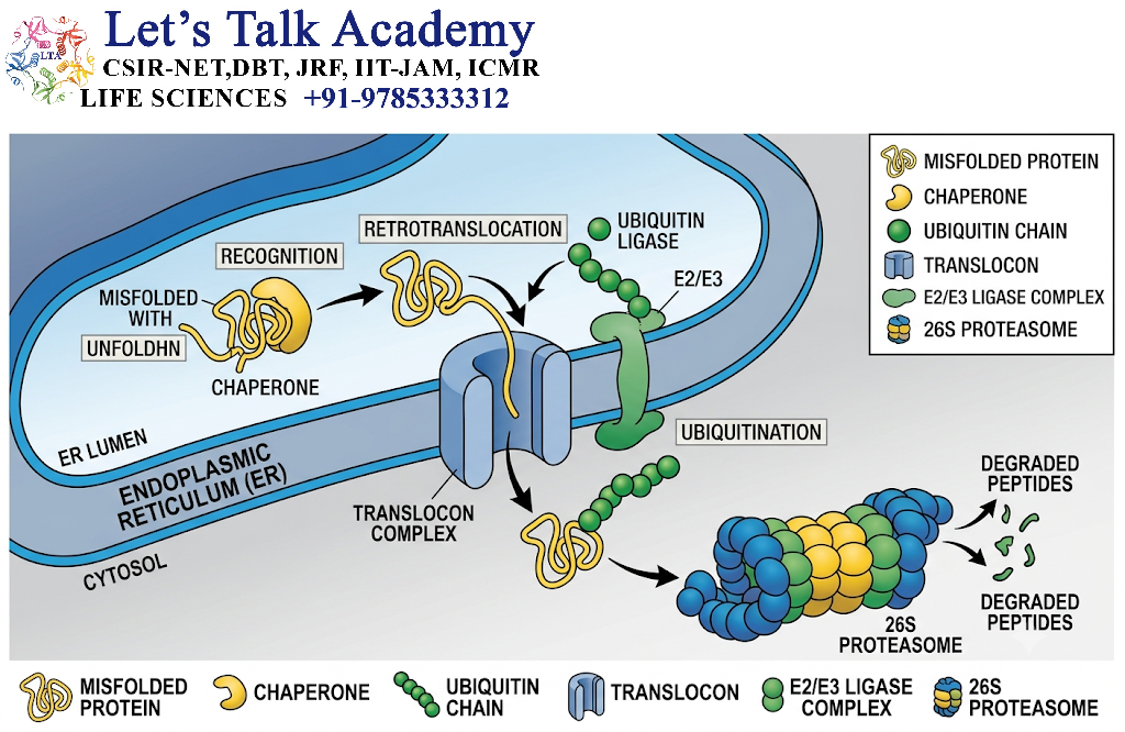
The endoplasmic reticulum is responsible for the synthesis and folding of many secreted and membrane proteins.
Proteins that fail to fold correctly in the endoplasmic reticulum can be recognized and removed through a process known as ER-associated degradation (ERAD).
In simplified terms:
Misfolded ER protein → recognition → retrotranslocation → ubiquitination → proteasomal degradation
ERAD is therefore an important component of cellular protein quality control.
19. Mitochondrial Protein Degradation
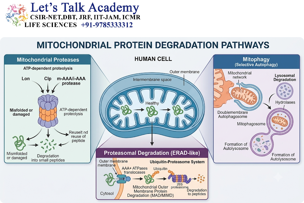
Mitochondria also possess specialized mechanisms for maintaining protein quality.
Mitochondrial proteins can become damaged through oxidative stress and other forms of cellular injury.
Protein quality-control mechanisms within mitochondria include:
- Mitochondrial proteases
- Ubiquitin-dependent pathways
- Selective autophagy of mitochondria, known as mitophagy
Mitophagy allows cells to remove damaged or dysfunctional mitochondria through autophagic degradation.
20. Ubiquitin–Proteasome System Versus Autophagy
The ubiquitin–proteasome system and autophagy have overlapping but distinct functions.
| Feature | Ubiquitin–Proteasome System | Autophagy–Lysosome System |
|---|---|---|
| Major machinery | Proteasome | Lysosome |
| Typical substrate | Individual proteins | Proteins, aggregates, organelles |
| Selectivity | Generally highly selective | Can be bulk or selective |
| Major role | Short-lived and regulatory proteins | Long-lived proteins and larger structures |
| Energy requirement | ATP-dependent | Energy-dependent cellular process |
| Ubiquitin involvement | Frequently central | Can be involved in selective autophagy |
| Final degradation site | Proteasome | Lysosome |
The distinction is not absolute. The two systems communicate extensively and can compensate for one another under certain conditions.
21. Crosstalk Between Proteasome and Autophagy
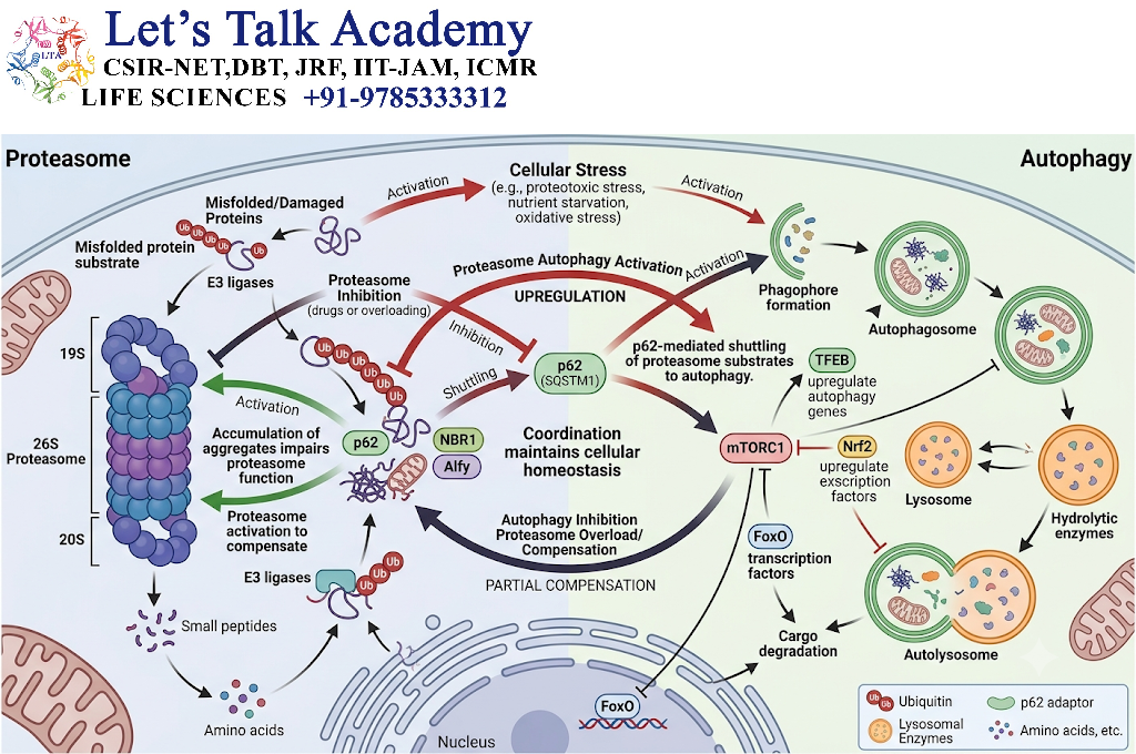
The ubiquitin–proteasome system and autophagy are increasingly understood as components of an interconnected protein-clearance network.
When one pathway becomes insufficient, the other may contribute to maintaining proteostasis.
For example, accumulation of protein aggregates that cannot efficiently enter the proteasome can stimulate selective autophagy.
Conversely, components of the proteasomal machinery can themselves become targets of autophagic degradation.
This functional cooperation becomes especially important during cellular stress.
22. Regulation of Protein Degradation
Protein degradation must be carefully regulated because inappropriate degradation can be as harmful as insufficient degradation.
Major regulatory factors include:
22.1 Substrate Recognition
Specific degradation signals determine which proteins are targeted.
22.2 E3 Ubiquitin Ligases
E3 ligases provide much of the substrate specificity within the ubiquitin system.
22.3 Deubiquitinating Enzymes
DUBs regulate ubiquitin attachment and remove ubiquitin from selected substrates.
22.4 Cellular Energy Status
Both proteasomal activity and autophagy are influenced by cellular energy availability.
22.5 Nutrient Availability
Nutrient deprivation strongly influences autophagy and promotes recycling of intracellular components.
22.6 Protein Folding Status
Misfolded and damaged proteins can be recognized by quality-control mechanisms and directed toward degradation.
23. Protein Degradation and Cell-Cycle Regulation
Protein degradation is particularly important during the cell cycle.
Cell-cycle regulators must appear and disappear at specific times.
The selective degradation of proteins such as cyclins and other regulatory factors helps ensure that cellular events occur in the correct sequence.
Thus, protein degradation acts as a molecular timing mechanism.
A protein does not necessarily need to be permanently inhibited. Instead, the cell can simply eliminate it when its function is no longer required.
24. Protein Degradation in Signal Transduction
Signal transduction pathways frequently depend on controlled protein turnover.
For example, degradation of inhibitory proteins can activate signaling pathways. A well-known example involves IκBα, whose ubiquitination and subsequent degradation allows NF-κB to become active and enter the nucleus.
This illustrates how degradation can function as an active component of signal transmission rather than merely as a disposal mechanism.
25. Protein Degradation and Apoptosis
Protein degradation contributes to programmed cell death.
During apoptosis, many cellular proteins are cleaved or degraded in a coordinated manner.
Proteolytic activity helps dismantle cellular structures and regulate the molecular pathways responsible for cell death.
At the same time, protein degradation systems can remove damaged proteins before they reach a level that triggers irreversible cellular damage.
26. Protein Degradation and Disease
Defects in protein degradation can disturb cellular homeostasis.
If damaged proteins are not efficiently removed, they may accumulate and form aggregates.
If proteins that should remain functional are degraded excessively, essential cellular pathways can also be disrupted.
Abnormal protein degradation has been associated with several broad disease processes, including:
- Neurodegenerative disorders
- Cancer
- Metabolic disorders
- Protein-folding diseases
- Immune dysfunction
- Disorders of cellular stress responses
Both excessive and insufficient degradation can therefore have pathological consequences.
27. Protein Aggregation and Cellular Toxicity
Protein aggregation occurs when abnormal or misfolded proteins associate to form larger assemblies.
Aggregation can be particularly harmful because aggregates may:
- Sequester functional proteins
- Disrupt organelles
- Interfere with intracellular transport
- Alter membrane function
- Overload quality-control systems
- Promote cellular stress
Cells therefore use molecular chaperones, the proteasome, and autophagy to prevent or remove harmful protein aggregates.
When these systems become overwhelmed, cellular damage can increase.
28. Role of Protein Degradation in Proteostasis
Proteostasis is the coordinated maintenance of the cellular protein environment.
It involves three major activities:
Protein synthesis + protein folding + protein degradation
Protein degradation ensures that proteins that cannot be properly folded, repaired, or appropriately maintained are removed.
Therefore, degradation is not the final step of protein biology. It is an integral part of the complete protein life cycle.
29. Protein Turnover
Protein turnover refers to the continuous process of protein synthesis and degradation.
A protein’s half-life depends on:
- Protein sequence
- Cellular location
- Structural stability
- Post-translational modifications
- Presence of degrons
- Cellular conditions
- Interaction with other proteins
- Activity of degradation machinery
Rapid turnover allows cells to respond quickly to environmental and developmental changes.
Slow turnover, in contrast, may be suitable for structural or long-lived proteins.
30. Energy Requirement in Protein Degradation
Protein degradation is often an energy-dependent process.
In the ubiquitin–proteasome pathway, ATP is required during ubiquitin activation and during ATP-dependent substrate processing and unfolding by the proteasome.
This energy requirement demonstrates that selective degradation is an actively regulated cellular process rather than simple spontaneous protein breakdown.
31. Biological Significance of Protein Degradation
Protein degradation contributes to almost every major aspect of cell biology.
Its major functions include:
- Maintenance of protein homeostasis
- Elimination of damaged proteins
- Removal of misfolded proteins
- Regulation of protein concentration
- Control of signaling pathways
- Regulation of the cell cycle
- Cellular adaptation to stress
- Recycling of amino acids
- Removal of damaged organelles
- Regulation of development and differentiation
- Maintenance of organelle quality
- Prevention of toxic protein accumulation
Protein degradation can therefore be considered one of the central mechanisms through which cells maintain their internal organization.
32. Integrated View of Cellular Protein Degradation
Protein degradation should not be understood as a single pathway.
Instead, cells possess an integrated network of degradation mechanisms.
A simplified model is:
Protein synthesis
↓
Protein folding and maturation
↓
Functional protein
↓
Normal turnover or regulatory degradation
↓
Ubiquitin–proteasome system / Autophagy–lysosome system
↓
Peptides and amino acids
↓
Recycling and metabolic utilization
For damaged proteins:
Misfolded or damaged protein
↓
Recognition by quality-control machinery
↓
Attempted refolding
↓
Successful refolding → Functional protein
or
Failed refolding → Protein degradation
This integrated system allows cells to continuously monitor and remodel their proteome.


