
1. Introduction
Proteins are synthesized at specific locations inside the cell, but they often perform their functions somewhere else. A protein synthesized in the cytoplasm may need to enter the nucleus, mitochondria, endoplasmic reticulum, lysosome, plasma membrane, or extracellular space.
The cellular processes that direct proteins to their correct destinations are collectively known as protein trafficking and transport.
Protein trafficking ensures that newly synthesized proteins are:
- transported to the correct cellular compartment,
- folded correctly,
- modified appropriately,
- delivered at the right time,
- retained in the correct location,
- or degraded when they are damaged or no longer required.
The cell therefore operates as a highly organized transport network.
A simplified pathway is:
Protein synthesis
↓
Targeting signal
↓
Recognition by transport machinery
↓
Transport
↓
Protein folding and modification
↓
Correct cellular destination
↓
Biological function
2. Definition of Protein Trafficking and Transport
2.1 Protein Transport
Protein transport is the movement of a protein from one cellular location to another through specific molecular mechanisms.
2.2 Protein Trafficking
Protein trafficking is the regulated process by which proteins are sorted, transported, modified, delivered, and maintained at their appropriate cellular destinations.
Transport is therefore one component of the broader trafficking process.
3. Importance of Protein Trafficking
Correct protein localization is essential because proteins usually function only in particular cellular compartments.
For example:
- DNA-associated proteins must reach the nucleus.
- Many metabolic proteins function in the cytoplasm.
- Respiratory proteins are present in mitochondria.
- Lysosomal enzymes must reach lysosomes.
- Many membrane receptors must reach the plasma membrane.
- Secreted proteins must pass through the secretory pathway.
If a protein reaches the wrong compartment, it may:
- become inactive,
- interact with inappropriate molecules,
- be degraded,
- disturb cellular processes,
- cause disease.
Thus:
Correct protein → Correct location → Correct function
4. Major Cellular Destinations of Proteins
Proteins may be targeted to:
- Cytoplasm
- Nucleus
- Nucleolus
- Mitochondria
- Endoplasmic reticulum
- Golgi apparatus
- Lysosomes
- Peroxisomes
- Plasma membrane
- Endosomes
- Extracellular space
5. Protein Targeting Signals
5.1 Definition
A protein targeting signal is a specific sequence or structural feature that directs a protein to a particular cellular destination.
These signals can occur:
- at the N-terminus,
- at the C-terminus,
- internally,
- as short amino acid sequences,
- as structural motifs.
Targeting signals are recognized by specific receptors or transport machinery.
6. Signal Sequences
Signal sequences act like molecular addresses.
For example:
Protein
↓
Targeting signal
↓
Recognition by receptor
↓
Transport machinery
↓
Specific organelle
Different organelles use different targeting signals.
7. Cytoplasmic Proteins
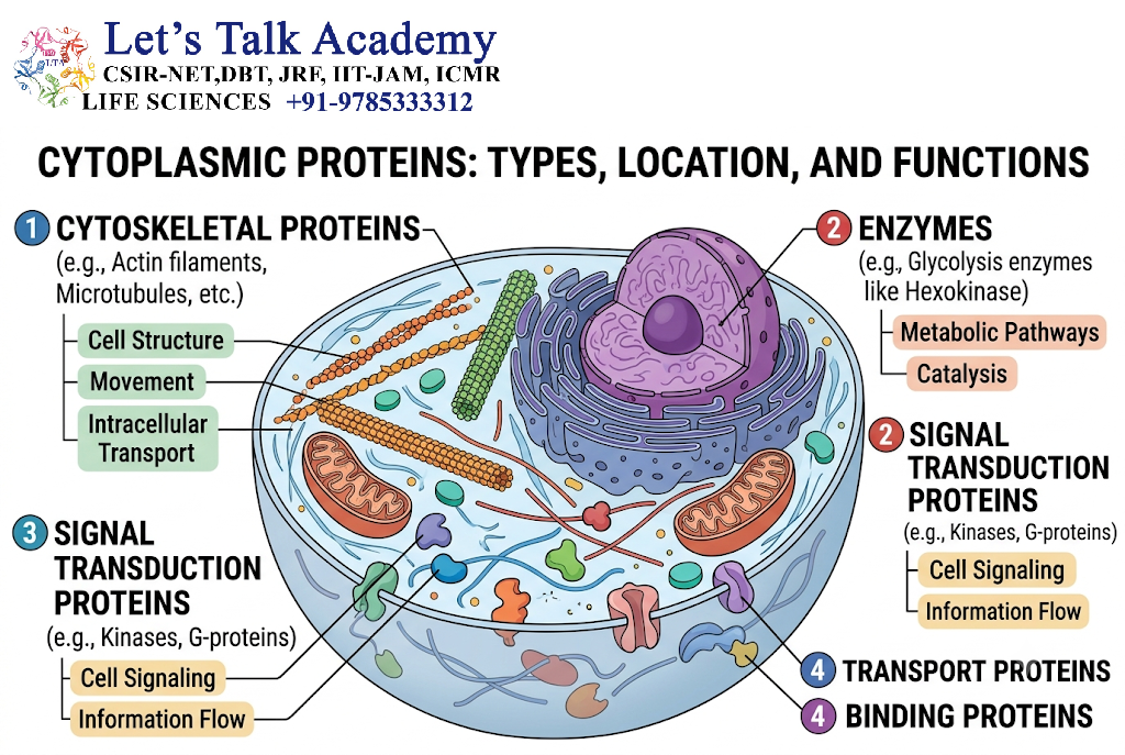
Many proteins remain in the cytoplasm after synthesis.
Such proteins generally lack signals that direct them to other organelles.
Examples include many:
- glycolytic enzymes,
- cytoskeletal proteins,
- metabolic enzymes.
Their synthesis occurs on free ribosomes in the cytoplasm.
8. Free and Bound Ribosomes
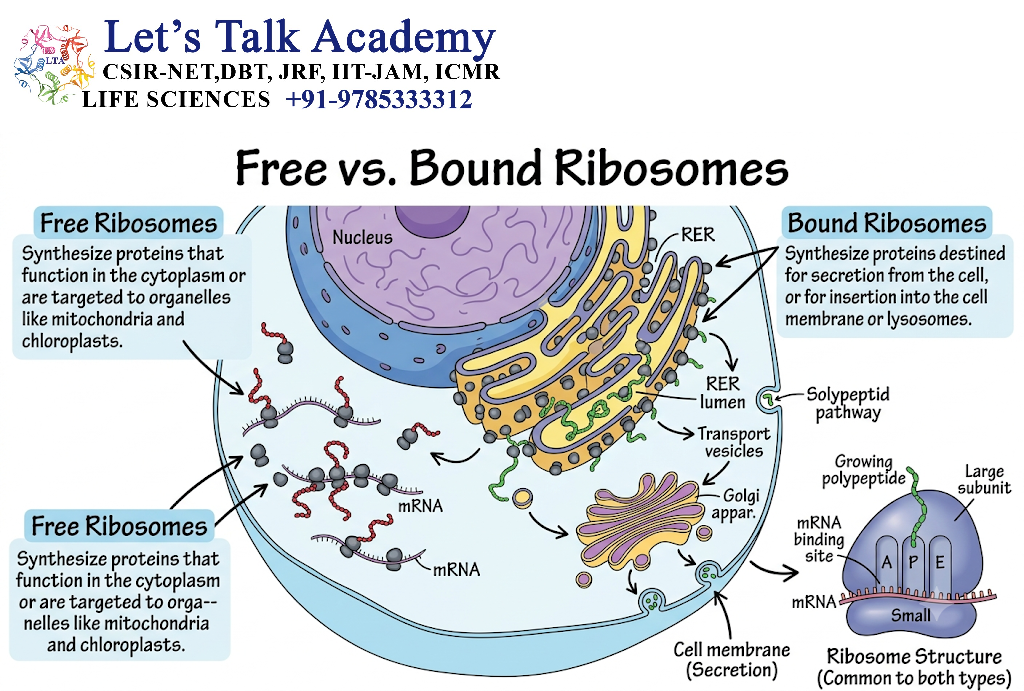
Ribosomes can be found as:
- free ribosomes in the cytosol,
- ribosomes associated with the rough ER.
An important point is that the same general ribosomal population can participate in different targeting pathways depending on the signals present on the newly synthesized protein.
Proteins synthesized on ribosomes associated with the ER generally enter the secretory pathway.
9. Two Major Protein Targeting Pathways
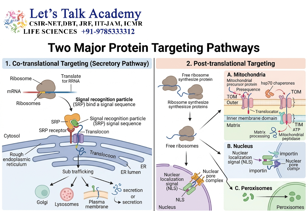
Protein transport can broadly be divided into:
9.1 Non-Vesicular Transport
Direct movement through:
- nuclear pores,
- protein translocators,
- membrane transport systems.
Examples:
- nuclear import,
- mitochondrial protein import,
- peroxisomal protein import.
9.2 Vesicular Transport
Proteins move between membrane-bound compartments inside vesicles.
Examples:
ER → Golgi
Golgi → plasma membrane
Golgi → lysosome
Endosome → lysosome
10. Endoplasmic Reticulum Targeting
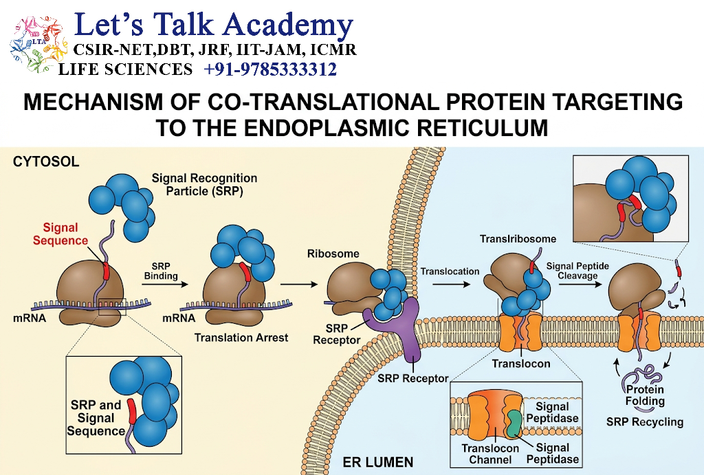
The endoplasmic reticulum (ER) is the entry point for proteins destined for the secretory pathway.
Proteins targeted to the ER commonly contain an N-terminal hydrophobic signal sequence.
These proteins include many:
- secreted proteins,
- membrane proteins,
- lysosomal proteins,
- Golgi proteins,
- ER-resident proteins.
11. Signal Recognition Particle
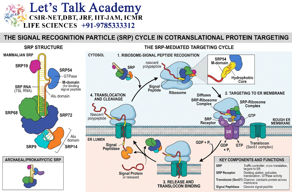
The signal recognition particle (SRP) recognizes the ER signal sequence on a newly synthesized protein.
The basic pathway is:
Nascent protein
↓
Signal sequence emerges
↓
SRP binds signal sequence
↓
Translation temporarily slows
↓
SRP-ribosome complex binds ER membrane
↓
Ribosome interacts with SRP receptor
↓
Protein is transferred to translocon
↓
Protein enters ER
12. SRP-Mediated Targeting
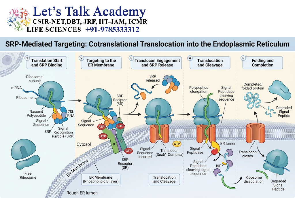
SRP is a ribonucleoprotein complex that plays a central role in targeting many newly synthesized proteins to the ER.
It recognizes both:
- the emerging signal sequence,
- the ribosome.
This ensures that the protein is delivered to the ER while it is being synthesized.
13. Translocon
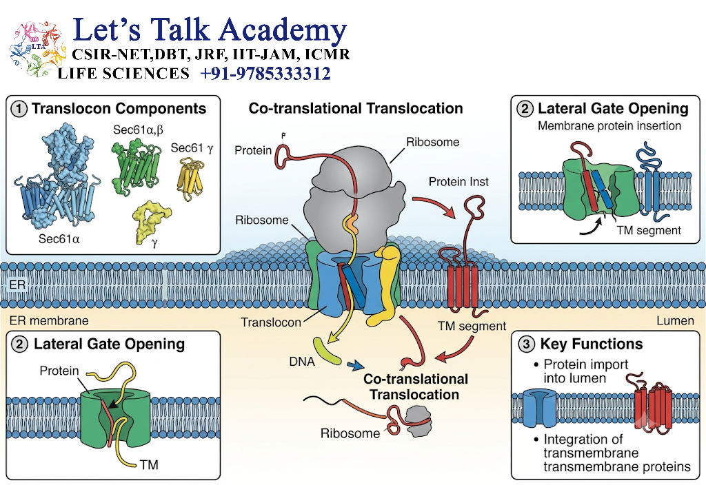
The translocon is a protein-conducting channel located in the ER membrane.
In eukaryotic cells, the central component is the:
Sec61 complex
The translocon allows a newly synthesized polypeptide to:
- cross the ER membrane,
- or become inserted into the membrane.
14. Co-Translational Translocation
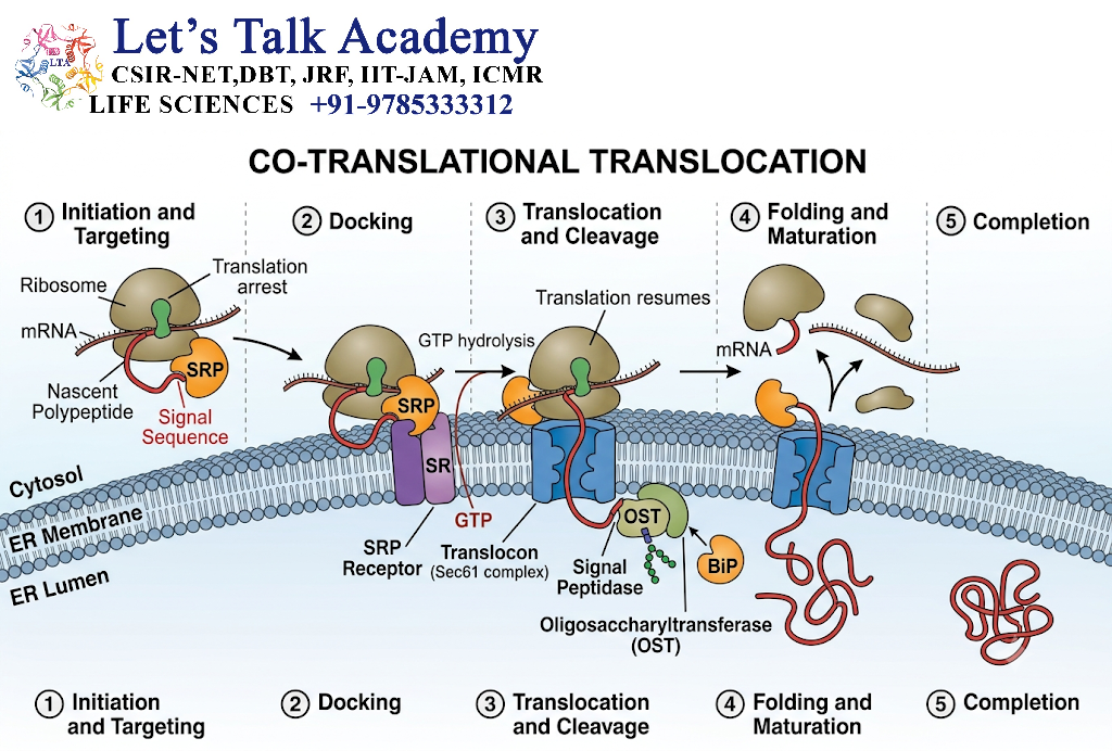
Many proteins enter the ER while translation is still occurring.
This is called:
Co-translational translocation
The process can be summarized as:
Ribosome
↓
Nascent protein
↓
Signal sequence recognized
↓
ER targeting
↓
Translocon
↓
Polypeptide enters ER
15. Post-Translational Translocation
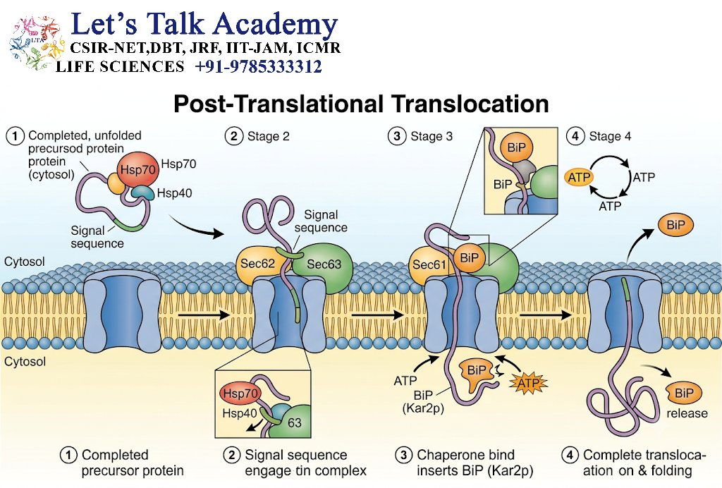
Some proteins are transported across membranes after translation is complete.
This is called:
Post-translational translocation
Different organisms and organelles use different molecular machinery for this process.
16.Signal Peptide Cleavage
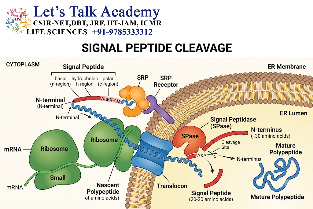
For many soluble secretory proteins, the ER signal peptide is removed after or during translocation.
The enzyme responsible is:
Signal peptidase
The pathway is:
Precursor protein
↓
Signal peptide
↓
ER translocation
↓
Signal peptide cleavage
↓
Mature protein
17. Membrane Protein Targeting
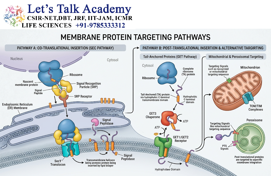
Not all ER-targeted proteins become soluble proteins.
Some are inserted into the ER membrane.
These proteins may eventually become:
- plasma membrane proteins,
- Golgi membrane proteins,
- lysosomal membrane proteins,
- ER membrane proteins.
18. Stop-Transfer Sequences
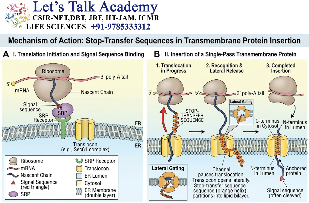
A stop-transfer sequence is a hydrophobic sequence that can stop the movement of a growing polypeptide through the translocon and promote membrane insertion.
The hydrophobic sequence remains embedded in the lipid bilayer.
19. Signal Anchor Sequences
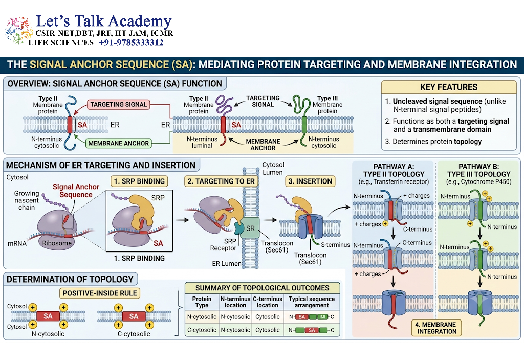
Some hydrophobic sequences function as both:
- targeting signals,
- membrane anchors.
These are called:
Signal-anchor sequences
They determine the insertion and orientation of membrane proteins.
20. Membrane Protein Topology
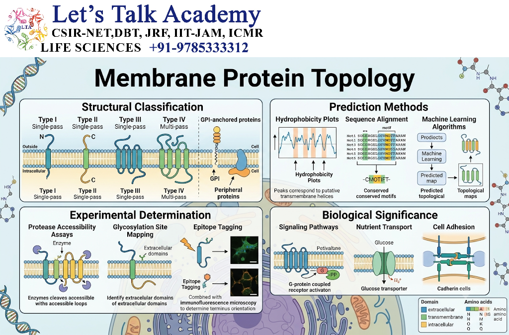
Membrane proteins can have different orientations.
Important categories include:
- single-pass membrane proteins,
- multipass membrane proteins,
- N-terminal lumenal proteins,
- C-terminal lumenal proteins.
The orientation is determined by:
- signal sequences,
- charged residues,
- translocon interactions,
- topology rules.
21. Endoplasmic Reticulum as a Protein Processing Center
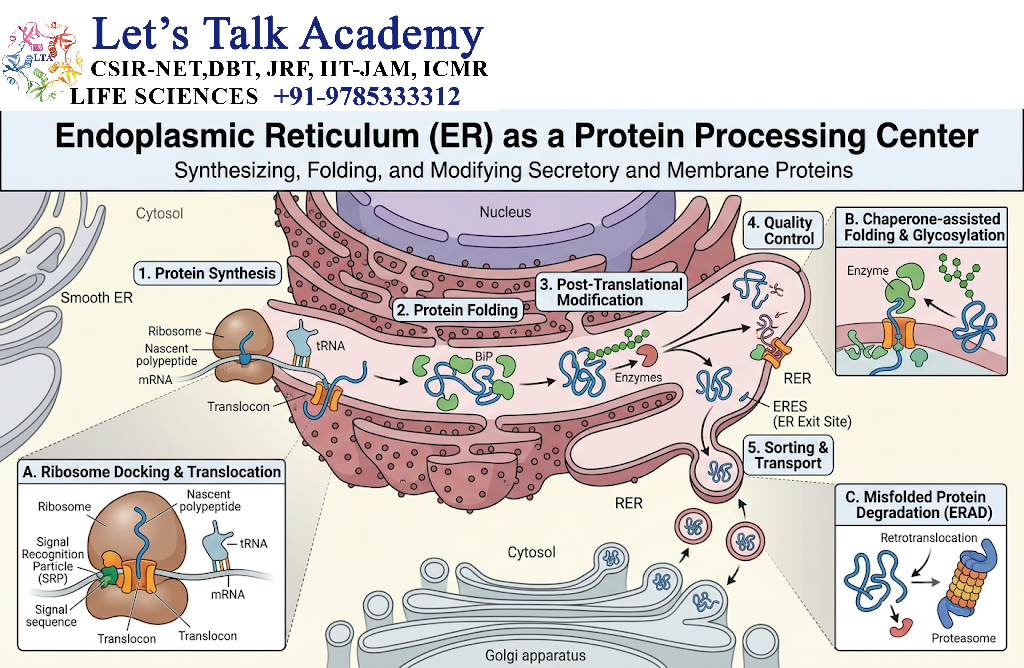
The ER is not simply a transport station.
It is also an important site for:
- protein folding,
- disulfide-bond formation,
- N-linked glycosylation,
- quality control.
Therefore:
ER targeting
↓
Transport + processing + quality control
22. Molecular Chaperones
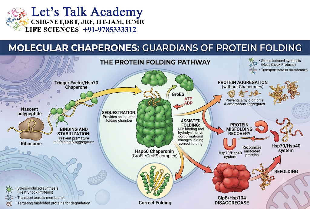
Molecular chaperones assist proteins in achieving proper folding.
Important ER chaperones include:
- BiP,
- calnexin,
- calreticulin.
They help prevent inappropriate protein aggregation and support proper folding.
23. Protein Quality Control in the ER
The ER monitors the folding state of proteins.
Correctly folded proteins:
↓
Continue through secretory pathway
Misfolded proteins:
↓
Retained
↓
Refolded or degraded
This process is called:
ER protein quality control
24. ER-Associated Degradation
Misfolded ER proteins can be removed through:
ER-associated degradation (ERAD)
The general pathway is:
Misfolded ER protein
↓
Recognition
↓
Retrotranslocation toward cytosol
↓
Ubiquitination
↓
Proteasomal degradation
This prevents accumulation of defective proteins.
25. Unfolded Protein Response
If misfolded proteins accumulate extensively in the ER, cells activate the:
Unfolded Protein Response (UPR)
The UPR attempts to restore protein homeostasis by:
- reducing protein synthesis,
- increasing protein-folding capacity,
- enhancing degradation of misfolded proteins.
If severe ER stress persists, cellular survival pathways can be affected.
26. Golgi Apparatus
The Golgi apparatus receives many proteins from the ER.
It performs:
- protein modification,
- glycan processing,
- sorting,
- packaging,
- trafficking.
The major direction is:
ER → Golgi → Final destination
27. Cis, Medial and Trans Golgi
The Golgi apparatus can be divided into functional regions:
- Cis-Golgi network
- Cis-Golgi cisternae
- Medial Golgi
- Trans-Golgi cisternae
- Trans-Golgi network
Proteins generally move through these compartments in an ordered manner while undergoing processing.
28. Vesicular Transport
A vesicle is a small membrane-bound structure that transports cargo between cellular compartments.
A typical vesicular transport pathway involves:
- Cargo selection
- Vesicle budding
- Vesicle transport
- Vesicle docking
- Membrane fusion
- Cargo delivery
29. Coat Proteins
Coat proteins help form transport vesicles.
Important coat systems include:
- COPII,
- COPI,
- clathrin.
30. COPII Vesicles
COPII-coated vesicles generally transport proteins:
ER → Golgi
They therefore participate in anterograde transport through the secretory pathway.
31. COPI Vesicles
COPI-coated vesicles are important for:
- Golgi → ER retrieval,
- intra-Golgi transport.
They contribute significantly to maintaining the correct composition of ER and Golgi compartments.
32. Clathrin-Coated Vesicles
Clathrin participates in several trafficking pathways.
Clathrin-coated vesicles can mediate transport from:
- trans-Golgi network,
- plasma membrane,
- endosomal compartments.
Clathrin is particularly important in:
Receptor-mediated endocytosis
and certain Golgi-to-endosome trafficking pathways.
33. Vesicle Budding
Vesicle formation begins when cargo is selected and a region of the membrane begins to curve.
Coat proteins help generate the budding vesicle.
The sequence is:
Cargo selection
↓
Coat assembly
↓
Membrane curvature
↓
Vesicle budding
↓
Coat removal
↓
Transport
34. Rab GTPases
Rab GTPases are important regulators of vesicular trafficking.
They help determine:
- vesicle identity,
- docking,
- target membrane recognition.
Different Rab proteins are associated with different trafficking steps.
35. SNARE Proteins
SNARE proteins are essential for membrane fusion.
They help bring:
- vesicle membrane,
- target membrane,
into close proximity.
The general pathway is:
Vesicle
↓
Target recognition
↓
SNARE pairing
↓
Membrane fusion
↓
Cargo release
36. Vesicle Docking
Before fusion, a vesicle must recognize and attach to the correct target membrane.
This involves:
- Rab proteins,
- tethering factors,
- SNARE proteins.
This provides specificity to intracellular transport.
37. Membrane Fusion
When vesicle and target-membrane SNAREs interact correctly, the membranes can fuse.
This releases the vesicle’s soluble cargo into the target compartment.
Membrane proteins become incorporated into the target membrane.
38. ER-to-Golgi Transport
Proteins leaving the ER are transported to the Golgi mainly through COPII-coated vesicles.
The pathway is:
ER
↓
COPII vesicle
↓
Golgi
↓
Protein modification and sorting
39. Golgi-to-ER Retrieval
Some ER-resident proteins accidentally escape to the Golgi.
They must be returned to the ER.
COPI-mediated transport contributes to this retrieval process.
Specific sorting signals help identify proteins that belong in the ER.
40. KDEL Signal
Many soluble ER-resident proteins contain a C-terminal:
KDEL
retrieval signal.
If these proteins escape to the Golgi, the KDEL receptor helps recognize them and promotes their return to the ER.
41. Lysosomal Targeting
Lysosomal proteins must be delivered from the secretory pathway to lysosomes.
Many soluble lysosomal hydrolases receive a carbohydrate-based sorting signal:
Mannose-6-phosphate (M6P)
42. Mannose-6-Phosphate Pathway
The pathway can be summarized as:
Lysosomal enzyme synthesized in ER
↓
ER processing
↓
Golgi modification
↓
Mannose-6-phosphate added
↓
M6P receptor recognition
↓
Transport to endosome
↓
Delivery to lysosome
43. Lysosomal Enzymes
Lysosomes contain hydrolytic enzymes capable of degrading:
- proteins,
- lipids,
- nucleic acids,
- carbohydrates,
- damaged cellular components.
Correct trafficking is therefore essential for lysosomal function.
44. Endosomal System
Endosomes are membrane-bound compartments involved in sorting internalized material.
Major stages include:
- early endosome,
- recycling endosome,
- late endosome.
Cargo can be:
- recycled to the plasma membrane,
- transported to lysosomes,
- sorted into other pathways.
45. Plasma Membrane Targeting
Many membrane proteins are transported:
ER
↓
Golgi
↓
Transport vesicles
↓
Plasma membrane
Examples include:
- receptors,
- ion channels,
- transporters,
- adhesion molecules.
46. Constitutive Secretion
Some proteins are continuously transported to the plasma membrane and extracellular space.
This is called:
Constitutive secretion
It does not require a specific external signal for every individual transport event.
47. Regulated Secretion
Some proteins are stored in secretory vesicles and released only after a specific signal.
This is called:
Regulated secretion
Examples include the release of:
- hormones,
- neurotransmitter-related cargo,
- digestive enzymes in specialized cells.
48. Nuclear Protein Transport
The nucleus is surrounded by the:
Nuclear envelope
It contains:
Nuclear pore complexes (NPCs)
These regulate movement between:
- cytoplasm,
- nucleus.
49. Nuclear Localization Signal
Proteins destined for the nucleus often contain a:
Nuclear localization signal (NLS)
NLS sequences are commonly enriched in basic amino acids such as:
- lysine,
- arginine.
50. Importins
Importins are transport receptors that recognize nuclear localization signals and facilitate nuclear import.
The general pathway is:
Cargo protein + NLS
↓
Importin binding
↓
Nuclear pore complex
↓
Transport into nucleus
↓
Cargo release
51. Ran GTPase
Nuclear transport depends heavily on the:
Ran GTPase system
Different Ran nucleotide states are distributed between the nucleus and cytoplasm.
This creates directionality in nuclear transport.
A simplified concept is:
Ran-GTP high in nucleus
Ran-GDP high in cytoplasm
This gradient helps determine whether cargo is imported or exported.
52. Nuclear Export
Proteins and RNA molecules can also move from the nucleus to the cytoplasm.
Export receptors called:
Exportins
participate in nuclear export.
Some cargoes contain:
Nuclear export signals (NESs)
53. Import Versus Export
| Feature | Nuclear import | Nuclear export |
|---|---|---|
| Direction | Cytoplasm → nucleus | Nucleus → cytoplasm |
| Major receptor | Importin | Exportin |
| Common signal | NLS | NES |
| Ran system | Required | Required |
54. Mitochondrial Protein Targeting
Mitochondria contain their own genome but most mitochondrial proteins are encoded by nuclear genes and synthesized in the cytoplasm.
These proteins must be imported into mitochondria.
Many mitochondrial precursor proteins contain an:
N-terminal mitochondrial targeting sequence
55. Mitochondrial Targeting Sequences
Mitochondrial targeting sequences are often:
- enriched in positively charged residues,
- capable of forming amphipathic helices,
- generally lacking long hydrophobic stretches characteristic of ER signal peptides.
They are recognized by mitochondrial import machinery.
56. TOM Complex
The outer mitochondrial membrane contains:
Translocase of the Outer Membrane (TOM)
The TOM complex recognizes and helps import many mitochondrial precursor proteins.
57. TIM Complex
The inner mitochondrial membrane contains:
Translocase of the Inner Membrane (TIM)
Different TIM complexes participate in the import and sorting of proteins into:
- mitochondrial matrix,
- inner membrane,
- intermembrane space.
58. Mitochondrial Protein Import Pathway
A simplified pathway is:
Cytosolic precursor protein
↓
Mitochondrial targeting sequence
↓
TOM complex
↓
Intermembrane space
↓
TIM complex
↓
Mitochondrial destination
59. Protein Import into the Mitochondrial Matrix
Many matrix proteins are transported across both mitochondrial membranes.
After reaching the matrix, the targeting sequence may be removed by mitochondrial processing peptidases.
The mature protein then folds into its functional form.
60. Peroxisomal Protein Targeting
Peroxisomes use a distinctive protein-import system.
Many peroxisomal matrix proteins contain a:
Peroxisomal targeting signal 1 (PTS1)
This is commonly associated with a C-terminal tripeptide:
SKL
or related sequences.
61. PEX Proteins
Peroxisomal protein import requires specialized proteins called:
Peroxins
or:
PEX proteins
They recognize targeting signals and participate in the transport of proteins into peroxisomes.
62. PTS2 Pathway
Some peroxisomal proteins use:
PTS2
which is generally located near the N-terminus.
Thus, peroxisomal targeting can involve:
- PTS1,
- PTS2.
63. Unique Feature of Peroxisomal Import
Peroxisomes have the unusual ability to import some folded proteins, and in some cases oligomeric proteins.
This differs from mitochondrial and ER import pathways, where proteins often need to be unfolded or maintained in specific conformations during translocation.
64. Protein Transport to Lysosomes
Lysosomal proteins can reach lysosomes through the secretory pathway.
The major route is:
Ribosome
↓
ER
↓
Golgi
↓
M6P-dependent sorting
↓
Endosome
↓
Lysosome
65. Protein Recycling
Cells continuously recycle proteins and membranes.
Recycling can occur through:
- endosomal recycling,
- lysosomal degradation,
- proteasomal degradation,
- autophagy.
This allows cellular resources to be reused.
66. Endocytosis
Endocytosis is the process by which material from the extracellular environment is brought into the cell.
Major forms include:
- receptor-mediated endocytosis,
- phagocytosis,
- pinocytosis.
67. Receptor-Mediated Endocytosis
In receptor-mediated endocytosis:
Ligand
↓
Receptor binding
↓
Clathrin-coated pit
↓
Vesicle formation
↓
Early endosome
↓
Cargo sorting
Cargo may then:
- return to the plasma membrane,
- move toward lysosomes,
- follow other intracellular pathways.
68. Protein Sorting at Endosomes
Endosomes act as major sorting stations.
A protein may be:
Recycled
or
Sent to lysosome
or
Transported elsewhere
This sorting is controlled by molecular signals and trafficking machinery.
69. Retrograde and Anterograde Transport
69.1 Anterograde Transport
Movement toward the forward direction of a pathway.
Example:
ER → Golgi
69.2 Retrograde Transport
Movement in the reverse direction.
Example:
Golgi → ER
Both are essential for maintaining organelle composition and protein distribution.
70. Protein Trafficking and Cytoskeleton
Long-distance intracellular transport often occurs along the cytoskeleton.
Major cytoskeletal tracks include:
- microtubules,
- actin filaments.
Motor proteins move cargo along these tracks.
71. Kinesin
Kinesins are motor proteins that generally move cargo along microtubules toward the plus end, although specific kinesins have different movement properties.
They participate in:
- vesicle transport,
- organelle movement,
- intracellular cargo distribution.
72. Dynein
Dynein generally transports cargo toward the minus end of microtubules.
It is important for movement toward microtubule-organizing regions and contributes to:
- vesicle transport,
- organelle positioning,
- retrograde transport.
73. Myosin
Myosins are motor proteins that move primarily along actin filaments.
They participate in:
- vesicle movement,
- membrane trafficking,
- cell migration,
- intracellular organization.
74. Role of ATP and GTP
Protein trafficking requires energy.
Important energy sources include:
ATP
and
GTP
ATP is important for:
- motor-protein activity,
- protein folding,
- some transport processes.
GTP is important for:
- Rab GTPases,
- Ran GTPase,
- coat assembly and disassembly,
- other trafficking events.
75. Protein Sorting Signals
Different destinations use different sorting signals.
| Destination | Major targeting signal/system |
|---|---|
| ER | ER signal sequence |
| Nucleus | NLS |
| Mitochondria | Mitochondrial targeting sequence |
| Peroxisome | PTS1/PTS2 |
| Lysosome | Mannose-6-phosphate |
| Plasma membrane | Secretory pathway + membrane sorting signals |
76. Protein Trafficking and Post-Translational Modification
Protein trafficking and PTMs are closely connected.
For example:
ER
→ N-linked glycosylation
Golgi
→ glycan processing and other modifications
Cytoplasm/Nucleus
→ phosphorylation, acetylation, ubiquitination
Therefore, a protein’s destination can determine which modifications it receives.
77. Protein Quality Control During Trafficking
Quality control operates at multiple stages.
It checks:
- protein folding,
- assembly,
- modification,
- localization.
Incorrectly folded proteins may be:
- retained,
- refolded,
- transported to degradation pathways.
78. Ubiquitin and Protein Trafficking
Ubiquitination can influence more than protein degradation.
It can regulate:
- endocytosis,
- membrane-protein sorting,
- trafficking,
- signaling.
Therefore:
Ubiquitination ≠ only degradation
Its function depends on the type and context of ubiquitin modification.
79. Retrieval Signals
Cells must retain proteins in their correct compartments.
For example:
KDEL
helps retrieve certain soluble ER proteins.
Other proteins contain different sorting signals that determine:
- retention,
- recycling,
- degradation,
- transport.
80. Protein Mislocalization
If a protein is transported to the wrong location, it may fail to perform its function.
Protein mislocalization can result from:
- mutation in targeting sequences,
- defective transport receptors,
- abnormal vesicle trafficking,
- membrane defects,
- altered processing.
81. Consequences of Protein Mislocalization
Protein mislocalization can cause:
- loss of normal protein function,
- inappropriate interactions,
- accumulation of proteins,
- cellular stress,
- tissue dysfunction.
Correct trafficking is therefore essential for cellular homeostasis.
82. Protein Trafficking and Disease
Defects in protein trafficking are associated with several human diseases.
Problems may arise from defects in:
- protein folding,
- ER export,
- vesicle transport,
- lysosomal targeting,
- mitochondrial import,
- peroxisomal import,
- membrane trafficking.
83. ER Protein-Misfolding Disorders
If misfolded proteins accumulate in the ER, they can cause chronic ER stress.
Persistent ER stress can interfere with cellular survival and contribute to disease processes.
84. Lysosomal Trafficking Disorders
Defects in lysosomal enzyme targeting can prevent enzymes from reaching lysosomes.
For example, defects in the mannose-6-phosphate pathway can result in abnormal lysosomal enzyme distribution and accumulation of undegraded material.
85. Peroxisomal Targeting Disorders
Mutations affecting PEX proteins can impair peroxisomal protein import.
This can disrupt peroxisomal metabolism and produce severe cellular and developmental abnormalities.
86. Mitochondrial Import Defects
Defects in mitochondrial protein targeting or import machinery can impair mitochondrial function.
Because mitochondria are essential for:
- energy metabolism,
- apoptosis,
- metabolic regulation,
mitochondrial protein-import defects can have widespread effects.
87. Protein Trafficking in Neurons
Neurons have particularly high demands for intracellular transport.
Proteins and organelles must move over long distances between:
- cell body,
- axon,
- dendrites,
- synaptic terminals.
Microtubules, kinesin, dynein and vesicular transport are therefore particularly important in neuronal cells.
88. Axonal Transport
Axonal transport moves proteins, vesicles and organelles along axons.
It can be:
Anterograde
or
Retrograde
Anterograde transport generally carries cargo away from the cell body.
Retrograde transport generally carries cargo toward the cell body.
89. Protein Trafficking in Secretory Cells
Cells specialized for secretion have highly developed:
- rough ER,
- Golgi apparatus,
- secretory vesicles.
The pathway is:
Protein synthesis
↓
ER
↓
Golgi
↓
Secretory vesicles
↓
Plasma membrane
↓
Extracellular environment
90. Protein Trafficking and Cell Surface Receptors
Many cell-surface receptors follow the secretory pathway.
They are synthesized in the ER and processed in the Golgi before reaching the plasma membrane.
Once at the cell surface, receptors may undergo:
- activation,
- internalization,
- recycling,
- degradation.
This allows cells to regulate receptor abundance.
91. Receptor Recycling
After internalization, some receptors are returned to the plasma membrane.
The pathway is:
Plasma membrane receptor
↓
Endocytosis
↓
Early endosome
↓
Sorting
↓
Recycling endosome
↓
Plasma membrane
This process allows repeated receptor use.
92. Receptor Degradation
Other receptors are directed toward lysosomal degradation.
The pathway is:
Plasma membrane
↓
Endocytosis
↓
Endosome
↓
Late endosome
↓
Lysosome
↓
Degradation
This helps control signaling intensity.
93. Secretory Pathway Overview
The secretory pathway can be summarized as:
Ribosome
↓
ER
↓
COPII vesicle
↓
Golgi
↓
Trans-Golgi network
↓
Sorting
↓
Plasma membrane / lysosome / extracellular space
94. Nuclear Transport Versus Secretory Transport
| Feature | Nuclear transport | Secretory transport |
|---|---|---|
| Major structure | Nuclear pore complex | ER and vesicular system |
| Direction | Nucleus ↔ cytoplasm | ER → Golgi → destinations |
| Signal | NLS/NES | Signal sequences/sorting signals |
| Receptors | Importins/exportins | SRP, cargo receptors, coat-associated machinery |
| Membrane crossing | Nuclear pore | ER translocon or vesicle fusion |
95. Protein Transport Versus Protein Trafficking
| Feature | Protein transport | Protein trafficking |
|---|---|---|
| Meaning | Movement of proteins | Complete process of sorting and movement |
| Scope | Relatively specific | Broader |
| Includes sorting | Sometimes | Yes |
| Includes modification | Not necessarily | Often |
| Includes recycling/degradation | Not necessarily | Can include these processes |
96. Stepwise Model of Protein Trafficking
A generalized trafficking process can be divided into:
Step 1: Protein synthesis
The protein is produced by ribosomes.
Step 2: Targeting signal recognition
The cell identifies the protein’s destination.
Step 3: Cargo selection
The protein interacts with the appropriate transport machinery.
Step 4: Transport
The protein moves through:
- translocons,
- vesicles,
- nuclear pores,
- organelle import complexes.
Step 5: Modification and folding
The protein may undergo:
- glycosylation,
- phosphorylation,
- cleavage,
- disulfide-bond formation.
Step 6: Sorting
The protein is directed to its final destination.
Step 7: Functional localization
The mature protein reaches the compartment where it performs its biological function.
97. Important Molecular Machinery
Major components involved in protein trafficking include:
- signal sequences,
- SRP,
- SRP receptor,
- Sec61 translocon,
- COPII,
- COPI,
- clathrin,
- Rab GTPases,
- tethering proteins,
- SNAREs,
- importins,
- exportins,
- Ran GTPase,
- TOM,
- TIM,
- PEX proteins,
- motor proteins.
98. Integrated Example: Lysosomal Enzyme
Consider a lysosomal enzyme.
The pathway is:
Gene
↓
mRNA
↓
Translation on ER-associated ribosome
↓
ER entry
↓
Protein folding
↓
N-linked glycosylation
↓
Golgi transport
↓
Mannose-6-phosphate tagging
↓
M6P receptor recognition
↓
Endosome
↓
Lysosome
↓
Functional enzyme
This example demonstrates how protein synthesis, PTMs, sorting and trafficking are interconnected.
99. Integrated Example: Plasma Membrane Protein
A membrane receptor can follow:
Translation
↓
ER targeting
↓
Insertion into ER membrane
↓
Golgi transport
↓
Protein/glycan processing
↓
Sorting at trans-Golgi network
↓
Secretory vesicle
↓
Plasma membrane
↓
Functional receptor
100. Integrated Example: Nuclear Protein
A nuclear protein can follow:
Translation in cytoplasm
↓
NLS recognition
↓
Importin binding
↓
Nuclear pore complex
↓
Nuclear import
↓
Cargo release
↓
Nuclear function
This pathway does not require ER or Golgi trafficking.
101. Integrated Example: Mitochondrial Protein
A mitochondrial protein can follow:
Translation in cytoplasm
↓
Mitochondrial targeting sequence
↓
TOM complex
↓
TIM complex
↓
Mitochondrial compartment
↓
Targeting sequence processing
↓
Protein folding
↓
Functional mitochondrial protein
102. Key Differences Among Organellar Targeting
| Destination | Protein synthesis site | Main targeting mechanism |
|---|---|---|
| Cytoplasm | Cytosolic ribosomes | No major targeting signal |
| ER | ER-associated ribosome during targeting | Signal sequence + SRP |
| Nucleus | Cytosolic ribosomes | NLS + importins |
| Mitochondria | Cytosolic ribosomes | Mitochondrial targeting sequence |
| Peroxisome | Cytosolic ribosomes | PTS1/PTS2 |
| Lysosome | ER-associated ribosome | Secretory pathway + M6P |
| Plasma membrane | ER-associated ribosome | Secretory pathway + membrane sorting |
103. Regulation of Protein Trafficking
Protein trafficking is tightly regulated.
Regulatory mechanisms include:
- phosphorylation,
- ubiquitination,
- GTPase activity,
- receptor availability,
- cargo concentration,
- membrane composition,
- cellular signaling.
Cells can therefore increase or decrease trafficking according to physiological conditions.
104. Protein Trafficking During Cellular Stress
Cellular stress can alter protein transport.
For example:
Stress
↓
Protein folding problems
↓
ER stress
↓
UPR activation
↓
Changes in protein synthesis and trafficking
This protects the cell from accumulation of damaged proteins.
105. Protein Trafficking and Autophagy
Autophagy is another major cellular pathway for handling proteins and organelles.
Autophagic pathways can deliver cellular material to lysosomes for degradation.
Thus:
Autophagy
↓
Cargo delivery
↓
Lysosome
↓
Degradation and recycling
106. Protein Trafficking and Cellular Homeostasis
Cellular homeostasis depends on maintaining the correct:
- amount,
- location,
- modification,
- activity,
- lifetime
of proteins.
Protein trafficking contributes to all of these processes.
107. Common Protein Sorting Errors
Important errors include:
- Failure to recognize targeting signal
- Defective translocation
- Incorrect vesicle formation
- Incorrect cargo selection
- Defective vesicle docking
- Failed membrane fusion
- Incorrect lysosomal targeting
- Defective nuclear import
- Defective mitochondrial import
- Defective peroxisomal import
108. Major Protein Trafficking Pathways at a Glance
| Pathway | Main direction |
|---|---|
| Secretory pathway | ER → Golgi → cell surface/lysosome/extracellular space |
| ER retrieval | Golgi → ER |
| Endocytosis | Plasma membrane → endosome |
| Lysosomal pathway | Endosome → lysosome |
| Nuclear import | Cytoplasm → nucleus |
| Nuclear export | Nucleus → cytoplasm |
| Mitochondrial import | Cytoplasm → mitochondria |
| Peroxisomal import | Cytoplasm → peroxisome |


