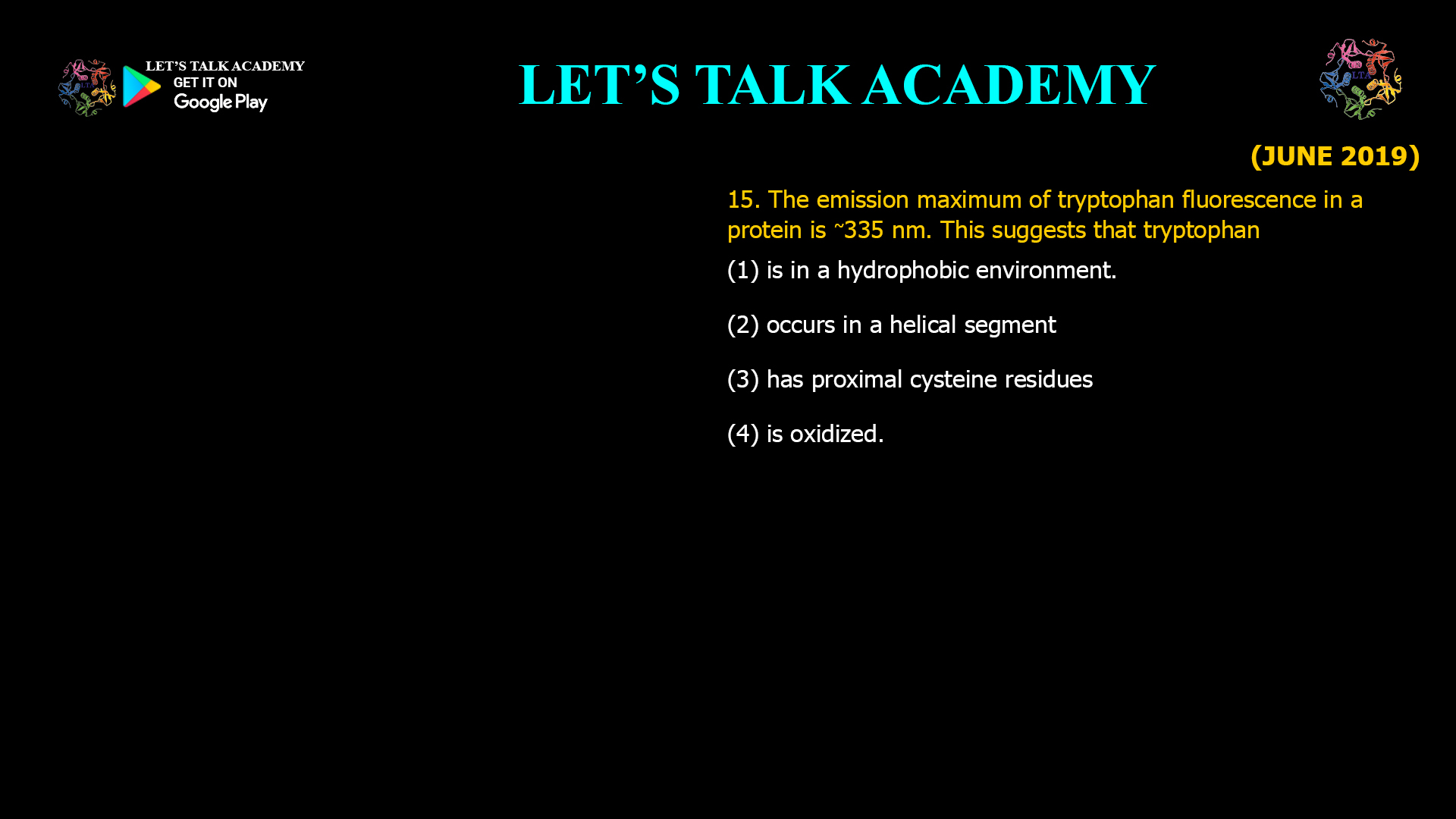
15. The emission maximum of tryptophan fluorescence in a protein is ~335 nm. This suggests that tryptophan
(1) is in a hydrophobic environment.
(2) occurs in a helical segment.
(3) has proximal cysteine residues.
(4) is oxidized.
Why Does Tryptophan Fluorescence at 335 nm Indicate a Hydrophobic Environment?
Intrinsic fluorescence is one of the most informative techniques used to investigate protein structure, folding, stability, and molecular interactions. Among the naturally occurring amino acids, tryptophan is the dominant intrinsic fluorophore because it possesses the highest quantum yield and exhibits remarkable sensitivity to its surrounding environment. The fluorescence emission wavelength of tryptophan changes according to the polarity of its local environment, making it an excellent probe for studying protein conformation.
Correct Answer
Option (1): Tryptophan is in a Hydrophobic Environment
The correct answer is Option (1). The fluorescence emission wavelength of tryptophan is highly sensitive to the polarity of its surroundings. When a tryptophan residue is buried within the hydrophobic core of a folded protein, it is shielded from surrounding water molecules. This non-polar environment stabilizes the excited electronic state differently from a polar solvent, causing fluorescence emission to occur at shorter wavelengths, typically between 330 and 335 nm.
In contrast, when tryptophan is exposed to the aqueous environment on the protein surface, solvent molecules interact strongly with the excited state. These interactions stabilize the excited state more effectively, reducing the energy difference between the excited and ground states. Consequently, fluorescence emission shifts toward longer wavelengths, generally between 350 and 355 nm. Therefore, an emission maximum around 335 nm strongly indicates that the tryptophan residue is buried inside a hydrophobic region of the protein.
Understanding Tryptophan Fluorescence
Tryptophan is the most important intrinsic fluorescent amino acid found in proteins because its indole ring absorbs ultraviolet light near 280 nm and emits fluorescence in the range of approximately 320–355 nm. Unlike tyrosine and phenylalanine, whose fluorescence signals are relatively weak, tryptophan fluorescence is extremely sensitive to environmental changes such as solvent polarity, hydrogen bonding, nearby charged groups, conformational changes, and ligand binding.
The exact emission wavelength depends on the microenvironment surrounding the indole ring. As proteins fold, many tryptophan residues become buried within hydrophobic cores, leading to a blue shift in fluorescence emission. Conversely, unfolding exposes these residues to water, resulting in a red shift toward longer wavelengths. This property allows researchers to monitor protein folding, denaturation, stability, and molecular interactions without introducing external fluorescent labels.
Relationship Between Solvent Polarity and Emission Wavelength
The emission wavelength of a fluorophore is determined by the energy difference between its excited and ground electronic states. Polar solvents such as water stabilize the excited state more strongly than the ground state through dipole interactions and hydrogen bonding. This stabilization decreases the energy gap, causing emitted photons to possess lower energy and longer wavelengths, a phenomenon known as a red shift.
Hydrophobic environments lack strong solvent interactions. As a result, the excited state is less stabilized, the energy gap remains larger, and fluorescence emission occurs at shorter wavelengths. Therefore, an emission maximum near 335 nm is characteristic of a tryptophan residue located within the hydrophobic interior of a properly folded protein.
Why Option (1) is Correct – Hydrophobic Environment
A fluorescence emission maximum around 335 nm is widely recognized as evidence that the tryptophan residue is buried inside a hydrophobic region of the protein. Such environments are typically formed by non-polar amino acid side chains within the protein core. Limited interaction with water prevents significant stabilization of the excited state, resulting in higher-energy fluorescence and consequently a shorter emission wavelength. This principle is routinely used to study protein folding, membrane proteins, and conformational changes.
Why Option (2) is Incorrect – Occurs in a Helical Segment
The secondary structure of a protein, whether α-helix, β-sheet, or random coil, does not directly determine the fluorescence emission wavelength of tryptophan. A tryptophan residue may be located within an α-helix yet remain fully exposed to solvent, or it may be positioned within a β-sheet while deeply buried inside the protein. The fluorescence emission depends primarily on solvent accessibility and local polarity rather than on secondary structural elements.
Why Option (3) is Incorrect – Has Proximal Cysteine Residues
Although nearby amino acid residues can influence fluorescence intensity through quenching or energy transfer mechanisms, the mere presence of cysteine residues does not specifically produce an emission maximum at 335 nm. Cysteine lacks intrinsic fluorescent properties capable of generating this characteristic wavelength shift. Therefore, proximity to cysteine cannot be used as a reliable indicator of fluorescence emission maxima.
Why Option (4) is Incorrect – Is Oxidized
Oxidation of tryptophan generally damages the indole ring, alters its photophysical properties, and often decreases fluorescence intensity rather than producing the characteristic emission maximum at 335 nm. Oxidized tryptophan residues frequently exhibit fluorescence quenching or altered spectral characteristics, making oxidation inconsistent with the observed emission wavelength.
Applications of Tryptophan Fluorescence in Protein Research
Intrinsic tryptophan fluorescence has become one of the most widely used techniques for investigating protein folding, unfolding, conformational dynamics, ligand binding, enzyme catalysis, membrane protein insertion, antibody-antigen interactions, and protein stability. By monitoring changes in fluorescence emission wavelength and intensity, researchers can detect structural alterations without chemically modifying the protein. A blue shift toward approximately 330–335 nm generally indicates burial of tryptophan residues within hydrophobic environments, whereas a red shift toward approximately 350–355 nm suggests solvent exposure during protein unfolding.
Final Answer
Correct Option: (1) Tryptophan is in a hydrophobic environment.
A fluorescence emission maximum near 335 nm indicates that the tryptophan residue is buried within the hydrophobic interior of the protein, where interaction with water molecules is minimal. The reduced polarity of this environment results in a larger energy gap between the excited and ground states, leading to emission at a shorter wavelength compared with solvent-exposed tryptophan residues. Consequently, an emission maximum of approximately 335 nm is a well-established indicator of a hydrophobic microenvironment, making Option (1) the correct answer.


