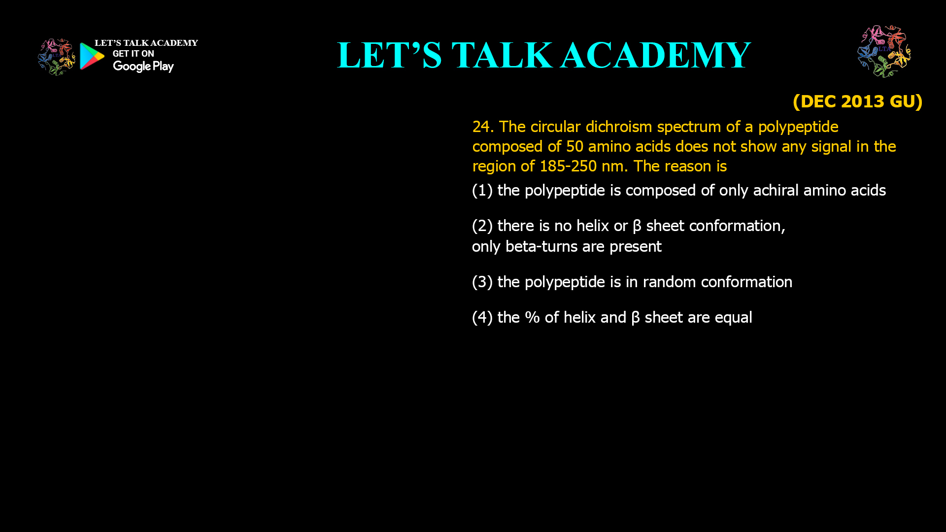
24. The circular dichroism spectrum of a polypeptide composed of 50 amino acids does not show any signal in the region of 185-250 nm. The reason is
(1) the polypeptide is composed of only achiral amino acids
(2) there is no helix or β sheet conformation, only beta-turns are present
(3) the polypeptide is in random conformation
(4) the % of helix and β sheet are equal
Why Does a Polypeptide Show No Circular Dichroism Signal Between 185–250 nm?
Circular Dichroism (CD) spectroscopy is one of the most powerful techniques for investigating protein secondary structure. In the far-ultraviolet region, typically between 185 and 250 nm, the peptide backbone absorbs ultraviolet light, and the resulting Circular Dichroism spectrum reflects the arrangement of peptide bonds into α-helices, β-sheets, β-turns, and random coils. Because each secondary structure possesses a unique three-dimensional arrangement, it generates a characteristic CD signature. Researchers routinely use this technique to study protein folding, denaturation, conformational transitions, ligand binding, and structural stability.
Correct Answer
Option (2): There Is No α-Helix or β-Sheet Conformation; Only β-Turns Are Present
The correct answer is Option (2). The far-UV Circular Dichroism spectrum primarily arises from the regular secondary structures of proteins, especially α-helices and β-sheets. These ordered conformations produce strong positive and negative ellipticity bands because of the highly organized arrangement of peptide bonds.
If a polypeptide lacks both α-helices and β-sheets and instead consists predominantly of β-turns, the characteristic far-UV CD signals become extremely weak. β-turns contribute comparatively little to the overall CD spectrum because they involve only short segments of the peptide chain and do not produce the extensive cooperative optical activity observed in long α-helices or β-sheets. Consequently, the measured spectrum may appear nearly featureless or exhibit almost no detectable signal.
Understanding Circular Dichroism in the Far-UV Region
Far-UV Circular Dichroism spectroscopy monitors the electronic transitions of peptide bonds. The principal transitions are the π→π* transition near 190 nm and the n→π* transition near 220 nm. The exact appearance of the spectrum depends upon how peptide bonds are arranged in three-dimensional space.
An α-helix produces characteristic negative bands near 208 nm and 222 nm together with a strong positive band near 190 nm. β-sheets generate a distinctly different spectral pattern, while random coils produce another recognizable spectrum. Because these conformations differ in the orientation of peptide bonds, each secondary structure exhibits a unique CD signature.
Why β-Turns Produce Very Weak CD Signals
β-turns involve only four or a few amino acid residues that reverse the direction of the peptide backbone. Unlike α-helices and β-sheets, β-turns do not extend over long distances and therefore lack the cooperative arrangement of peptide bonds responsible for strong Circular Dichroism signals.
Although β-turns are chiral structures, their contribution to the overall far-UV CD spectrum is relatively small. When a peptide consists predominantly of β-turns without significant α-helical or β-sheet content, the characteristic CD bands become very weak and may appear almost absent in experimental measurements.
Why Option (1) is Incorrect
This option assumes that the peptide is composed only of achiral amino acids. In biological proteins, this situation is practically impossible because almost all naturally occurring amino acids except glycine are chiral. Even if several glycine residues are present, a 50-residue polypeptide cannot realistically consist entirely of achiral amino acids under normal biological conditions.
Moreover, the peptide backbone itself adopts chiral conformations that contribute to the CD spectrum. Therefore, the absence of a signal cannot be explained simply by amino acid chirality.
Why Option (2) is Correct
The strongest far-UV Circular Dichroism signals arise from α-helices and β-sheets because these structures contain highly ordered arrays of peptide bonds. If neither of these secondary structures is present and the peptide contains predominantly β-turns, the cooperative optical activity becomes extremely small. As a result, the CD spectrum between 185 and 250 nm may exhibit almost no measurable ellipticity, making this the most appropriate explanation.
Why Option (3) is Incorrect
A random coil does not produce a zero CD spectrum. Instead, random coils generate a characteristic far-UV CD signature with a strong negative band near approximately 195–200 nm and relatively weak positive ellipticity at longer wavelengths. Therefore, a completely random conformation still produces a measurable Circular Dichroism spectrum rather than no signal at all.
Why Option (4) is Incorrect
Equal percentages of α-helix and β-sheet structures do not eliminate the Circular Dichroism signal because their spectra are not exact mirror images of one another. Each secondary structure contributes unique positive and negative bands with different wavelengths and intensities. Consequently, combining equal amounts of α-helix and β-sheet does not result in complete cancellation of ellipticity.
Importance of Circular Dichroism in Protein Structural Analysis
Circular Dichroism spectroscopy is widely employed to estimate secondary structure content, investigate protein folding pathways, monitor thermal denaturation, evaluate conformational stability, characterize recombinant proteins, and examine protein-ligand interactions. Computational algorithms frequently compare experimental CD spectra with reference databases to estimate the proportions of α-helices, β-sheets, turns, and random coils within unknown proteins. Because CD measurements require only small amounts of sample and can be performed rapidly in solution, the technique remains one of the most valuable tools in structural biology.
Relationship Between Secondary Structure and CD Signals
The intensity of the far-UV Circular Dichroism spectrum depends strongly on the degree of structural organization within the protein. Long α-helices and extended β-sheets generate intense signals because their peptide bonds are arranged in highly ordered geometries. In contrast, isolated β-turns contribute relatively little optical activity, while random coils produce a distinct but weaker characteristic spectrum. Therefore, the overall CD spectrum provides a direct fingerprint of protein secondary structure.
Final Answer
Correct Option: (2) There is no α-helix or β-sheet conformation; only β-turns are present.
The far-UV Circular Dichroism spectrum is dominated by the ordered secondary structures of proteins, particularly α-helices and β-sheets. When these structural elements are absent and the peptide consists predominantly of β-turns, the characteristic cooperative optical activity becomes extremely weak. As a result, the spectrum recorded between 185 and 250 nm may show almost no measurable signal. Therefore, Option (2) is the correct answer.


