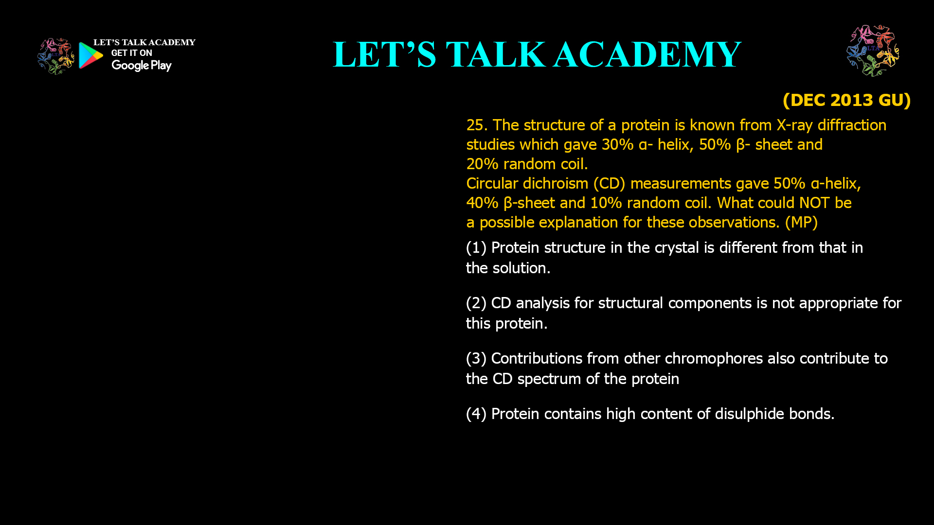
25. The structure of a protein is known from X-ray diffraction studies which gave 30% α-helix, 50% β-sheet and 20% random coil. Circular dichroism (CD) measurements gave 50% α-helix, 40% β-sheet and 10% random coil. What could NOT be a possible explanation for these observations. (MP)
(1) Protein structure in the crystal is different from that in the solution.
(2) CD analysis for structural components is not appropriate for this protein.
(3) Contributions from other chromophores also contribute to the CD spectrum of the protein.
(4) Protein contains high content of disulphide bonds.
Why Do Circular Dichroism and X-ray Diffraction Give Different Protein Secondary Structures?
Determining the three-dimensional structure of proteins is one of the most important objectives in structural biology. Different experimental techniques provide complementary information about protein architecture, stability, and conformational dynamics. Among these techniques, X-ray diffraction and Circular Dichroism (CD) spectroscopy are widely used to investigate protein structure. Although both methods provide valuable structural information, they differ fundamentally in what they measure, the environment in which the protein is analyzed, and the level of structural detail they provide. Consequently, it is not unusual for the two techniques to produce slightly different estimates of secondary structure content.
Correct Answer
Option (4): Protein Contains a High Content of Disulfide Bonds
The correct answer is Option (4). A high disulfide bond content by itself does not provide a convincing explanation for the substantial disagreement between the secondary structure estimates obtained from X-ray diffraction and Circular Dichroism spectroscopy. Disulfide bonds stabilize protein structure by covalently linking different parts of the polypeptide chain, but they do not directly alter the percentages of α-helices, β-sheets, or random coils reported by these techniques. While disulfide bonds may contribute weakly to the near-UV CD spectrum or influence protein stability, they are not expected to produce the large discrepancy observed in secondary structure estimation.
In contrast, the other three explanations are scientifically plausible because they involve either genuine structural differences between crystal and solution states or limitations in the interpretation of CD spectra.
Understanding X-ray Diffraction and Circular Dichroism
X-ray diffraction determines protein structure at atomic or near-atomic resolution by analyzing how X-rays are diffracted by protein crystals. It provides a detailed three-dimensional model from which the proportions of α-helices, β-sheets, and loops can be calculated with high accuracy. However, the protein must first be crystallized, and the resulting structure represents the conformation adopted within the crystal lattice.
Circular Dichroism spectroscopy, on the other hand, examines proteins in solution. It measures the differential absorption of left-handed and right-handed circularly polarized light by the peptide backbone. The far-ultraviolet CD spectrum reflects the average secondary structure of the protein in its native solution environment. Computational algorithms compare the experimental spectrum with reference datasets to estimate the proportions of α-helices, β-sheets, turns, and random coils.
Why Option (1) is a Possible Explanation
Crystal Structure and Solution Structure May Differ
Proteins are dynamic molecules that continuously undergo conformational fluctuations in solution. During crystallization, proteins become packed into an ordered crystal lattice where intermolecular contacts may stabilize conformations that differ slightly from those present in solution. Flexible loops may become ordered, secondary structural elements may shift slightly, and some regions may adopt alternative conformations because of crystal packing forces.
Since Circular Dichroism measures the protein directly in solution, while X-ray diffraction examines the crystallized protein, differences in the observed secondary structure are entirely possible. Therefore, Option (1) represents a reasonable explanation for the discrepancy.
Why Option (2) is a Possible Explanation
CD Structural Analysis Has Certain Limitations
The estimation of secondary structure from Circular Dichroism spectra depends on computational deconvolution methods that compare experimental spectra with reference protein datasets. Some proteins possess unusual folds, uncommon amino acid compositions, membrane-associated regions, intrinsically disordered segments, or rare structural motifs that are poorly represented in these reference databases.
In such cases, the mathematical algorithms used to estimate α-helical and β-sheet content may produce less accurate results. Consequently, the CD-derived percentages may differ from those obtained through X-ray crystallography. Therefore, Option (2) is also a plausible explanation.
Why Option (3) is a Possible Explanation
Additional Chromophores Can Influence the CD Spectrum
Although the far-ultraviolet Circular Dichroism spectrum primarily originates from peptide bond electronic transitions, other chromophores present within the protein may also contribute to the measured signal. Aromatic amino acids, prosthetic groups, cofactors, metal-binding centers, or unusual chemical modifications may introduce additional spectral features that complicate interpretation.
If these contributions are not properly accounted for during spectral analysis, the calculated percentages of secondary structure may deviate from the actual values. Therefore, Option (3) provides another scientifically reasonable explanation for the discrepancy.
Why Option (4) is NOT a Possible Explanation
Disulfide Bonds Stabilize Structure but Do Not Explain the Discrepancy
Disulfide bonds are covalent linkages formed between the sulfur atoms of two cysteine residues. Their principal biological role is to stabilize the folded structure of proteins by reducing conformational flexibility. While this stabilization may increase resistance to thermal or chemical denaturation, it does not inherently change the fraction of α-helices, β-sheets, or random coils measured by X-ray diffraction or Circular Dichroism.
Furthermore, the presence of numerous disulfide bonds does not invalidate CD-based secondary structure estimation. Although disulfide bonds can contribute weak signals, particularly in the near-ultraviolet region, these effects are generally insufficient to explain the substantial differences reported in the question. Therefore, Option (4) cannot adequately account for the observed disagreement.
Why X-ray Diffraction and CD Spectroscopy Complement Each Other
X-ray diffraction and Circular Dichroism are complementary rather than competing techniques. X-ray crystallography provides detailed atomic coordinates and precise structural models but requires protein crystallization. Circular Dichroism rapidly evaluates proteins in solution under physiological conditions and is particularly useful for monitoring folding, unfolding, ligand binding, thermal stability, and conformational changes. Researchers frequently use both methods together to obtain a more complete understanding of protein structure and dynamics.
Importance in Protein Structural Biology
Understanding the strengths and limitations of structural techniques is essential in modern biochemistry and molecular biology. Differences between solution-state and crystal-state structures, computational limitations of spectral deconvolution, and contributions from additional chromophores are well-recognized sources of disagreement between experimental methods. Appreciating these factors allows researchers to interpret structural data critically rather than assuming that one technique is always correct.
Final Answer
Correct Option: (4) Protein contains a high content of disulfide bonds.
Differences between X-ray diffraction and Circular Dichroism measurements can reasonably arise because proteins may adopt different conformations in crystals and in solution, because CD secondary structure estimation has computational limitations, or because additional chromophores influence the CD spectrum. However, simply having a high content of disulfide bonds does not explain the significant disagreement in the measured percentages of α-helices, β-sheets, and random coils. Therefore, Option (4) is the explanation that could NOT account for the observations.


