
Introduction
Cells are highly organized systems in which thousands of proteins, lipids, nucleic acids, and other molecules must reach specific locations at the correct time. A newly synthesized protein may need to remain in the cytosol, enter the nucleus, become part of a membrane, move into the endoplasmic reticulum, reach the Golgi apparatus, or be delivered to lysosomes, mitochondria, peroxisomes, or the plasma membrane.
This precise distribution of cellular components is achieved through intracellular transport. Intracellular transport is not simply the movement of molecules from one place to another. It is a highly regulated process involving molecular recognition, sorting signals, transport carriers, cytoskeletal tracks, motor proteins, tethering factors, membrane fusion machinery, and regulatory proteins.
The accuracy of intracellular transport is essential for maintaining cellular organization and function. Errors in protein targeting or vesicle trafficking can interfere with secretion, metabolism, signaling, membrane composition, degradation, and cellular homeostasis.
This chapter describes the major mechanisms by which proteins and membranes are sorted, transported, targeted, and regulated within eukaryotic cells.
1. Introduction to Intracellular Transport
The interior of a eukaryotic cell is divided into specialized compartments called organelles. Each organelle possesses a characteristic molecular composition and performs specific functions.
For example, the nucleus stores genetic information, mitochondria participate in energy metabolism, the endoplasmic reticulum synthesizes proteins and lipids, the Golgi apparatus modifies and sorts proteins, lysosomes degrade macromolecules, and peroxisomes perform specialized oxidative reactions.
Because these compartments have different functions, proteins must be delivered to their correct destinations.
A protein synthesized in the cytosol may follow several possible pathways:
Cytosol → Nucleus
Cytosol → Mitochondria
Cytosol → Peroxisome
Cytosol → Endoplasmic reticulum → Golgi apparatus → Lysosome
Cytosol → Endoplasmic reticulum → Golgi apparatus → Plasma membrane
Thus, intracellular transport establishes and maintains the internal organization of the cell.
1.1 Importance of Protein Sorting
Protein sorting determines where a protein will function after synthesis.
A protein cannot normally reach its destination randomly. Instead, it contains specific molecular information that allows the cellular transport machinery to recognize it.
These molecular signals are commonly called sorting signals or targeting signals.
A targeting signal can be present as:
- a short amino acid sequence,
- a particular structural region,
- a transmembrane segment,
- a carbohydrate modification,
- or a combination of molecular features.
The transport machinery recognizes these signals and directs the protein toward the appropriate cellular compartment.
2. Major Mechanisms of Intracellular Transport
Intracellular transport can be broadly divided into three major mechanisms:
- Gated transport
- Transmembrane transport
- Vesicular transport
2.1 Gated Transport
Gated transport occurs between the cytosol and the nucleus through nuclear pore complexes.
The nuclear envelope contains large protein assemblies called nuclear pore complexes (NPCs). These pores regulate the movement of proteins and RNA between the nucleus and cytoplasm.
Small molecules can pass through nuclear pores relatively easily, whereas larger proteins generally require specific transport receptors.
2.2 Transmembrane Transport
In transmembrane transport, proteins move across a membrane through specialized protein complexes.
Examples include:
- protein transport into the endoplasmic reticulum,
- protein import into mitochondria,
- protein import into peroxisomes.
The transported protein interacts with receptors and translocation machinery located in the target membrane.
2.3 Vesicular Transport
Vesicular transport moves proteins and lipids between membrane-bound compartments.
A portion of one membrane buds outward to form a transport vesicle. The vesicle then moves through the cytoplasm and eventually fuses with another membrane.
The general pathway is:
Cargo selection → Vesicle budding → Vesicle transport → Tethering → Docking → Membrane fusion
Vesicular transport is particularly important in communication among:
- endoplasmic reticulum,
- Golgi apparatus,
- endosomes,
- lysosomes,
- plasma membrane.
3. Protein Sorting Signals
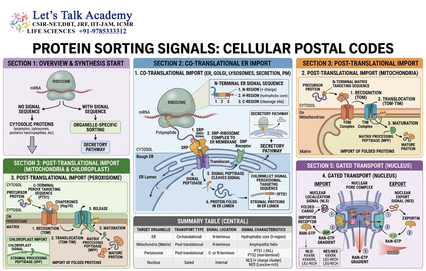
Protein sorting depends on molecular signals that act as intracellular addresses.
These signals are recognized by specific receptors or transport proteins.
3.1 Signal Peptides
A signal peptide is a short amino acid sequence that directs a newly synthesized protein toward the endoplasmic reticulum.
Signal peptides generally contain:
- a positively charged region,
- a hydrophobic core,
- and a cleavage region.
The hydrophobic region allows recognition by the signal recognition particle and interaction with the ER membrane.
3.2 Nuclear Localization Signals
Proteins destined for the nucleus often contain a nuclear localization signal (NLS).
NLS sequences are frequently enriched in positively charged amino acids such as lysine and arginine.
The NLS is recognized by nuclear transport receptors called importins.
3.3 Nuclear Export Signals
Proteins leaving the nucleus may contain a nuclear export signal (NES).
Many NES sequences contain hydrophobic amino acids and are recognized by export receptors called exportins.
3.4 Mitochondrial Targeting Signals
Many mitochondrial proteins contain targeting sequences that direct them toward mitochondrial membranes.
These sequences are commonly enriched in positively charged amino acids and can form amphipathic α-helices.
3.5 Peroxisomal Targeting Signals
Proteins transported into peroxisomes may contain peroxisomal targeting signals (PTS).
A well-known PTS1 signal is frequently located at the extreme C-terminus of the protein and commonly contains the sequence SKL or related variants.
4. Nuclear Protein Transport
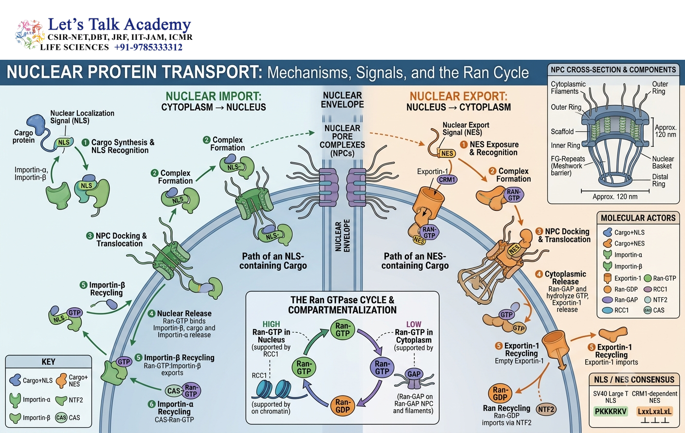
The nucleus is separated from the cytoplasm by the nuclear envelope.
The nuclear envelope contains nuclear pore complexes that regulate molecular exchange between the two compartments.
4.1 Nuclear Import
Proteins containing an NLS are recognized by importin receptors.
The general process is:
Cargo protein + NLS → Importin recognition → Nuclear pore complex → Nuclear entry → Cargo release
Importin-cargo complexes interact with components of the nuclear pore complex and move through the pore.
4.2 Role of Ran GTPase
Nuclear transport is strongly regulated by the small GTPase Ran.
Ran exists in two major forms:
- Ran-GTP
- Ran-GDP
A high concentration of Ran-GTP is maintained inside the nucleus, whereas Ran-GDP predominates in the cytoplasm.
This gradient provides directionality to nuclear transport.
4.3 Nuclear Export
During nuclear export, exportins recognize cargo proteins containing suitable export signals.
Ran-GTP participates in the formation of the export complex.
The complex moves through the nuclear pore into the cytoplasm, where GTP hydrolysis contributes to complex disassembly.
Thus, the Ran-GTP gradient is central to maintaining the direction of nuclear transport.
5. Protein Targeting to the Endoplasmic Reticulum
The endoplasmic reticulum is a major entry point into the secretory pathway.
Proteins destined for secretion, lysosomes, the plasma membrane, or many endomembrane compartments generally enter the ER.
5.1 Signal Recognition Particle
Many proteins entering the ER possess an N-terminal signal peptide.
During translation, the signal peptide is recognized by the signal recognition particle (SRP).
SRP temporarily interacts with the ribosome and slows translation.
The SRP-ribosome complex then interacts with the SRP receptor located on the ER membrane.
5.2 Sec61 Translocation Channel
The ribosome is positioned near the Sec61 translocon, a protein-conducting channel in the ER membrane.
Translation resumes, and the growing polypeptide passes through the translocon into the ER lumen or becomes inserted into the ER membrane.
This process is called co-translational translocation.
5.3 Soluble and Membrane Proteins
Proteins can enter the ER lumen as soluble proteins or become membrane proteins.
Membrane proteins contain hydrophobic sequences that function as:
- signal-anchor sequences,
- stop-transfer sequences,
- or internal targeting signals.
These sequences determine how the protein is oriented and inserted into the membrane.
6. Protein Targeting to Mitochondria
Mitochondria contain their own genome but also depend heavily on proteins encoded by nuclear genes.
Most mitochondrial proteins are synthesized in the cytosol and subsequently imported into mitochondria.
6.1 Mitochondrial Targeting Sequence
Many mitochondrial proteins possess an N-terminal targeting sequence.
This sequence often forms an amphipathic α-helix containing positively charged residues.
6.2 TOM Complex
The outer mitochondrial membrane contains the TOM complex, or translocase of the outer membrane.
The TOM complex recognizes and translocates precursor proteins across the outer membrane.
6.3 TIM Complex
Proteins destined for the mitochondrial matrix are subsequently transported through complexes in the inner membrane called TIM complexes, or translocases of the inner membrane.
Different TIM pathways help direct proteins toward different mitochondrial compartments.
Thus, mitochondrial protein import involves coordinated recognition and translocation across mitochondrial membranes.
7. Protein Targeting to Peroxisomes
Peroxisomes contain enzymes involved in oxidation, lipid metabolism, and detoxification reactions.
Many peroxisomal proteins are synthesized on free ribosomes in the cytosol and subsequently imported into peroxisomes.
7.1 PTS1 and PTS2
Two important targeting systems are:
- PTS1
- PTS2
PTS1 is commonly located at the C-terminus of a protein.
PTS2 is generally associated with an N-terminal targeting sequence.
7.2 Peroxins
Peroxisomal protein import requires proteins known as peroxins, encoded by PEX genes.
Peroxins participate in:
- cargo recognition,
- membrane docking,
- translocation,
- and recycling of transport receptors.
An important feature of peroxisomal import is that certain proteins can be imported in a folded or oligomeric state.
8. Vesicular Transport
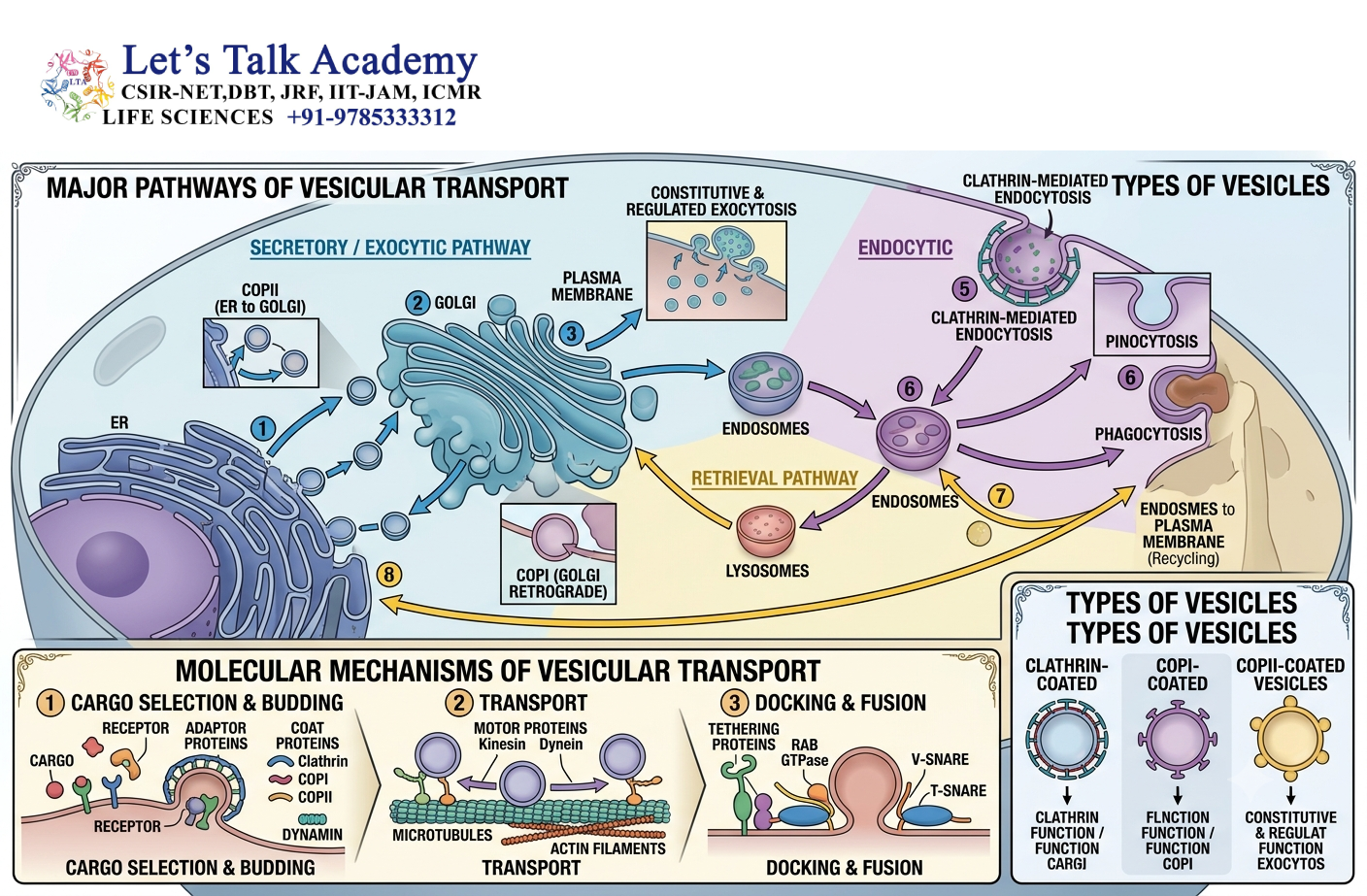
Vesicular transport is one of the major mechanisms of communication between intracellular membrane compartments.
A transport vesicle is a small membrane-bound carrier that transports selected proteins and lipids.
The basic stages are:
- Cargo selection
- Coat assembly
- Membrane budding
- Vesicle scission
- Uncoating
- Transport
- Tethering
- Docking
- Membrane fusion
Each stage is regulated by specific molecular components.
9. Cargo Selection
Transport vesicles do not generally contain random portions of the donor membrane.
Specific cargo molecules are concentrated into the budding region.
Cargo selection may occur through:
- direct interaction with coat proteins,
- cargo receptors,
- adaptor proteins,
- sorting signals.
9.1 Cargo Receptors
Some soluble proteins cannot directly interact with coat proteins.
In such cases, a membrane-associated cargo receptor binds the soluble cargo and connects it with the vesicle formation machinery.
Cargo receptors therefore function as molecular bridges between cargo and transport machinery.
10. Coat Proteins
Coat proteins help select cargo and shape the donor membrane during vesicle formation.
Three major coat systems are:
- COPII
- COPI
- Clathrin
Each system participates in distinct transport pathways.
10.1 COPII-Coated Vesicles
COPII vesicles generally transport cargo from:
Endoplasmic reticulum → Golgi apparatus
10.2 COPI-Coated Vesicles
COPI vesicles participate mainly in:
Golgi apparatus → ER
and in transport between Golgi cisternae.
10.3 Clathrin-Coated Vesicles
Clathrin-coated vesicles participate in several pathways, including:
Plasma membrane → Endosome
and:
Trans-Golgi network → Endosome
11. COPII-Mediated Transport
COPII vesicle formation begins at specialized ER regions known as ER exit sites.
11.1 Sar1 GTPase
The small GTPase Sar1 initiates COPII assembly.
When Sar1 binds GTP, it undergoes a conformational change that exposes a membrane-interacting region.
Sar1-GTP associates with the ER membrane and recruits coat components.
11.2 Sec23/Sec24 Complex
The Sar1-associated membrane recruits the Sec23/Sec24 complex.
Sec24 plays an important role in cargo recognition.
11.3 Sec13/Sec31 Complex
The outer coat consists primarily of Sec13/Sec31.
It helps generate the membrane curvature required for vesicle budding.
The simplified sequence is:
Sar1 activation → Sec23/Sec24 recruitment → Cargo selection → Sec13/Sec31 assembly → Budding → Vesicle release
12. COPI-Mediated Transport
COPI vesicles are involved in retrograde transport and intra-Golgi trafficking.
A major function of COPI is to return proteins and membrane components from the Golgi apparatus to the ER.
This retrieval pathway is essential because some ER proteins accidentally escape into the secretory pathway.
COPI also contributes to recycling between Golgi cisternae.
12.1 ARF1 GTPase
The small GTPase ARF1 participates in COPI coat recruitment.
ARF1-GTP associates with the Golgi membrane and promotes recruitment of coatomer components.
13. Clathrin-Mediated Vesicle Formation
Clathrin is a major coat protein involved in vesicle formation at the plasma membrane and trans-Golgi network.
Clathrin does not usually recognize cargo directly.
Instead, adaptor proteins connect cargo receptors to clathrin.
13.1 Adaptor Proteins
Adaptor proteins perform two major functions:
- recognize sorting signals on cargo or cargo receptors,
- recruit clathrin.
This produces a protein coat that helps shape the membrane.
13.2 Dynamin and Vesicle Scission
At the plasma membrane, dynamin participates in the final separation of the budding vesicle from the membrane.
Dynamin is a GTPase that forms a ring-like structure around the neck of the budding vesicle.
GTP hydrolysis contributes to membrane scission.
14. Vesicle Uncoating
After a vesicle separates from the donor membrane, its coat is generally removed.
Uncoating is important because the vesicle must expose its targeting and fusion machinery before interacting efficiently with the target membrane.
The released coat components can then be reused in subsequent rounds of vesicle formation.
15. Rab GTPases and Vesicle Targeting
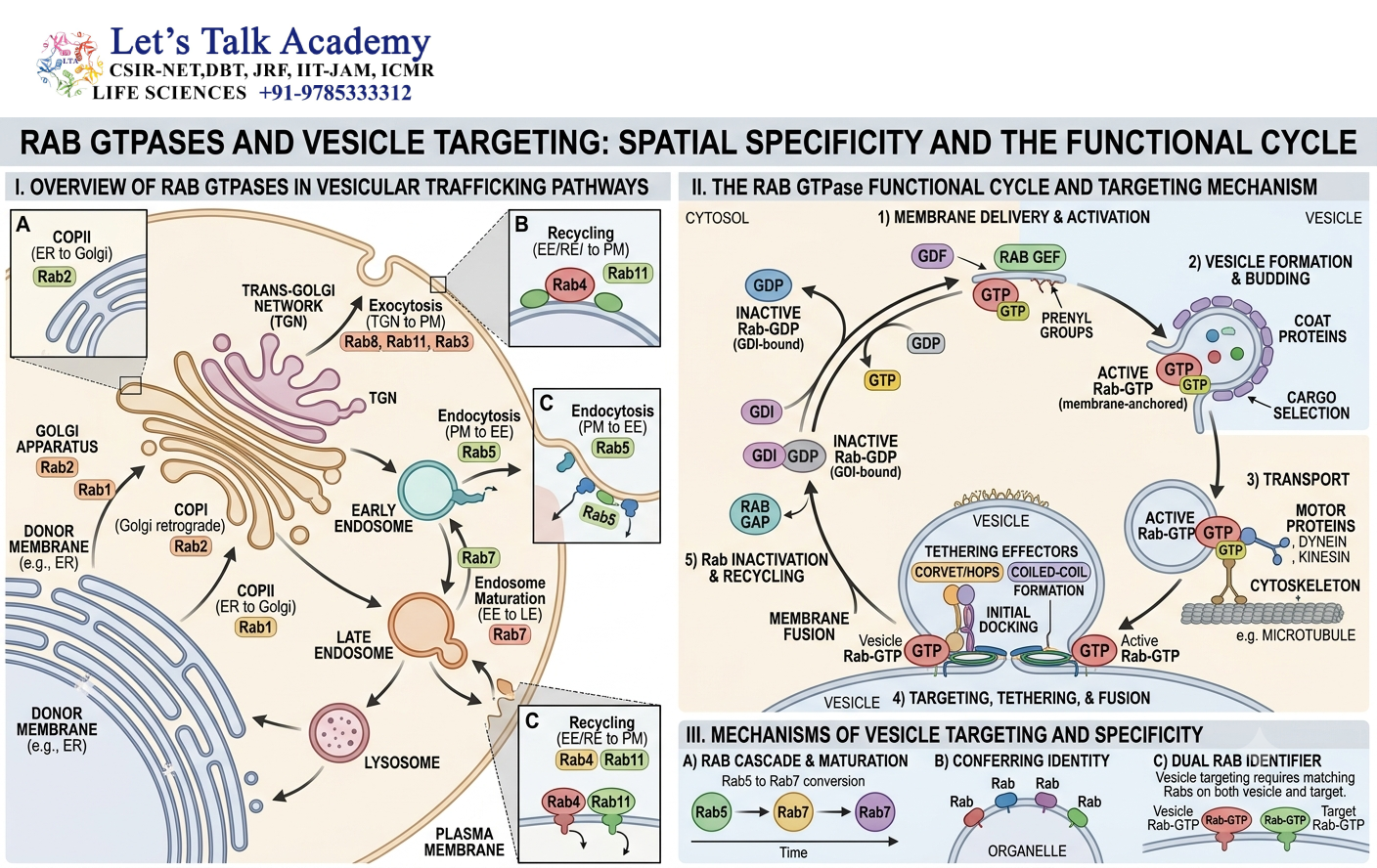
A transport vesicle must reach the correct target membrane.
One of the major systems responsible for organelle-specific targeting involves Rab GTPases.
Rab proteins function as molecular identity markers for membrane compartments.
15.1 GTP-Bound and GDP-Bound Rab
Rab proteins cycle between:
- inactive GDP-bound form,
- active GTP-bound form.
Active Rab proteins recruit specific effector proteins.
15.2 Rab Effectors
Rab effectors participate in:
- vesicle tethering,
- motor recruitment,
- membrane organization,
- coordination with fusion machinery.
Different organelles contain characteristic combinations of Rab proteins, helping vesicles identify their correct destination.
16. Vesicle Tethering
Before membrane fusion, a vesicle must be captured near its target membrane.
This initial interaction is called tethering.
Tethering proteins can extend from the target membrane and interact with the incoming vesicle.
Rab proteins and their effectors frequently participate in this process.
Tethering provides an important layer of specificity before the final membrane docking and fusion event.
17. SNARE Proteins and Membrane Fusion
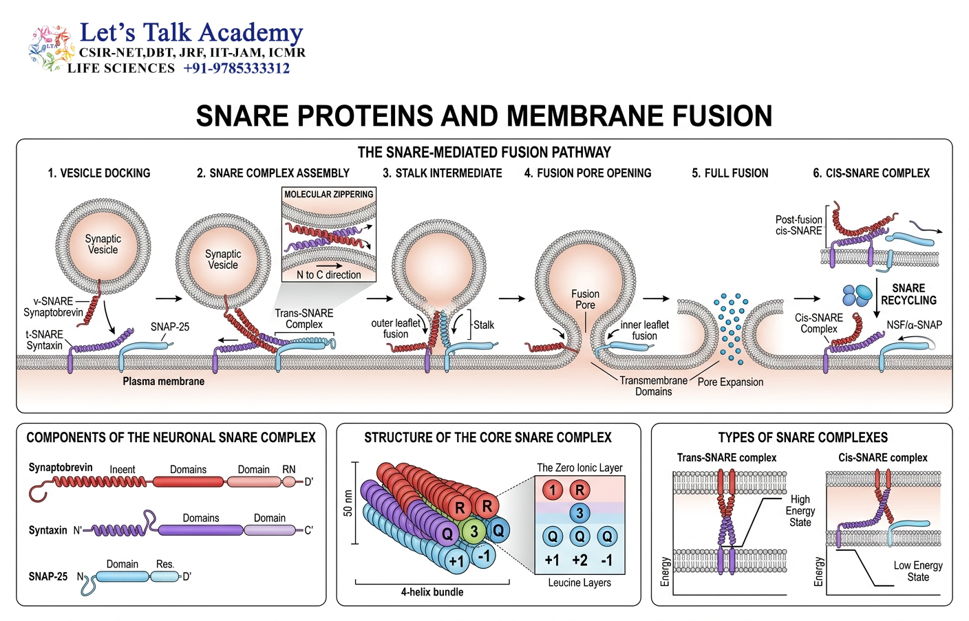
After tethering, the vesicle must fuse with the target membrane.
The major proteins responsible for membrane fusion are SNAREs.
SNAREs are located on both transport vesicles and target membranes.
Historically, these were commonly described as:
- v-SNAREs on vesicles,
- t-SNAREs on target membranes.
SNARE proteins form a tightly associated complex that brings the two membranes close together.
The energy released during SNARE complex formation helps overcome the energetic barrier to membrane fusion.
The process can be represented as:
Tethering → SNARE pairing → Membrane approximation → Fusion pore formation → Complete fusion
18. Cytoskeleton and Intracellular Transport
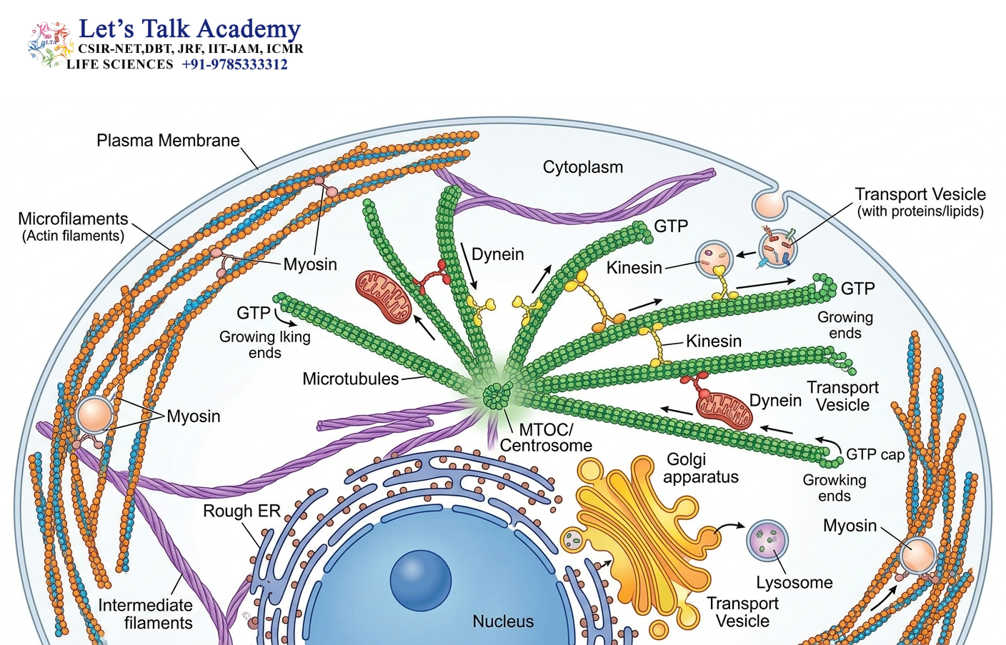
Transport vesicles often travel along cytoskeletal tracks.
The two major cytoskeletal systems involved are:
- microtubules,
- actin filaments.
18.1 Microtubules
Microtubules provide long-distance tracks for vesicle movement.
They are particularly important for transport between the cell center and peripheral regions.
18.2 Kinesin
Kinesins are motor proteins that generally move cargo toward the plus end of microtubules.
In many cells, this corresponds to movement toward the cell periphery.
18.3 Dynein
Dynein generally moves cargo toward the minus end of microtubules.
This commonly corresponds to movement toward the centrosomal region of the cell.
18.4 Myosin
Myosin motors move along actin filaments.
They are particularly important for short-range transport and transport near the plasma membrane.
19. Golgi Apparatus as a Sorting Center
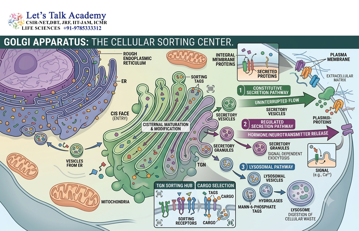
The Golgi apparatus is an important station in the secretory pathway.
It consists of a series of flattened membrane-bound compartments called cisternae.
These are commonly divided into:
- cis-Golgi network,
- cis cisternae,
- medial cisternae,
- trans cisternae,
- trans-Golgi network.
Proteins entering the Golgi undergo chemical modifications and are subsequently sorted toward different destinations.
20. Protein Processing in the Golgi
Proteins passing through the Golgi may undergo:
- glycosylation,
- glycan modification,
- phosphorylation,
- proteolytic processing,
- sulfation.
These modifications can influence protein stability, activity, and destination.
The trans-Golgi network functions as a major sorting station.
From this region, cargo can be directed toward:
- plasma membrane,
- secretory vesicles,
- endosomes,
- lysosomes.
21. Lysosomal Protein Sorting
Lysosomes contain hydrolytic enzymes responsible for degradation of cellular and extracellular material.
Many lysosomal enzymes are synthesized in the ER and transported through the Golgi apparatus.
21.1 Mannose-6-Phosphate
A major sorting signal for lysosomal enzymes is mannose-6-phosphate (M6P).
The lysosomal enzyme is modified in the Golgi so that M6P can be recognized by M6P receptors.
21.2 Lysosomal Targeting Pathway
The general pathway is:
ER → Golgi → M6P modification → M6P receptor binding → Transport vesicle → Endosome → Lysosome
At acidic endosomal pH, lysosomal enzymes dissociate from their receptors.
The enzymes continue toward lysosomes, while receptors can be recycled.
22. Endocytosis and Endosomal Sorting
Endocytosis allows the cell to internalize material from the extracellular environment.
Several forms of endocytosis exist, including:
- receptor-mediated endocytosis,
- phagocytosis,
- pinocytosis.
22.1 Early Endosome
Internalized material initially reaches the early endosome.
The early endosome functions as a sorting station.
Cargo may be directed toward:
- recycling back to the plasma membrane,
- transport toward late endosomes,
- lysosomal degradation.
22.2 Recycling Pathway
Some receptors and membrane components return to the plasma membrane.
This recycling allows the cell to reuse important membrane proteins and maintain surface composition.
22.3 Degradation Pathway
Other cargo molecules are transported through late endosomes and eventually delivered to lysosomes for degradation.
23. Exocytosis
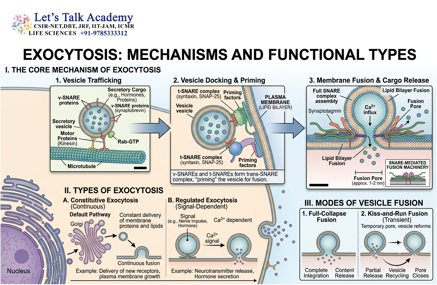
Exocytosis is the process by which intracellular vesicles fuse with the plasma membrane and release their contents outside the cell.
There are two broad forms.
23.1 Constitutive Exocytosis
Constitutive secretion occurs continuously.
It contributes to:
- plasma membrane growth,
- extracellular matrix secretion,
- delivery of membrane proteins.
23.2 Regulated Exocytosis
Regulated secretion occurs in response to specific signals.
Examples include secretion of:
- neurotransmitters,
- peptide hormones,
- digestive enzymes.
23.3 Calcium as a Trigger
In many regulated secretory systems, an increase in cytosolic Ca²⁺ triggers rapid membrane fusion.
Proteins such as synaptotagmin function as calcium-sensitive components of the fusion machinery in many secretory pathways.
24. Endoplasmic Reticulum Quality Control
Protein folding begins during and after protein synthesis.
The ER contains molecular chaperones and quality-control systems that help newly synthesized proteins achieve their correct structures.
Proteins that fail to fold properly may be retained within the ER.
24.1 Molecular Chaperones
Chaperones assist protein folding and prevent inappropriate aggregation.
Important ER chaperones include:
- BiP,
- calnexin,
- calreticulin.
24.2 ER-Associated Degradation
Misfolded proteins can be transported from the ER back into the cytosol.
This process is known as ER-associated degradation (ERAD).
The proteins are subsequently ubiquitinated and degraded by the proteasome.
Thus:
Misfolded protein → Recognition → ER export → Ubiquitination → Proteasomal degradation
25. Unfolded Protein Response
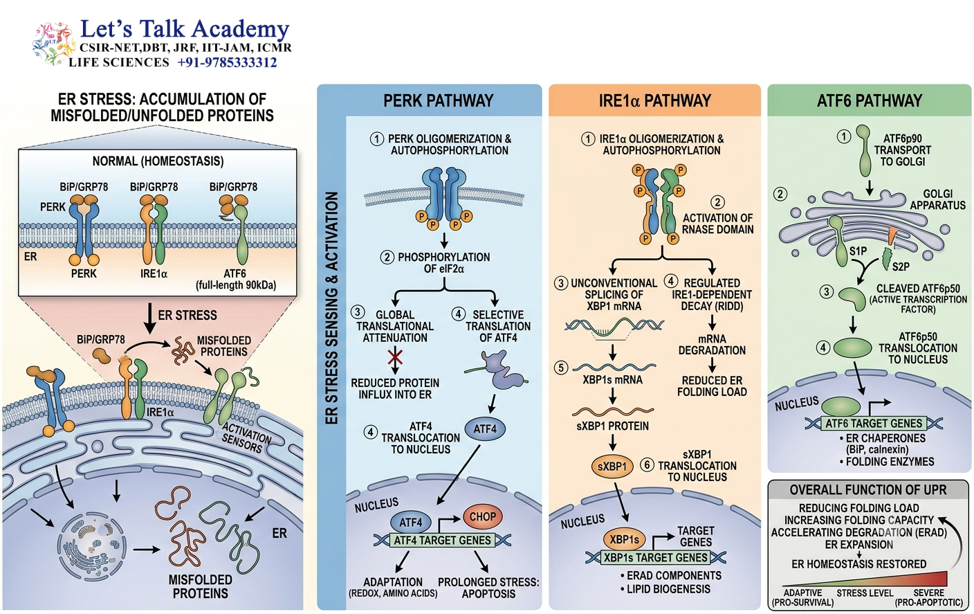
Accumulation of unfolded or misfolded proteins in the ER produces ER stress.
Cells respond through the unfolded protein response (UPR).
The UPR helps restore protein-folding capacity by:
- reducing protein synthesis,
- increasing expression of chaperones,
- enhancing degradation of misfolded proteins,
- modifying ER function.
If ER stress becomes prolonged or severe, cellular survival pathways may be compromised.
26. Autophagy and Intracellular Transport
Autophagy is a cellular degradation pathway that delivers cytoplasmic components to lysosomes.
A major form is macroautophagy.
26.1 Formation of the Autophagosome
During macroautophagy, a membrane structure called a phagophore begins to surround cytoplasmic material.
The phagophore expands and eventually closes to form a double-membrane structure called an autophagosome.
26.2 Fusion with Lysosomes
The autophagosome subsequently fuses with lysosomal compartments.
The enclosed material is degraded by lysosomal enzymes.
The resulting molecules can be recycled by the cell.
27. Regulation of Intracellular Transport
Intracellular transport must be precisely regulated.
Regulation occurs at multiple stages.
27.1 Regulation of Vesicle Formation
Vesicle formation depends on:
- small GTPases,
- coat proteins,
- adaptor proteins,
- cargo receptors,
- membrane lipid composition.
27.2 Regulation of Vesicle Movement
Vesicle movement is influenced by:
- motor proteins,
- cytoskeletal organization,
- phosphorylation,
- cellular energy availability.
27.3 Regulation of Tethering and Fusion
Rab proteins, tethering factors, SNAREs, and calcium-dependent proteins regulate the interaction between vesicles and target membranes.
27.4 Regulation by Phosphorylation
Protein kinases and phosphatases can regulate the activity of:
- motor proteins,
- trafficking proteins,
- coat proteins,
- tethering factors,
- fusion machinery.
This allows transport to respond rapidly to changes in cellular conditions.
28. Directionality of Intracellular Transport
Intracellular transport must be directional.
A vesicle leaving one organelle must generally reach a particular target rather than randomly fuse with any membrane.
Directionality is established through several mechanisms.
These include:
- organelle-specific Rab proteins,
- specific SNARE combinations,
- sorting signals,
- coat proteins,
- cargo receptors,
- cytoskeletal tracks,
- motor proteins,
- tethering factors,
- GTPase cycles.
Together, these systems create a highly selective transport network.
29. Protein Retrieval and Recycling
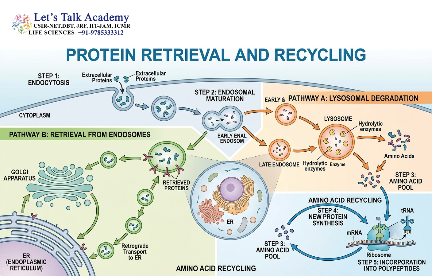
Intracellular transport is not always unidirectional.
Proteins can be retrieved from downstream compartments and returned to their original locations.
This is essential for maintaining organelle identity.
29.1 KDEL Retrieval Signal
Many soluble ER-resident proteins contain a C-terminal KDEL sequence.
If such proteins escape into the Golgi, KDEL receptors recognize them and facilitate their return to the ER.
29.2 KKXX Retrieval Signal
Certain ER membrane proteins contain cytosolic retrieval signals such as KKXX.
These signals can promote COPI-dependent retrieval from the Golgi back to the ER.
30. Maintenance of Organelle Identity
Every membrane-bound organelle possesses a characteristic molecular composition.
This includes specific:
- proteins,
- lipids,
- enzymes,
- receptors,
- Rab proteins,
- SNAREs.
Transport pathways must preserve these differences.
For example, the ER must maintain its characteristic protein composition even though proteins continuously leave the ER.
Retrieval pathways and selective transport help maintain this balance.
31. Energy Requirements of Intracellular Transport
Intracellular transport requires energy at several stages.
Energy is used for:
- GTPase activation,
- vesicle formation,
- motor-driven transport,
- membrane fusion,
- protein translocation,
- receptor recycling.
ATP is particularly important for motor proteins and several remodeling processes, whereas GTP is central to many regulatory GTPases such as Rab, Sar1, and ARF.
32. Small GTPases as Molecular Switches
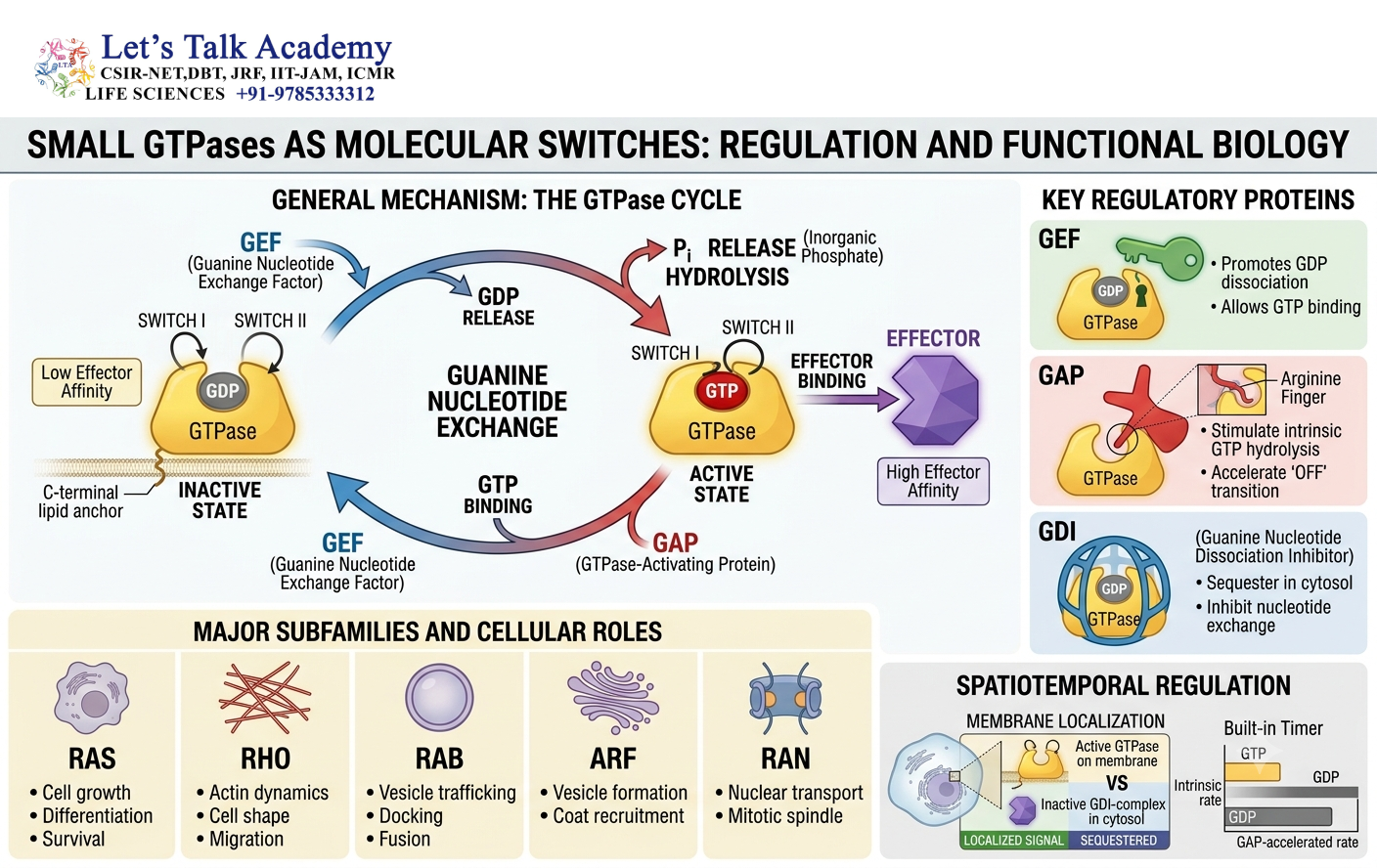
Small GTPases are central regulators of intracellular transport.
They function by switching between two states:
GTP-bound → Active
GDP-bound → Inactive
Three important transport-related GTPase systems include:
32.1 Sar1
Sar1 regulates COPII vesicle formation at the ER.
32.2 ARF
ARF proteins regulate membrane recruitment of coat components, particularly in Golgi-associated transport.
32.3 Rab
Rab proteins regulate vesicle identity, targeting, tethering, and interactions with transport effectors.
The cycling of these GTPases provides temporal and spatial control over intracellular transport.
33. Intracellular Transport During Cellular Stress
Transport pathways are modified when cells experience stress.
Changes in nutrient availability, protein-folding conditions, energy state, or membrane organization can alter intracellular trafficking.
Cells may:
- reduce certain secretory activities,
- increase degradation pathways,
- activate autophagy,
- modify ER function,
- alter vesicle transport,
- increase protein quality-control mechanisms.
This flexibility helps maintain cellular homeostasis.
34. Intracellular Transport and Cellular Homeostasis
Cellular homeostasis depends on continuous coordination between synthesis, transport, recycling, and degradation.
For example:
Protein synthesis → Folding → Sorting → Transport → Function → Recycling or degradation
A disturbance at any stage can affect the entire cellular system.
Efficient transport therefore contributes to:
- membrane integrity,
- protein quality,
- nutrient uptake,
- secretion,
- signaling,
- degradation,
- organelle maintenance.
35. Intracellular Transport and Disease
Defects in intracellular transport can produce severe cellular dysfunction.
Problems may arise from mutations or abnormalities affecting:
- coat proteins,
- Rab proteins,
- SNAREs,
- motor proteins,
- lysosomal enzymes,
- trafficking receptors,
- mitochondrial import machinery,
- peroxisomal import machinery.
35.1 Lysosomal Storage Disorders
Defective lysosomal enzymes or lysosomal targeting can cause accumulation of undegraded material.
One example is I-cell disease, which results from a defect in the pathway responsible for adding mannose-6-phosphate to lysosomal enzymes.
35.2 Neurodegenerative Disorders
Neurons are particularly dependent on efficient long-distance intracellular transport.
Defects in axonal transport can interfere with the movement of proteins, organelles, and vesicles between the cell body and axon terminals.
35.3 Peroxisomal Disorders
Defects in peroxisomal targeting or import can prevent appropriate enzymes from entering peroxisomes.
This can disrupt lipid metabolism and other essential cellular processes.
36. Experimental Approaches to Studying Intracellular Transport
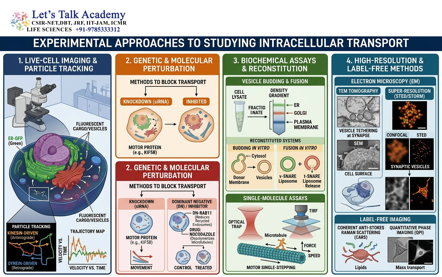
Intracellular transport can be investigated using several experimental approaches.
36.1 Fluorescence Microscopy
Fluorescent proteins can be attached to cargo molecules or trafficking proteins.
This allows researchers to observe their localization and movement inside living cells.
36.2 Live-Cell Imaging
Live-cell microscopy can reveal:
- vesicle movement,
- organelle dynamics,
- cargo transport,
- fusion events,
- recycling pathways.
36.3 Electron Microscopy
Electron microscopy provides high-resolution information about:
- vesicles,
- membranes,
- organelles,
- budding structures,
- membrane contact sites.
36.4 Biochemical Fractionation
Cellular compartments can be separated by biochemical fractionation.
The presence of specific proteins in different fractions can then be analyzed.
36.5 Mutational Analysis
Mutations in targeting signals or trafficking proteins can reveal their functions.
For example, removing a signal peptide may prevent a protein from entering the ER.
37. Comparison of Major Vesicle Coats
| Feature | COPII | COPI | Clathrin |
|---|---|---|---|
| Major direction | ER → Golgi | Golgi → ER and intra-Golgi | Plasma membrane/TGN → endosomal compartments |
| Important GTPase | Sar1 | ARF1 | ARF-family GTPases in many pathways |
| Main role | Forward secretory transport | Retrieval and recycling | Endocytosis and sorting |
| Major coat | Sec23/24 and Sec13/31 | Coatomer | Clathrin with adaptor proteins |
38. Comparison of Major Transport GTPases
| GTPase | Major function |
|---|---|
| Ran | Nuclear transport |
| Sar1 | COPII vesicle formation |
| ARF | Coat recruitment and Golgi trafficking |
| Rab | Vesicle identity, targeting, tethering |
Although these proteins belong to related families of molecular switches, they operate in different cellular pathways.
39. Comparison of Major Motor Proteins
| Motor | Cytoskeletal track | General direction |
|---|---|---|
| Kinesin | Microtubules | Usually toward plus end |
| Dynein | Microtubules | Toward minus end |
| Myosin | Actin | Direction depends on myosin type |
Motor proteins convert chemical energy into mechanical movement.
40. Integrated Pathway of a Secretory Protein
Consider a protein that must be secreted outside the cell.
The protein is synthesized on a ribosome associated with the ER through SRP-mediated targeting.
It enters the ER lumen through the Sec61 translocon.
Inside the ER, it undergoes folding and quality control.
The protein is then packaged into COPII vesicles.
The vesicle travels to the Golgi apparatus.
After processing in the Golgi, the protein reaches the trans-Golgi network.
It is sorted into a secretory carrier.
Finally, the carrier fuses with the plasma membrane and releases the protein outside the cell.
The complete pathway is:
Ribosome → ER → COPII vesicle → Golgi → Trans-Golgi network → Secretory vesicle → Plasma membrane → Extracellular space


