
1. Introduction to Membrane Proteins
1.1 What Are Membrane Proteins?
Membrane proteins are proteins that are associated with biological membranes and are essential for almost every major membrane-associated process in a cell. Although the lipid bilayer forms the basic structural framework of a membrane, many of the membrane’s specific functions are carried out by proteins.
The plasma membrane, for example, acts as a selective barrier between the inside and outside of the cell. However, the lipid bilayer alone cannot efficiently perform functions such as selective transport of ions, reception of extracellular signals, cell adhesion, or transmission of information into the cytoplasm. These functions are largely performed by membrane proteins.
Membrane proteins may be embedded within the lipid bilayer, attached to one side of the membrane, or connected to membrane lipids. Their location and orientation are closely related to their function.
For example, a protein that transports glucose across the plasma membrane must have a structure that allows it to interact with both the hydrophobic membrane interior and the hydrophilic environment on either side of the membrane. Similarly, a receptor must have a region capable of recognizing a specific extracellular signal and another region capable of communicating that information to the cell interior.
Thus, membrane proteins are not simply structural components. They are functional molecules that allow cells to interact with, respond to, and control their environment.
1.2 Why Are Membrane Proteins Important?
Membrane proteins are involved in a wide range of cellular activities.
The major functions include:
- Transport – movement of ions, nutrients, water, and other molecules across membranes.
- Signal reception – detection of hormones, neurotransmitters, growth factors, and other signaling molecules.
- Signal transduction – conversion of an extracellular signal into an intracellular response.
- Enzymatic activity – catalysis of biochemical reactions associated with membranes.
- Cell adhesion – attachment of one cell to another or to the extracellular matrix.
- Cell recognition – identification of particular cells or molecules.
- Energy conversion – participation in processes such as oxidative phosphorylation and photosynthesis.
- Maintenance of cell shape – interaction with the cytoskeleton.
- Cellular communication – formation of structures that allow neighboring cells to communicate.
A single membrane may therefore contain hundreds of different types of proteins, each performing a specific function.
2. The Membrane Environment
To understand membrane proteins properly, we first need to understand the environment in which these proteins function.
2.1 Structure of the Lipid Bilayer
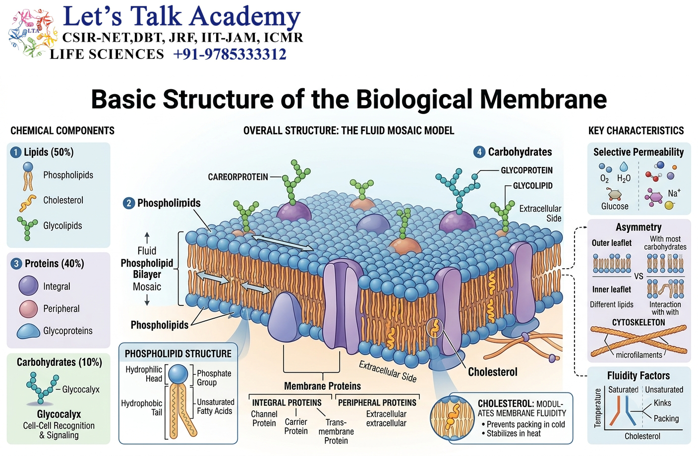
The biological membrane is primarily composed of a phospholipid bilayer.
Each phospholipid molecule contains two major regions:
- A hydrophilic head
- Hydrophobic tails
The hydrophilic head interacts with water, whereas the hydrophobic tails avoid water.
When phospholipids are placed in an aqueous environment, they spontaneously arrange themselves into a bilayer. The hydrophilic heads face the aqueous surroundings, while the hydrophobic tails point toward the interior.
This creates three important regions:
Extracellular aqueous environment → hydrophilic membrane surface → hydrophobic membrane core → hydrophilic membrane surface → cytoplasmic aqueous environment
The hydrophobic core creates a strong barrier against the free movement of ions and most polar molecules.
Membrane proteins overcome this barrier by providing specialized pathways.
2.2 Hydrophobic and Hydrophilic Regions
The distribution of amino acids within a membrane protein is strongly influenced by the chemical environment.
Amino acids with hydrophobic side chains are commonly found in membrane-spanning regions because they interact favorably with the hydrophobic fatty-acid tails of membrane lipids.
Examples include:
- Leucine
- Isoleucine
- Valine
- Phenylalanine
- Methionine
- Alanine
On the other hand, amino acids with polar or charged side chains are commonly found in regions exposed to water.
This difference in amino-acid composition is extremely important when studying membrane protein structure.
3. Classification of Membrane Proteins
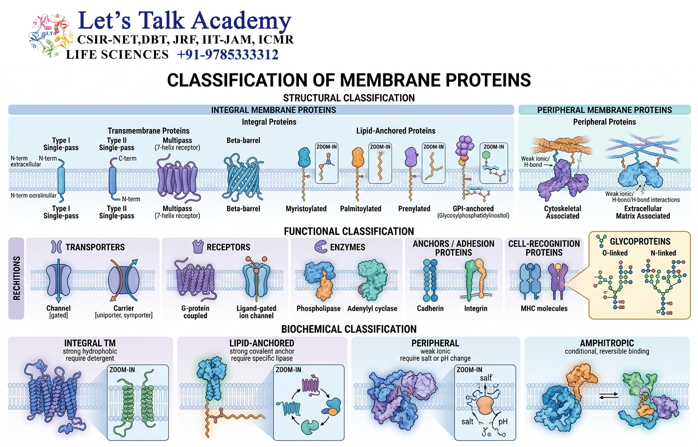
Membrane proteins can be classified according to how they are associated with the membrane.
The three major categories are:
3.1 Integral Membrane Proteins
These are firmly associated with the membrane and generally contain regions that penetrate the lipid bilayer.
3.2 Peripheral Membrane Proteins
These are associated with the membrane surface but do not normally penetrate the hydrophobic core of the bilayer.
3.3 Lipid-Anchored Proteins
These proteins are covalently attached to lipid groups that anchor them to the membrane.
4. Integral Membrane Proteins
4.1 Definition
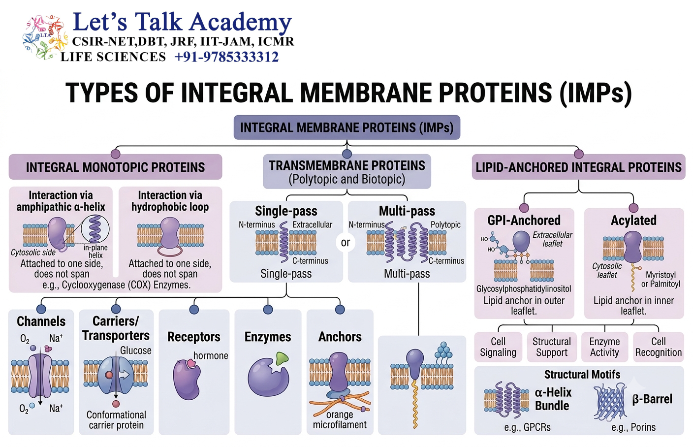
Integral membrane proteins are proteins that are embedded within the lipid bilayer.
Some integral proteins extend only partially into the membrane, whereas others span the entire membrane.
Most transmembrane proteins contain hydrophobic amino-acid sequences that interact with the membrane’s hydrophobic core.
Because these proteins are strongly associated with the lipid bilayer, they usually require detergents or other membrane-disrupting methods for extraction.
4.2 Transmembrane Proteins
A transmembrane protein crosses the entire lipid bilayer at least once.
A simple transmembrane protein can therefore have three major regions:
4.2.1 Extracellular Domain
This portion faces the extracellular environment.
It may:
- Bind ligands
- Interact with neighboring cells
- Participate in recognition
- Interact with extracellular matrix components
4.2.2 Transmembrane Domain
This portion lies within the membrane and usually contains hydrophobic amino acids.
4.2.3 Cytoplasmic Domain
This region faces the cytoplasm and may interact with:
- Enzymes
- Signaling proteins
- Cytoskeletal proteins
- Regulatory proteins
This arrangement allows a membrane protein to connect events occurring outside the cell with processes occurring inside the cell.
5. Transmembrane α-Helices
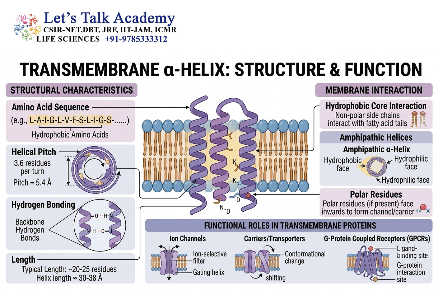
One of the most common structural arrangements used by proteins to cross biological membranes is the transmembrane α-helix.
5.1 Structure of a Transmembrane α-Helix
An α-helix is a common secondary structure of proteins. In membrane proteins, a stretch of approximately 20 hydrophobic amino acids can form an α-helix capable of spanning the hydrophobic region of a lipid bilayer.
The hydrophobic side chains generally face outward toward the lipid molecules.
The peptide backbone is stabilized by hydrogen bonding within the helix.
This arrangement is particularly favorable because the polar peptide backbone is shielded from direct exposure to the hydrophobic membrane interior.
5.2 Single-Pass Membrane Proteins
A single-pass membrane protein crosses the lipid bilayer once.
A simple example is a receptor containing:
Extracellular ligand-binding region → one transmembrane helix → intracellular signaling region
Such proteins are commonly involved in cell signaling and adhesion.
5.3 Multipass Membrane Proteins
Multipass membrane proteins cross the membrane multiple times.
They contain several transmembrane α-helices.
These proteins are especially important in:
- Ion transport
- Nutrient transport
- Signal transduction
- Electron transport
- Maintenance of ion gradients
Many transport proteins have multiple membrane-spanning helices that form a pathway through which molecules can move.
6. Transmembrane β-Barrels
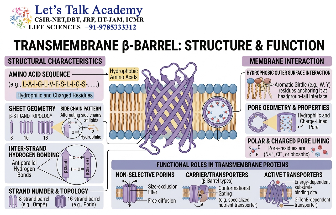
Not every membrane-spanning protein is built from α-helices.
Some membrane proteins form β-barrels.
6.1 Structure of β-Barrels
A β-barrel consists of multiple β-strands arranged in a cylindrical structure.
The alternating orientation of amino-acid side chains allows:
- Hydrophobic residues to interact with the membrane
- Hydrophilic residues to face the interior of the barrel
The interior may therefore form a water-filled channel.
6.2 Location of β-Barrel Proteins
β-barrel membrane proteins are particularly common in the outer membranes of Gram-negative bacteria.
They are also found in the outer membranes of mitochondria and chloroplasts.
These proteins can function as:
- Channels
- Transport proteins
- Receptors
- Structural components
7. Peripheral Membrane Proteins
Peripheral membrane proteins are associated with the membrane surface without being deeply embedded in the hydrophobic core.
7.1 How Do They Associate With Membranes?
They may interact with:
- Integral membrane proteins
- Polar lipid head groups
- Other peripheral proteins
- Cytoskeletal proteins
Because they do not usually penetrate deeply into the lipid bilayer, they can often be removed using relatively mild treatments that disrupt non-covalent interactions.
8. Lipid-Anchored Proteins
Some proteins are attached to membranes through covalently attached lipid groups.
These proteins are called lipid-anchored proteins.
The protein itself may not contain a transmembrane domain. Instead, the lipid modification provides the membrane attachment.
Important examples include:
8.1 GPI-Anchored Proteins
GPI stands for glycosylphosphatidylinositol.
GPI anchors attach proteins to the extracellular surface of the plasma membrane.
8.2 Prenylated Proteins
Certain proteins are attached to membranes through prenyl groups.
Examples include:
- Farnesyl groups
- Geranylgeranyl groups
8.3 Fatty-Acid-Anchored Proteins
Some proteins are attached to membranes through fatty acids such as:
- Myristate
- Palmitate
The type of lipid modification can influence where the protein is located and how it interacts with other molecules.
9. Membrane Protein Topology
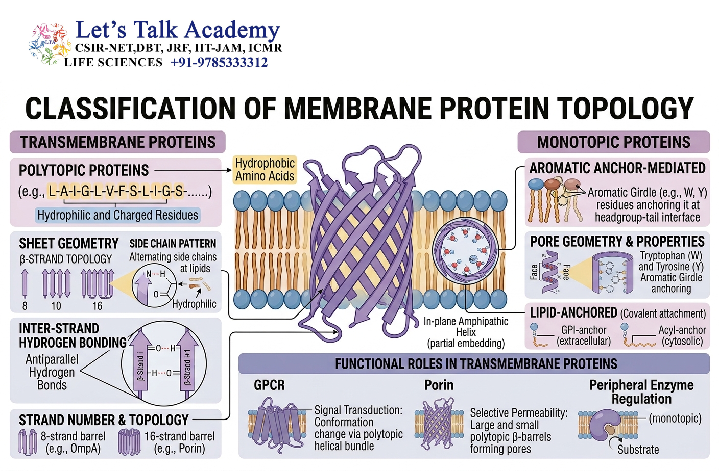
Membrane protein topology describes the orientation and arrangement of different regions of a membrane protein relative to the membrane.
This includes determining:
- Which part faces the cytoplasm
- Which part faces the extracellular space
- How many times the protein crosses the membrane
- Where the N-terminus is located
- Where the C-terminus is located
Topology is extremely important because the location of a protein domain determines which molecules it can interact with.
9.1 N-Terminal and C-Terminal Orientation
Proteins have two ends:
- N-terminus
- C-terminus
In a membrane protein, these termini can be located on either the cytoplasmic or extracellular side.
The orientation is determined during protein synthesis and membrane insertion.
9.2 Positive-Inside Rule
A commonly observed principle in membrane protein topology is the positive-inside rule.
According to this principle, positively charged amino acids such as lysine and arginine tend to be more abundant on the cytoplasmic side of transmembrane proteins.
This contributes to the correct orientation of many membrane proteins.
10. Hydropathy Analysis
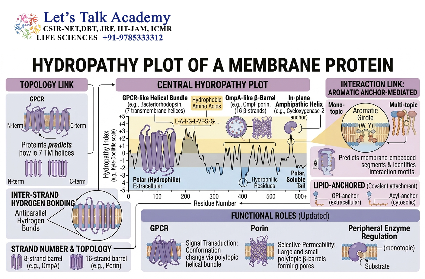
Hydropathy analysis is an important method for identifying potential membrane-spanning regions in a protein sequence.
10.1 Basic Principle
Different amino acids have different degrees of hydrophobicity.
A stretch of amino acids containing many hydrophobic residues may represent a transmembrane region.
A hydropathy plot represents the hydrophobic or hydrophilic character of different regions of a protein.
A strong hydrophobic region extending for an appropriate length can indicate a possible transmembrane helix.
11. Functions of Membrane Proteins
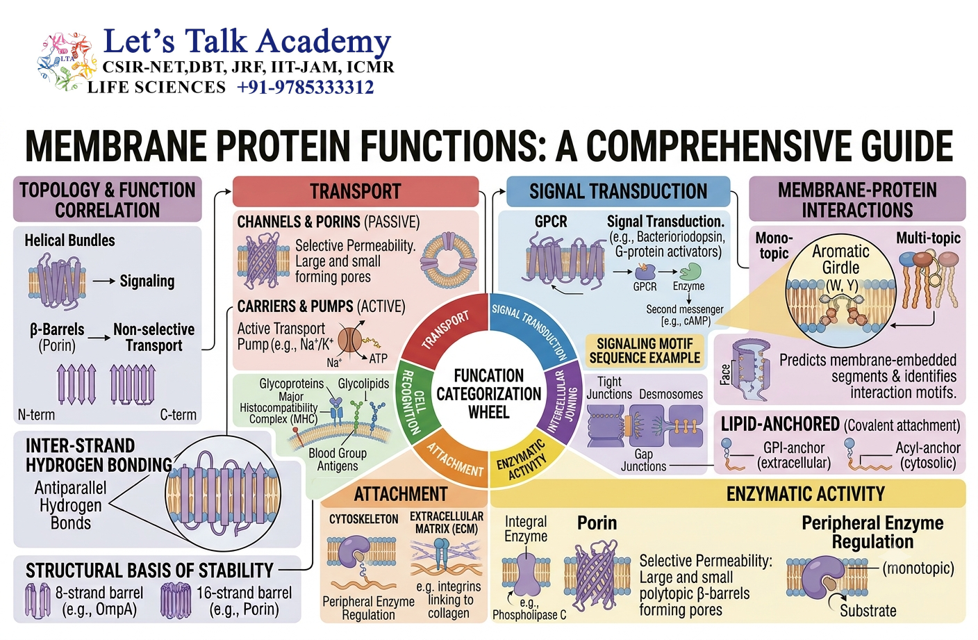
Membrane proteins perform diverse functions.
11.1 Transport
Transport proteins regulate the movement of substances across membranes.
These include:
- Channels
- Carriers
- Pumps
11.2 Receptors
Receptor proteins detect extracellular signals.
Examples of signals include:
- Hormones
- Neurotransmitters
- Growth factors
- Cytokines
11.3 Enzymes
Some membrane proteins have enzymatic activity and catalyze reactions at the membrane surface.
11.4 Cell Adhesion
Membrane proteins help cells attach to:
- Other cells
- Extracellular matrix
- Basement membranes
11.5 Cell Recognition
Specific proteins and glycoproteins on the cell surface contribute to recognition between cells.
11.6 Energy Conversion
Membrane proteins are central to energy-producing systems.
For example, proteins of the electron transport chain are embedded in the inner mitochondrial membrane and participate in establishing a proton gradient used for ATP production.
12. Membrane Proteins Involved in Transport
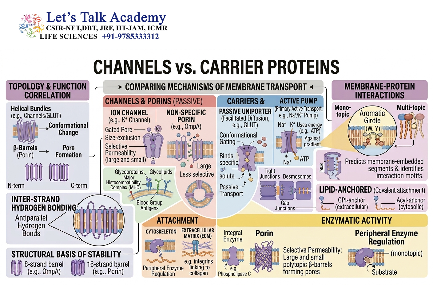
Transport is one of the most important functions of membrane proteins.
The lipid bilayer is selectively permeable, meaning that different substances cross it with different degrees of difficulty.
Membrane proteins provide controlled routes for substances that cannot readily cross the lipid bilayer.
12.1 Channels
Channels form hydrophilic pathways through the membrane.
They allow specific ions or molecules to move down their electrochemical gradient.
Examples include:
- Potassium channels
- Sodium channels
- Calcium channels
- Chloride channels
- Aquaporins
Channels generally transport substances rapidly because the transported molecules move through an open pathway rather than binding and being carried individually through a large conformational cycle.
12.2 Carrier Proteins
Carrier proteins bind specific molecules and undergo conformational changes to move them across the membrane.
The molecule first binds to the carrier on one side.
The protein then changes its conformation.
The binding site becomes accessible to the opposite side, where the molecule is released.
This is different from a channel, which provides a continuous aqueous pathway through the membrane.
13. Passive and Active Transport
13.1 Passive Transport
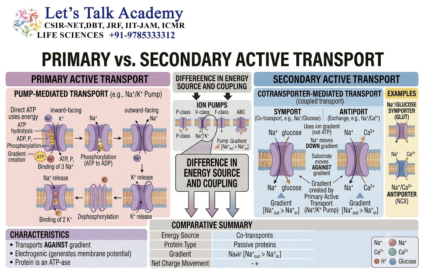
Passive transport does not directly require cellular energy in the form of ATP.
The substance moves down its electrochemical gradient.
Examples include:
- Simple diffusion
- Facilitated diffusion
- Ion movement through channels
13.2 Active Transport
Active transport moves substances against their electrochemical gradients.
It requires an energy source.
Active transport can be divided into:
Primary Active Transport
Uses energy directly, commonly from ATP hydrolysis.
Secondary Active Transport
Uses the energy stored in an electrochemical gradient established by another transport process.
14. Ion Channels
Ion channels are membrane proteins that allow selected ions to cross the membrane.
14.1 Ion Selectivity
Ion channels are selective.
A potassium channel, for example, does not simply allow every positively charged ion to pass freely. The structure of the channel determines which ions can enter and move through it.
Selectivity depends on factors such as:
- Ion size
- Charge
- Coordination with amino-acid side chains
- Hydration state
- Structure of the selectivity filter
14.2 Channel Gating
Channels can switch between different functional states.
They may be:
- Closed
- Open
- Inactivated
The transition between these states is called gating.
Channels can be regulated by different stimuli.
14.3 Voltage-Gated Channels
Voltage-gated channels respond to changes in membrane potential.
They are particularly important in:
- Neurons
- Muscle cells
- Electrical signaling
14.4 Ligand-Gated Channels
Ligand-gated channels open or close when a specific molecule binds to the channel or its associated receptor.
They provide a direct connection between chemical signaling and changes in membrane permeability.
14.5 Mechanically Gated Channels
These channels respond to mechanical forces such as:
- Stretch
- Pressure
- Membrane deformation
They are important in processes such as touch and mechanosensation.
15. Membrane Pumps
Pumps are membrane proteins that use energy to move substances across membranes.
15.1 Sodium-Potassium Pump
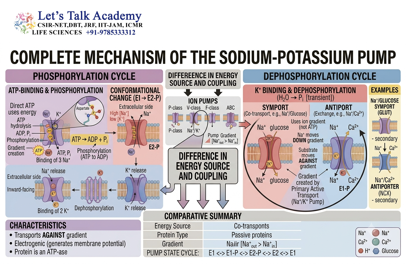
The Na⁺/K⁺-ATPase is an important P-type ATPase found in the plasma membrane of animal cells.
For each ATP molecule hydrolyzed, it typically transports:
- 3 Na⁺ out of the cell
- 2 K⁺ into the cell
This contributes to the maintenance of cellular ion gradients and membrane potential.
The pump undergoes conformational changes during its transport cycle.
16. Membrane Receptors
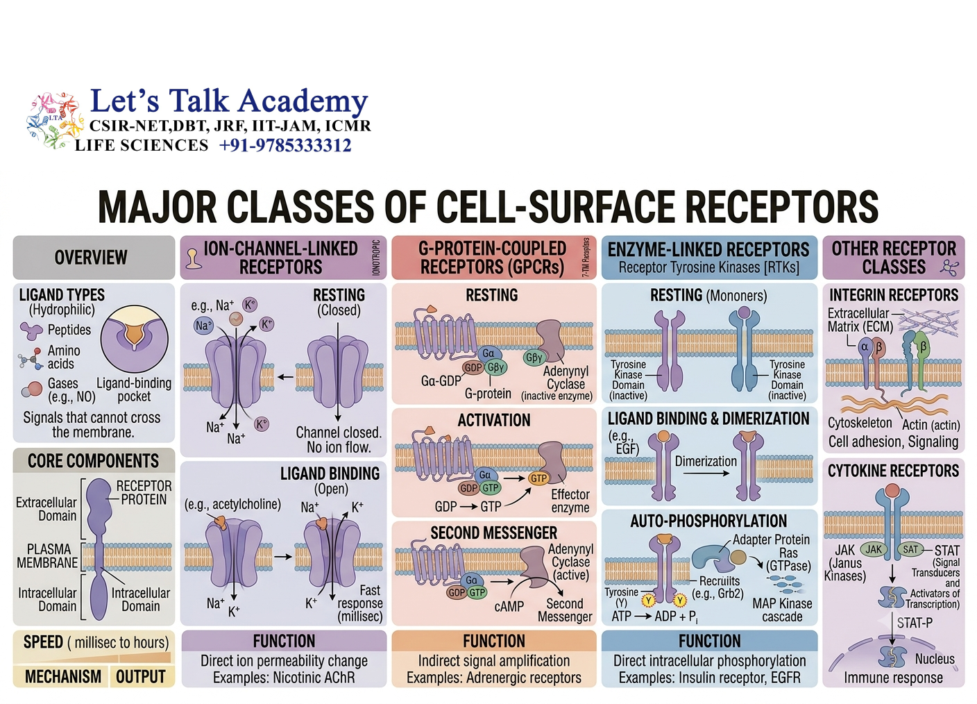
Receptors are membrane proteins that allow cells to detect extracellular signals.
A receptor generally contains a region that recognizes a signaling molecule and a mechanism that communicates the signal to the cell interior.
The overall process can be summarized as:
Signal → Receptor → Signal transduction → Cellular response
Different receptors use different mechanisms to transmit information.
17. G-Protein-Coupled Receptors
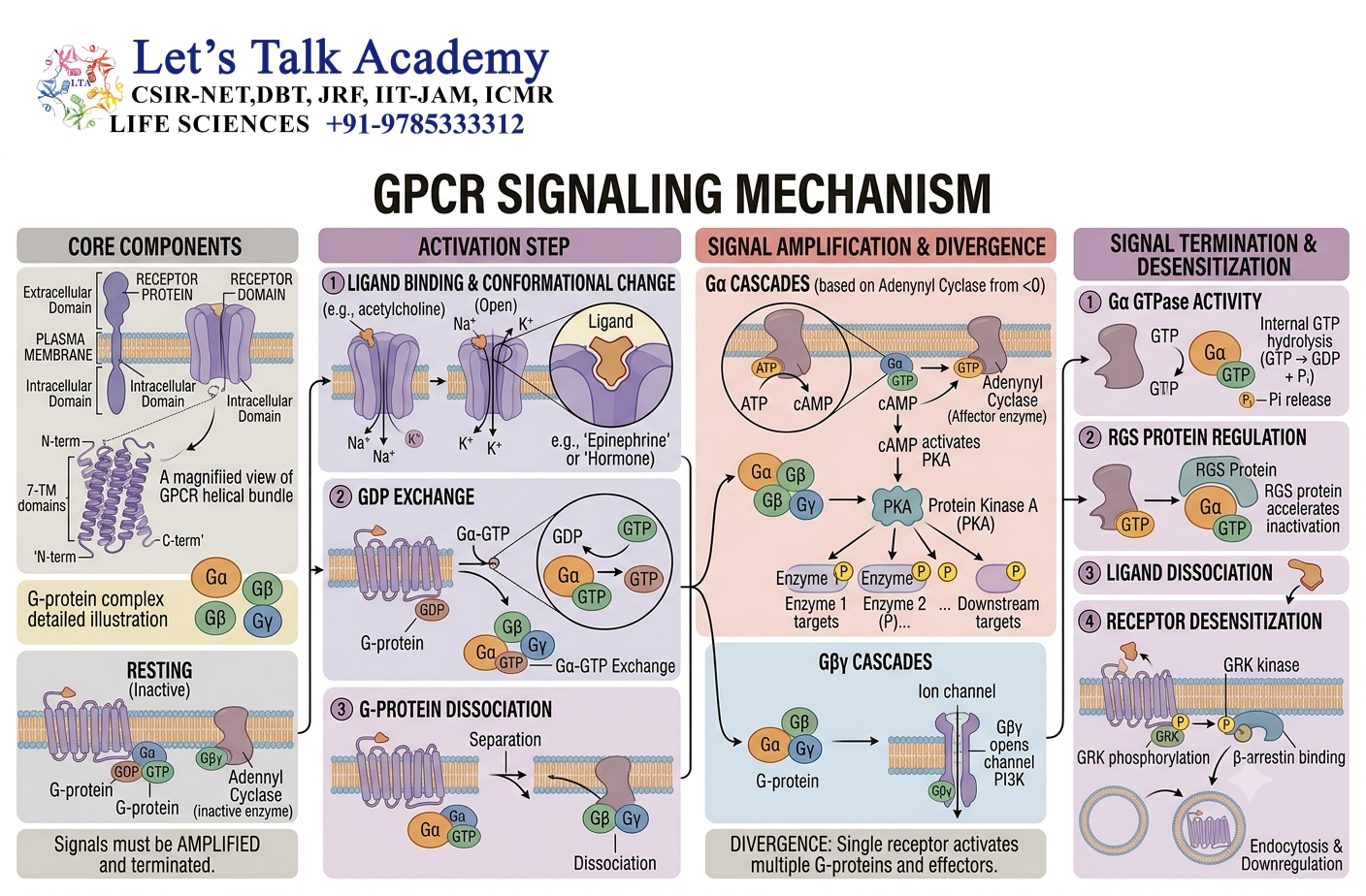
G-protein-coupled receptors, commonly called GPCRs, are a large family of cell-surface receptors.
17.1 Structure
GPCRs characteristically contain seven transmembrane α-helices.
The extracellular portion participates in ligand recognition, while the intracellular regions interact with heterotrimeric G proteins.
17.2 Activation
When a ligand binds to a GPCR, the receptor undergoes a conformational change.
This promotes activation of an associated G protein.
The G protein contains:
- α subunit
- β subunit
- γ subunit
The α subunit undergoes nucleotide exchange, replacing GDP with GTP.
The activated components can then regulate downstream effectors.
18. Receptor Tyrosine Kinases
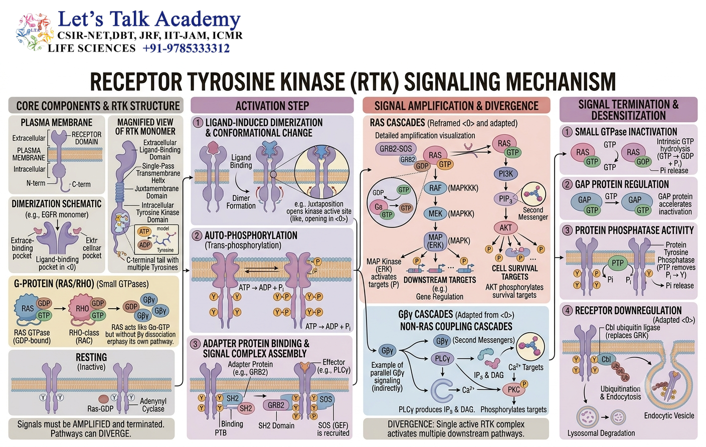
Receptor tyrosine kinases, or RTKs, are another major class of membrane receptors.
18.1 General Structure
A typical RTK contains:
- Extracellular ligand-binding domain
- Single transmembrane segment
- Intracellular tyrosine kinase domain
18.2 Activation Mechanism
Ligand binding commonly promotes receptor dimerization or rearrangement of receptor molecules.
The intracellular kinase domains become activated and phosphorylate tyrosine residues.
These phosphorylated residues can serve as docking sites for signaling proteins.
This initiates intracellular signaling pathways that can regulate:
- Cell growth
- Cell division
- Differentiation
- Survival
- Metabolism
19. Membrane Proteins and Cell Junctions
Membrane proteins are essential components of cell junctions.
Different junctions perform different functions.
19.1 Tight Junctions
Tight junctions help seal the space between adjacent epithelial cells and regulate paracellular movement.
19.2 Adherens Junctions
Adherens junctions connect neighboring cells and are associated with the actin cytoskeleton.
19.3 Desmosomes
Desmosomes provide strong mechanical attachment between cells and are associated with intermediate filaments.
19.4 Gap Junctions
Gap junctions allow direct communication between neighboring cells by providing pathways for small molecules and ions.
19.5 Hemidesmosomes
Hemidesmosomes help attach cells to the underlying extracellular matrix.
20. Membrane Protein Biosynthesis
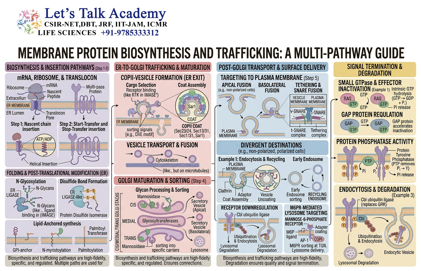
Membrane proteins must be synthesized and inserted into membranes in a coordinated manner.
Many membrane proteins destined for the secretory pathway begin synthesis on ribosomes associated with the rough endoplasmic reticulum.
20.1 Signal Sequences
Specific amino-acid sequences help direct proteins to the correct cellular compartment.
20.2 Signal Recognition Particle
The signal recognition particle recognizes appropriate signal sequences and helps direct the translating ribosome to the endoplasmic reticulum.
20.3 Protein Translocation
The growing protein is inserted into or transported across the endoplasmic reticulum membrane through specialized protein-conducting machinery.
20.4 Membrane Insertion
Hydrophobic sequences can function as membrane-spanning segments and become embedded in the lipid bilayer.
21. Membrane Protein Folding and Quality Control
Correct folding is essential for membrane protein function.
A newly synthesized membrane protein must achieve the correct:
- Secondary structure
- Tertiary structure
- Membrane orientation
- Oligomeric state
The endoplasmic reticulum contains quality-control mechanisms that identify improperly folded proteins.
Misfolded proteins may be retained, refolded, or targeted for degradation.
22. Membrane Protein Trafficking
After synthesis and processing, many membrane proteins are transported through the secretory pathway.
A typical pathway is:
Endoplasmic reticulum → Golgi apparatus → Transport vesicles → Plasma membrane or other destination
During this process, proteins may undergo:
- Folding
- Glycosylation
- Proteolytic processing
- Sorting
- Quality control
The correct delivery of membrane proteins is essential for normal cellular function.
23. Glycosylation of Membrane Proteins
Many membrane proteins are glycoproteins.
Carbohydrate groups can be attached to specific amino-acid residues.
Two major types are:
23.1 N-Linked Glycosylation
Carbohydrates are attached to the nitrogen atom of the side chain of asparagine.
23.2 O-Linked Glycosylation
Carbohydrates are attached to oxygen-containing groups of amino acids such as serine or threonine.
Glycosylation can influence:
- Protein folding
- Stability
- Recognition
- Cell adhesion
- Protein trafficking
Importantly, carbohydrate groups of plasma membrane glycoproteins are generally exposed on the extracellular surface, contributing to the glycocalyx.
24. Membrane Protein Degradation
Membrane proteins have different lifetimes.
Some remain functional for long periods, whereas others are rapidly removed and degraded.
A cell must carefully regulate protein degradation because excessive or defective membrane proteins can disturb cellular signaling and transport.
Membrane proteins may be internalized through endocytosis and transported through endosomal compartments.
Depending on cellular requirements, they may be:
- Returned to the plasma membrane
- Stored or redirected
- Transported to lysosomes for degradation
Ubiquitination can also function as an important signal in the sorting and degradation of many membrane proteins.
25. Membrane Proteins and the Cytoskeleton
The plasma membrane is closely connected to the cytoskeleton.
Membrane proteins can interact directly or indirectly with:
- Actin filaments
- Intermediate filaments
- Microtubule-associated systems
These interactions help control:
- Cell shape
- Membrane stability
- Protein localization
- Cell migration
- Cell adhesion
For example, membrane-associated proteins can act as bridges between transmembrane proteins and the underlying cytoskeleton.
26. Membrane Protein Dynamics
Membrane proteins are dynamic molecules.
They can:
- Move laterally within the membrane
- Rotate
- Change conformation
- Associate with other proteins
- Form clusters
- Move into specialized membrane domains
This dynamic behavior is essential for many cellular processes.
For example, receptors may cluster after ligand binding, bringing signaling molecules together and increasing the efficiency of signal transduction.
27. Membrane Microdomains and Lipid Rafts
Certain regions of membranes can differ in lipid and protein composition.
These regions are often referred to as membrane microdomains.
The concept of lipid rafts describes relatively ordered membrane domains enriched in particular lipids, including cholesterol and sphingolipids.
Certain signaling proteins can become concentrated in these domains, potentially facilitating interactions between components of signaling pathways.
The precise properties and biological significance of lipid rafts remain an area of active research.
28. Membrane Proteins in Mitochondria
Mitochondria contain specialized membrane proteins that are essential for energy production.
The inner mitochondrial membrane contains major components of the electron transport chain.
These proteins participate in electron transfer and proton pumping.
The resulting proton electrochemical gradient is used by ATP synthase to produce ATP.
Intermembrane space
I
II
III
IV
Mitochondrial matrix
Inner mitochondrial membrane
The electron transport chain and ATP synthase sit in the inner mitochondrial membrane.
This illustrates an important principle: membrane proteins can convert an electrochemical gradient into usable chemical energy.
29. Experimental Study of Membrane Proteins
Membrane proteins can be difficult to study because they exist in association with lipids.
Several experimental approaches are therefore used.
29.1 Cell Fractionation
Cell fractionation separates cellular components based on their physical properties.
It can be used to enrich membrane fractions before further analysis.
29.2 Detergents
Detergents can disrupt lipid-lipid interactions and solubilize membrane proteins.
Different detergents have different effects on protein structure and protein-protein interactions.
Ionic detergents
These can strongly disrupt protein interactions and may denature proteins.
Non-ionic detergents
These are generally milder and can sometimes preserve protein structure and protein complexes.
30. SDS-PAGE and Membrane Proteins
SDS-PAGE is widely used to analyze proteins according to their electrophoretic mobility.
SDS binds to proteins and helps give them a relatively uniform negative charge-to-mass ratio.
The proteins are then separated primarily according to molecular size.
For membrane proteins, appropriate sample preparation is particularly important because hydrophobic proteins may be difficult to solubilize without detergents.
31. Western Blotting
Western blotting can be used to detect a specific membrane protein.
The general workflow involves:
- Protein extraction
- Electrophoretic separation
- Transfer to a membrane
- Blocking
- Incubation with a primary antibody
- Detection using a labeled secondary antibody or other detection system
This technique provides information about the presence and approximate molecular size of a target protein.
32. Structural Study of Membrane Proteins
Understanding the three-dimensional structure of membrane proteins is essential for understanding their function.
Important structural approaches include:
32.1 X-Ray Crystallography
Can provide high-resolution structural information when suitable crystals can be obtained.
32.2 Cryo-Electron Microscopy
Cryo-EM has become particularly important for studying large membrane protein complexes and proteins that are difficult to crystallize.
32.3 Nuclear Magnetic Resonance
NMR can provide structural and dynamic information for suitable proteins and protein domains.
33. Functional Importance of Membrane Proteins
The importance of membrane proteins becomes especially clear when their function is disrupted.
Altered membrane proteins can affect:
- Ion balance
- Cellular signaling
- Nutrient uptake
- Cell adhesion
- Electrical activity
- Energy production
- Immune responses
Changes in membrane protein structure or abundance can therefore have major consequences for cell physiology.
34. Structure–Function Relationship
One of the central ideas in membrane biology is that structure determines function.
A channel requires a suitable pore.
A transporter requires conformational flexibility.
A receptor requires a ligand-binding region and a mechanism for transmitting information.
An enzyme requires a properly organized active site.
Thus, the position, sequence, topology, and three-dimensional structure of a membrane protein are directly related to what the protein can do.
35. Comparison of Major Types of Membrane Proteins
| Feature | Integral | Peripheral | Lipid-Anchored |
|---|---|---|---|
| Association | Embedded in membrane | Surface-associated | Attached through lipid |
| Transmembrane region | Often present | Usually absent | Usually absent |
| Interaction with bilayer | Strong | Indirect/surface | Through covalent lipid |
| Removal | Often requires detergent | Relatively mild treatment | Requires disruption of lipid attachment |
| Typical functions | Transport, receptors | Signaling, cytoskeletal interactions | Signaling, membrane localization |


