
1.1 Introduction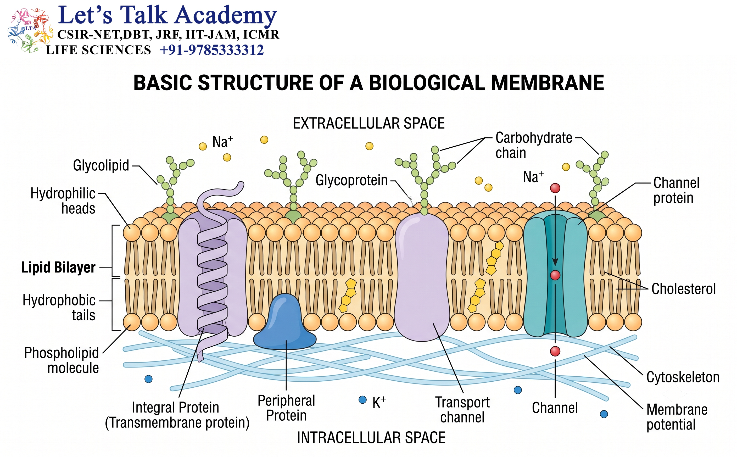
Every living cell must maintain a boundary between its internal environment and the surrounding environment. This boundary is provided primarily by the biological membrane. Although membranes are extremely thin at the cellular scale, they are structurally complex and perform a remarkable variety of functions.
A membrane is not simply a passive covering surrounding a cell. It is a dynamic molecular system that controls the movement of substances, receives and transmits signals, provides attachment sites for proteins, participates in energy conversion, and helps maintain the internal conditions required for life.
The fundamental structure of most biological membranes is a lipid bilayer. Within this bilayer, proteins are embedded or associated with the membrane, while carbohydrates are attached to certain lipids and proteins, particularly on the extracellular surface of eukaryotic plasma membranes.
The physical organization of these components determines important properties such as:
- Membrane fluidity
- Selective permeability
- Mechanical stability
- Membrane thickness
- Curvature
- Electrical properties
- Protein mobility
- Molecular recognition
- Signal transmission
Although the basic principle of membrane organization is shared by prokaryotes and eukaryotes, their membranes differ in lipid composition, protein organization, membrane-associated structures, and degree of compartmentalization.
Understanding these similarities and differences provides a foundation for understanding cellular organization and function.
1.2 The Concept of a Model Membrane
A model membrane is a simplified experimental system used to study the physical and chemical properties of biological membranes.
Real biological membranes contain hundreds or even thousands of different molecules. Studying such complex systems directly can be difficult. Researchers therefore use simplified membrane systems containing selected lipids and proteins.
Common model membrane systems include:
- Lipid monolayers
- Lipid bilayers
- Liposomes
- Large unilamellar vesicles
- Small unilamellar vesicles
- Supported lipid bilayers
- Giant unilamellar vesicles
These systems help researchers understand fundamental properties such as:
- Lipid organization
- Membrane fluidity
- Permeability
- Phase behavior
- Protein–lipid interactions
- Membrane curvature
- Fusion and fission
Model membranes do not reproduce every feature of a living cell. Instead, they allow individual physical principles to be studied under controlled conditions.
2. Basic Architecture of Biological Membranes
2.1 The Lipid Bilayer
The lipid bilayer is the fundamental structural framework of most cellular membranes.
It consists of two layers of amphipathic lipid molecules.
Each lipid has:
- A hydrophilic region
- A hydrophobic region
The hydrophilic portions interact with water, whereas the hydrophobic portions avoid contact with water.
In a typical bilayer, the hydrophilic heads face the aqueous environment on either side of the membrane, while the hydrophobic tails point toward the center.
The arrangement can be represented as:
Aqueous environment → Hydrophilic heads → Hydrophobic core → Hydrophilic heads → Aqueous environment
This organization produces a membrane with a hydrophobic interior and hydrophilic surfaces.
2.2 Why Does a Bilayer Form?
The spontaneous formation of a lipid bilayer is primarily driven by the hydrophobic effect.
Water molecules form an organized network around hydrophobic substances. When hydrophobic surfaces are exposed to water, this organization can be energetically unfavorable.
When amphipathic lipids assemble into a bilayer, their hydrophobic regions become shielded from water.
At the same time, their hydrophilic head groups remain exposed to water.
This makes the bilayer a thermodynamically favorable arrangement under appropriate conditions.
The stability of the membrane also involves:
- Van der Waals interactions between hydrocarbon chains
- Hydrogen bonding
- Electrostatic interactions
- Interactions between lipids and proteins
2.3 Thickness of the Bilayer
Biological membranes are extremely thin compared with the overall dimensions of a cell.
The hydrophobic core of a typical phospholipid bilayer is only a few nanometers thick.
Membrane thickness depends on several factors, including:
- Fatty-acid chain length
- Degree of saturation
- Lipid packing
- Sterol concentration
- Lipid head groups
- Associated proteins
Membrane thickness is biologically important because transmembrane proteins must interact appropriately with the hydrophobic region of the bilayer.
3. Amphipathic Lipids
3.1 Meaning of Amphipathic
The word amphipathic refers to a molecule that contains both hydrophilic and hydrophobic regions.
This property is central to membrane organization.
A typical phospholipid contains a polar head group and nonpolar hydrocarbon chains.
The polar region interacts with water.
The nonpolar region interacts with other hydrocarbon chains.
3.2 Phospholipid Structure
A common glycerophospholipid contains:
- Glycerol backbone
- Two fatty-acid chains
- Phosphate group
- Polar head group
The identity of the head group can vary.
Important phospholipids include:
- Phosphatidylcholine
- Phosphatidylethanolamine
- Phosphatidylserine
- Phosphatidylglycerol
- Phosphatidylinositol
- Cardiolipin
Different lipids produce different physical properties.
3.3 Fatty-Acid Chain Length
The length of hydrocarbon chains affects membrane behavior.
Longer hydrocarbon chains generally have stronger van der Waals interactions and can pack more tightly.
Shorter chains generally have weaker interactions and can increase membrane fluidity.
Therefore, changing chain length is one mechanism by which cells can modify membrane physical properties.
3.4 Saturated and Unsaturated Fatty Acids
Fatty-acid chains can be saturated or unsaturated.
Saturated fatty acids contain no carbon-carbon double bonds.
Their relatively straight chains can pack closely together.
Unsaturated fatty acids contain one or more carbon-carbon double bonds.
Many naturally occurring cis double bonds introduce bends into the hydrocarbon chain.
These bends interfere with close packing and generally increase membrane fluidity.
Thus:
More saturation → tighter packing → lower fluidity
More unsaturation → looser packing → higher fluidity
4. Major Lipid Classes in Biological Membranes
4.1 Glycerophospholipids
Glycerophospholipids are major components of bacterial and eukaryotic membranes.
They have a glycerol backbone attached to two hydrophobic chains and a phosphate-containing polar group.
Their chemical diversity contributes to membrane complexity.
4.2 Sphingolipids
Sphingolipids contain a sphingoid backbone.
They are especially important in eukaryotic membranes.
Sphingolipids participate in:
- Membrane organization
- Cell recognition
- Cell signaling
- Cell adhesion
- Formation of specialized membrane domains
4.3 Sterols
Sterols are important membrane components in many eukaryotic organisms.
Animal membranes contain cholesterol.
Plant membranes contain various phytosterols.
Fungal membranes characteristically contain ergosterol.
Sterols can influence:
- Membrane fluidity
- Membrane permeability
- Lipid packing
- Membrane thickness
- Formation of membrane domains
4.4 Cardiolipin
Cardiolipin is a distinctive phospholipid with an important role in energy-related membranes.
It is particularly abundant in:
- Bacterial membranes
- Mitochondrial inner membranes
Cardiolipin can influence membrane curvature and the organization of protein complexes involved in energy metabolism.
5. Membrane Fluidity
5.1 Meaning of Membrane Fluidity
A biological membrane is dynamic rather than rigid.
Lipids can move within the membrane, and many proteins can also move laterally.
This dynamic property is called membrane fluidity.
Fluidity is necessary for:
- Membrane growth
- Cell division
- Vesicle formation
- Membrane fusion
- Protein movement
- Signal transduction
- Transport
- Membrane repair
5.2 Types of Lipid Movement
Lipids can undergo several types of movement.
5.2.1 Lateral Diffusion
A lipid moves within the same leaflet of the membrane.
This movement is relatively rapid.
5.2.2 Rotation
A lipid can rotate around its molecular axis.
5.2.3 Flexion
Hydrocarbon chains can bend and change their conformations.
5.2.4 Transverse Diffusion
A lipid moves from one leaflet to the opposite leaflet.
This is often called flip-flop.
It is much slower spontaneously because the polar head group must cross the hydrophobic interior.
Specialized proteins can facilitate controlled lipid movement between leaflets.
6. Factors That Regulate Membrane Fluidity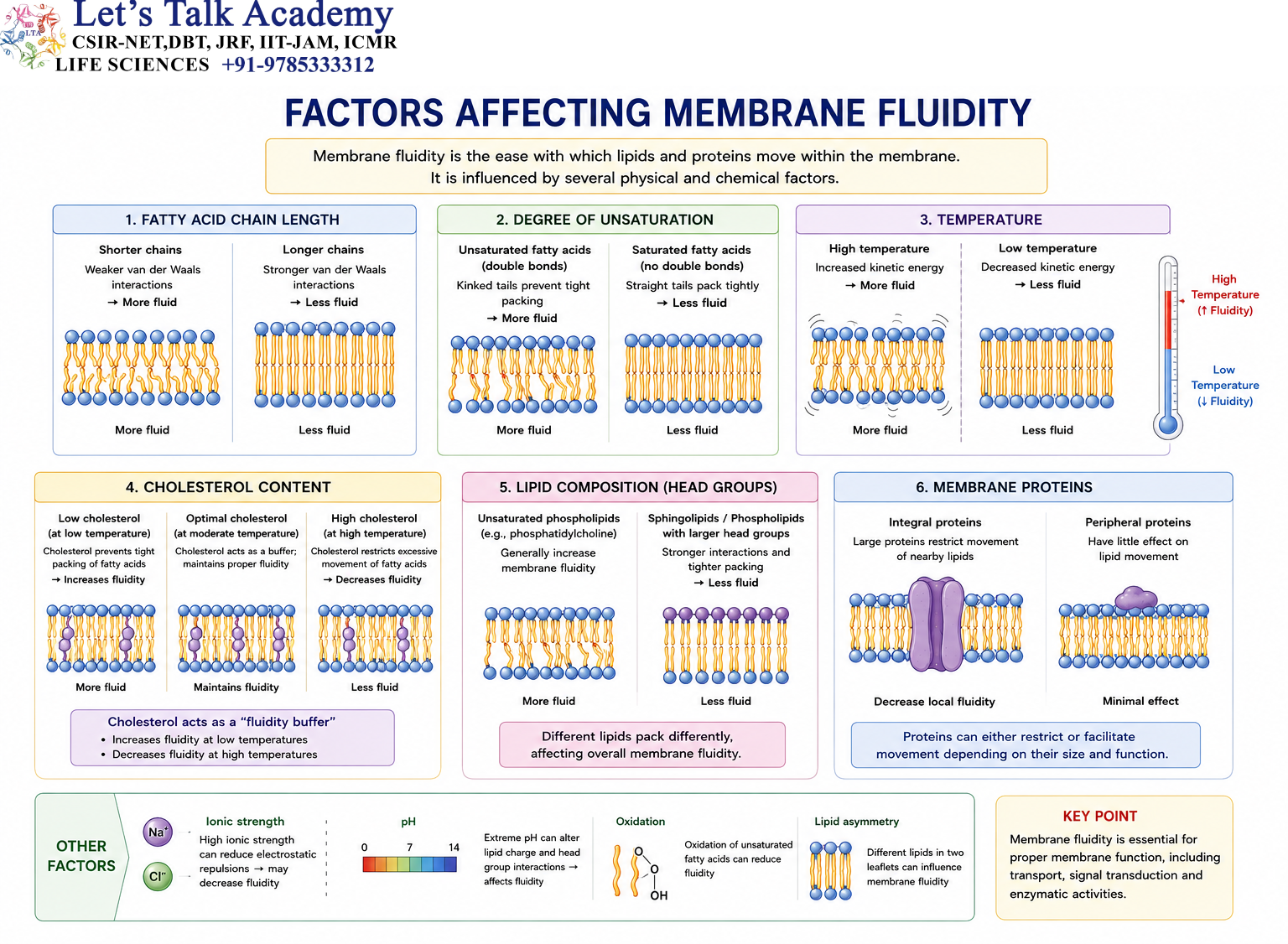
6.1 Temperature
Temperature strongly influences membrane fluidity.
At higher temperatures, molecular movement increases and membranes tend to become more fluid.
At lower temperatures, molecular movement decreases and membranes tend to become more rigid.
Cells regulate membrane composition to prevent excessive changes in fluidity.
6.2 Fatty-Acid Unsaturation
Increasing unsaturated fatty acids generally prevents tight packing.
This helps maintain fluidity at lower temperatures.
6.3 Fatty-Acid Chain Length
Shorter chains generally increase fluidity.
Longer chains generally increase intermolecular interactions and reduce fluidity.
6.4 Sterols
Sterols act as important regulators of membrane physical properties.
Cholesterol, for example, can reduce excessive lipid movement at high temperatures while interfering with tight lipid packing at lower temperatures.
It also decreases the permeability of the membrane to some small polar substances.
6.5 Homeoviscous Adaptation
The ability of organisms to adjust membrane composition in response to environmental conditions is called homeoviscous adaptation.
This is particularly important in microorganisms exposed to changing temperatures.
For example, a microorganism experiencing lower temperature may modify its lipid composition to prevent the membrane from becoming excessively rigid.
7. Membrane Proteins
7.1 Importance of Membrane Proteins
Lipids form the basic structural framework of the membrane, but proteins provide many of its specialized functions.
Membrane proteins can act as:
- Channels
- Transporters
- Pumps
- Receptors
- Enzymes
- Adhesion molecules
- Electron-transfer components
- Structural anchors
The exact protein composition differs greatly between different membranes.
7.2 Integral Membrane Proteins
Integral membrane proteins are embedded within the lipid bilayer.
Many span the entire membrane and are known as transmembrane proteins.
Their hydrophobic regions interact with the membrane interior.
Their hydrophilic regions interact with the surrounding aqueous environments.
7.3 Peripheral Membrane Proteins
Peripheral membrane proteins are associated primarily with one surface of the membrane.
They may interact with:
- Integral membrane proteins
- Lipid head groups
- Cytoskeletal proteins
- Signaling complexes
They can often be removed without disrupting the lipid bilayer itself.
7.4 Lipid-Anchored Proteins
Some proteins are attached to membranes through covalently attached lipid groups.
The lipid anchor inserts into the membrane while the protein remains on the membrane surface.
These proteins are important in:
- Signal transduction
- Cell recognition
- Membrane organization
8. The Fluid Mosaic Model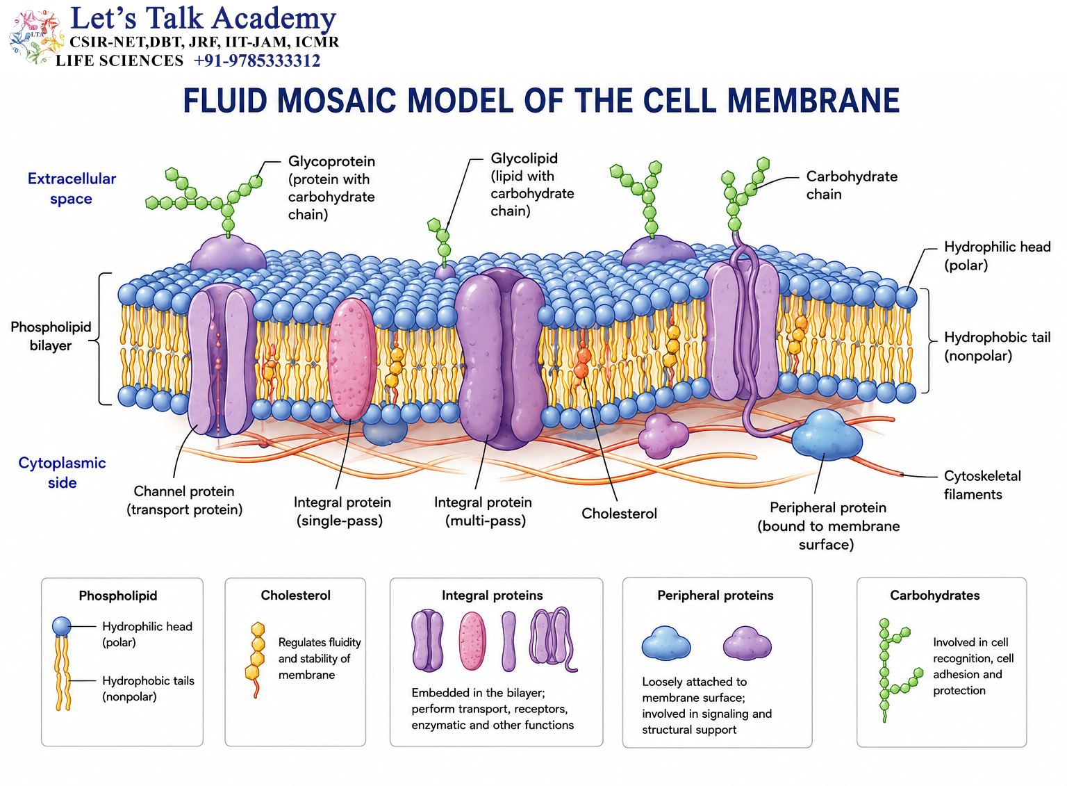
8.1 Development of the Model
The Fluid Mosaic Model was proposed by Singer and Nicolson in 1972.
The model described the membrane as a fluid lipid matrix containing proteins distributed within it.
The term fluid reflects the mobility of many membrane components.
The term mosaic reflects the heterogeneous distribution of proteins and other components.
8.2 Main Features
The model emphasizes that:
- Lipids form a bilayer.
- Proteins are embedded or associated with the bilayer.
- Many membrane components can move laterally.
- Membrane proteins have different orientations.
- Carbohydrates are associated with certain membrane lipids and proteins.
- Membranes are dynamic structures.
8.3 Modern View of the Fluid Mosaic Model
The modern understanding of membranes is more complex than the original model.
Membrane components do not always move freely without restriction.
Their movement can be influenced by:
- Cytoskeletal structures
- Extracellular matrix
- Protein–protein interactions
- Lipid domains
- Membrane curvature
- Cell junctions
Therefore, membranes can be described as fluid but organized.
9. Membrane Asymmetry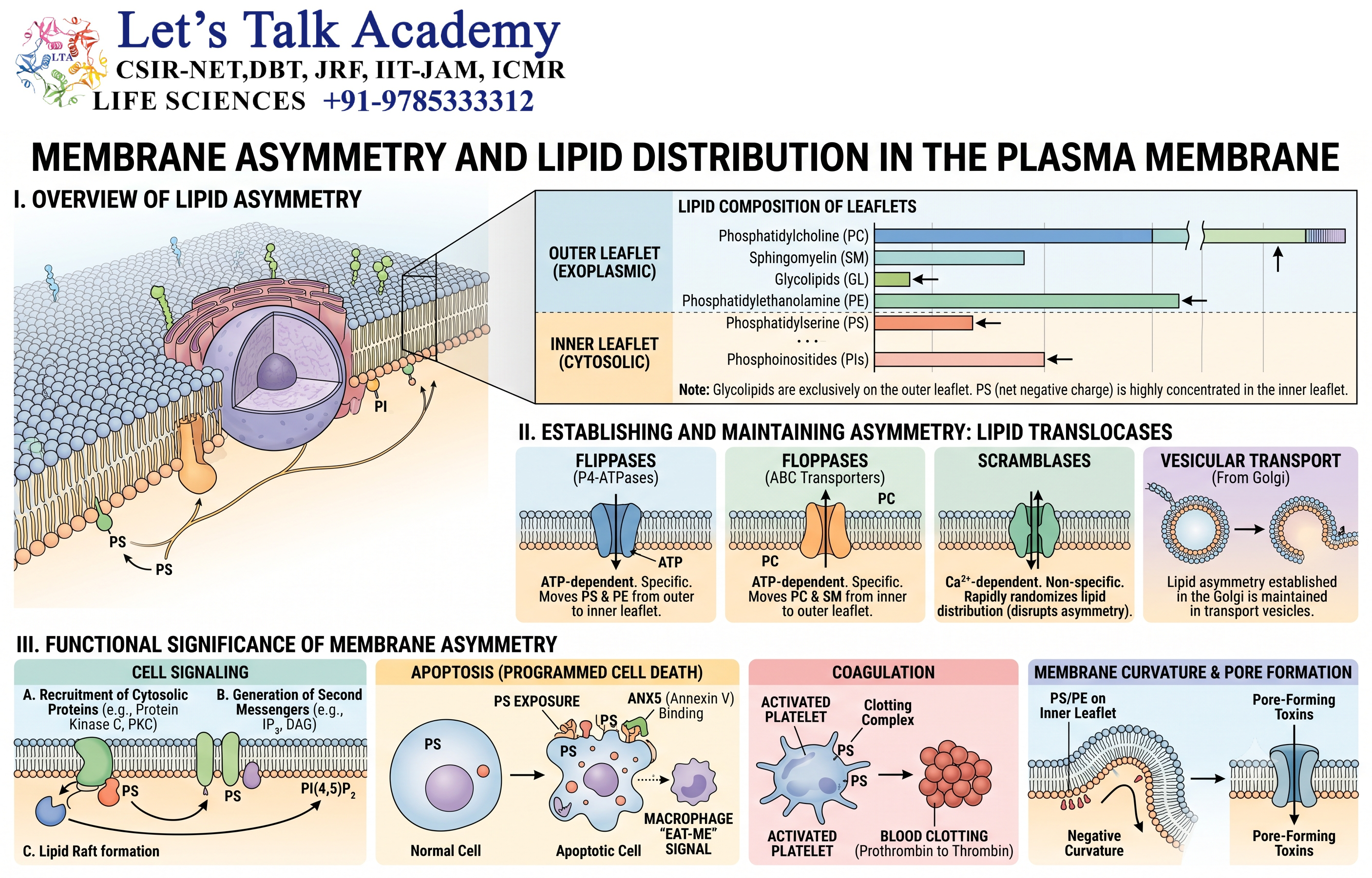
9.1 Definition
The two leaflets of a lipid bilayer are chemically and functionally different.
This unequal distribution is known as membrane asymmetry.
The inner and outer surfaces of a membrane therefore cannot always be considered equivalent.
9.2 Lipid Distribution
In many eukaryotic plasma membranes, different phospholipids show characteristic distributions.
For example:
- Phosphatidylcholine is commonly enriched in the outer leaflet.
- Sphingomyelin is commonly enriched in the outer leaflet.
- Phosphatidylethanolamine is commonly enriched in the inner leaflet.
- Phosphatidylserine is normally concentrated on the cytosolic side.
This distribution is actively maintained.
9.3 Biological Importance
Membrane asymmetry is important for:
- Cell signaling
- Membrane curvature
- Recognition
- Vesicle trafficking
- Protein recruitment
- Cell death signaling
A change in lipid distribution can itself act as a biological signal.
10. Membrane Carbohydrates and the Glycocalyx
10.1 Membrane-Associated Carbohydrates
Carbohydrates are attached to some membrane proteins and lipids.
These structures are found predominantly on the extracellular surface of the plasma membrane.
They form an important part of the glycocalyx.
10.2 Glycoproteins
Glycoproteins contain carbohydrate chains covalently attached to proteins.
They participate in:
- Cell recognition
- Cell adhesion
- Receptor activity
- Immune interactions
10.3 Glycolipids
Glycolipids contain carbohydrate groups attached to lipids.
They are particularly important in the outer leaflet of many eukaryotic plasma membranes.
10.4 Functions of the Glycocalyx
The glycocalyx contributes to:
- Cell protection
- Cell recognition
- Adhesion
- Molecular interactions
- Surface charge
- Communication between cells
11. Physical Structure of Prokaryotic Membranes
11.1 General Organization
Prokaryotic cells include bacteria and archaea.
A typical prokaryotic cell does not possess the extensive endomembrane system characteristic of eukaryotic cells.
However, its plasma or cytoplasmic membrane is highly organized and performs many essential functions.
In bacteria, the cytoplasmic membrane is generally a phospholipid bilayer containing numerous proteins.
11.2 Bacterial Cytoplasmic Membrane
The bacterial cytoplasmic membrane separates the cytoplasm from the external environment.
It performs functions including:
- Nutrient transport
- Ion transport
- Signal transduction
- Energy generation
- Cell-wall synthesis
- Protein secretion
Because bacteria do not have mitochondria, the plasma membrane is particularly important for energy metabolism.
11.3 Membrane-Bound Energy Conversion
In many bacteria, components of the electron transport chain are located in the cytoplasmic membrane.
Electron transfer can generate a proton gradient across the membrane.
The resulting electrochemical gradient can drive ATP synthesis through ATP synthase.
Therefore, the bacterial plasma membrane functions as both:
A selective barrier
and
An energy-converting platform
11.4 Bacterial Membrane Composition
Bacterial membranes commonly contain:
- Phosphatidylethanolamine
- Phosphatidylglycerol
- Cardiolipin
The exact lipid composition varies between bacterial species and environmental conditions.
Some bacteria also contain specialized lipids that help maintain membrane integrity under particular environmental stresses.
12. Gram-Positive Membrane and Cell Envelope
12.1 Basic Organization
Gram-positive bacteria generally have a cytoplasmic membrane surrounded by a thick cell wall.
The cell envelope can be represented as:
Cytoplasm → Cytoplasmic membrane → Thick peptidoglycan layer → External environment
12.2 Peptidoglycan Layer
The thick peptidoglycan layer provides:
- Mechanical strength
- Protection against osmotic stress
- Maintenance of cell shape
The membrane itself remains responsible for selective transport and many metabolic functions.
12.3 Teichoic Acids
Many Gram-positive bacteria contain teichoic acids associated with their cell wall.
These molecules contribute to:
- Cell-wall organization
- Surface properties
- Ion interactions
- Cell-envelope functions
13. Gram-Negative Membrane Organization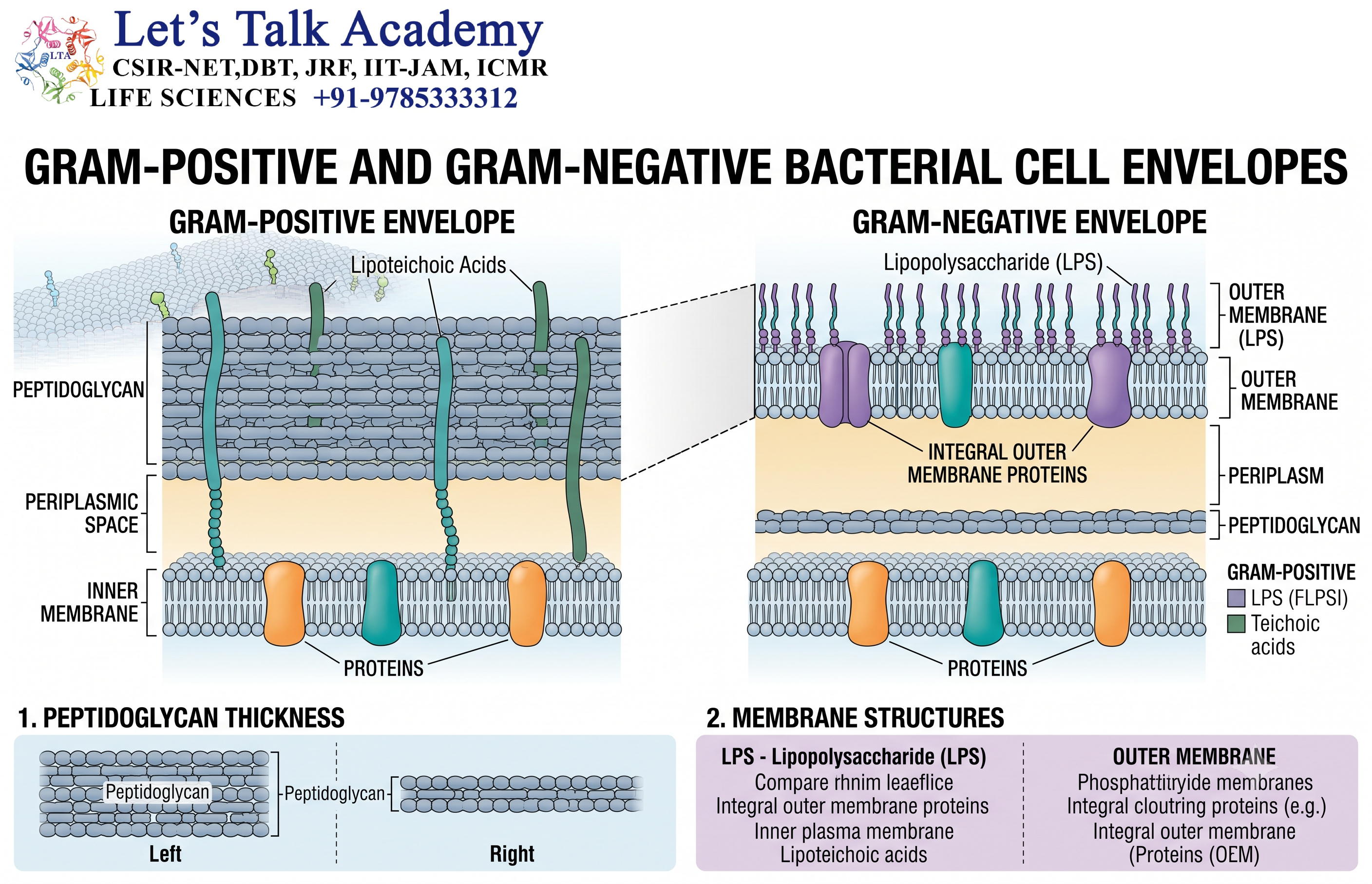
13.1 Two-Membrane System
Gram-negative bacteria possess a more complex cell envelope.
They generally contain:
- Inner cytoplasmic membrane
- Periplasmic region
- Outer membrane
The peptidoglycan layer is relatively thin and lies within the periplasmic region.
13.2 Outer Membrane
The outer membrane is an important permeability barrier.
Its outer leaflet contains lipopolysaccharide (LPS).
The inner leaflet primarily contains phospholipids.
This asymmetry gives the outer membrane distinctive physical and biological properties.
13.3 Porins
The outer membrane contains proteins called porins.
Porins form aqueous channels that permit the movement of certain relatively small hydrophilic molecules.
They contribute to the selective permeability of the bacterial envelope.
14. Archaeal Membranes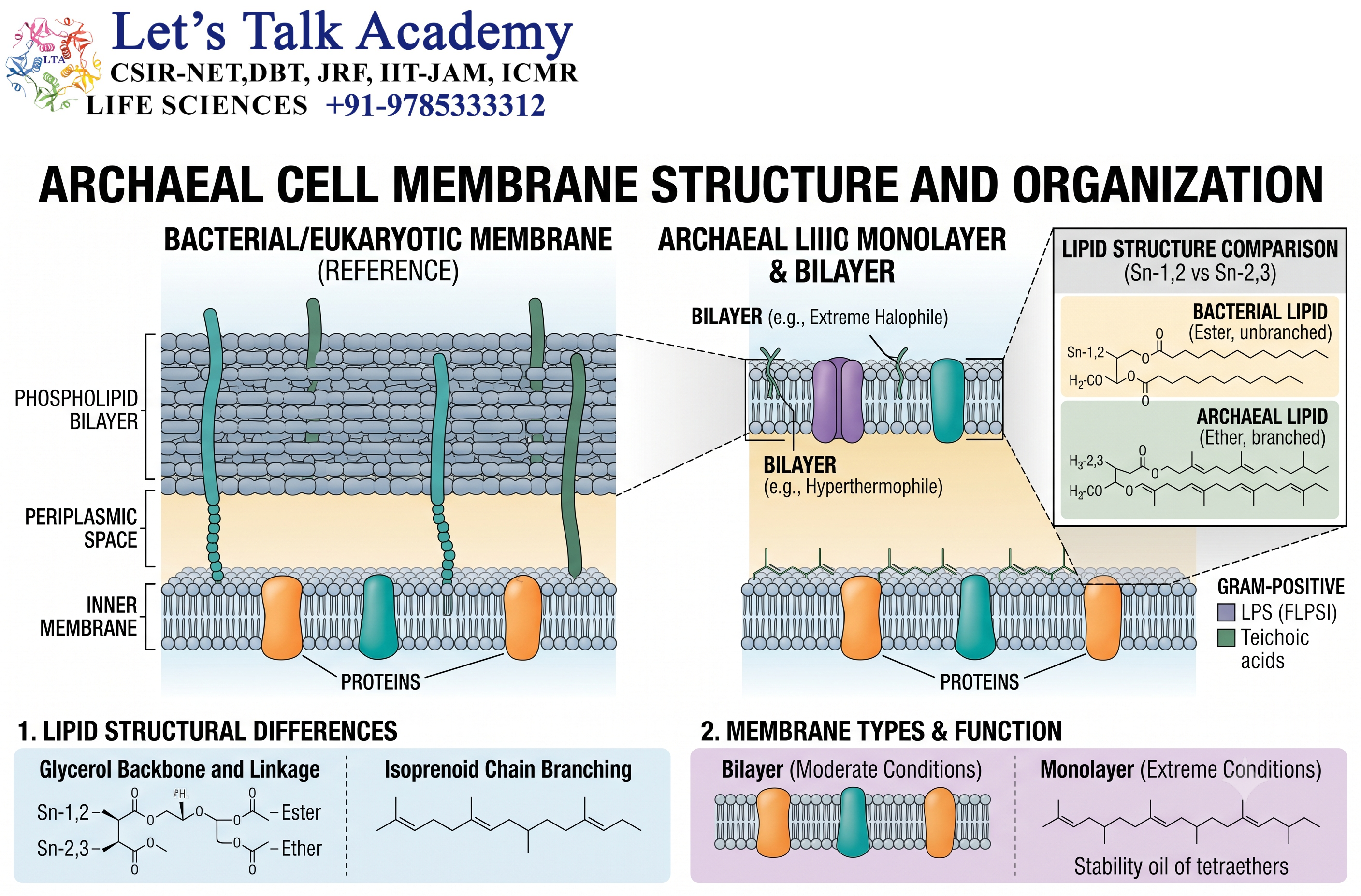
14.1 Distinctive Membrane Chemistry
Archaeal membranes are chemically distinct from typical bacterial and eukaryotic membranes.
A major difference is the nature of their lipid linkages.
Bacterial and eukaryotic glycerolipids commonly contain ester bonds connecting fatty acids to glycerol.
Archaeal membrane lipids characteristically contain ether bonds and isoprenoid hydrocarbon chains.
14.2 Isoprenoid Chains
Archaeal lipids often contain branched isoprenoid chains.
These chains can provide distinctive physical properties.
They can contribute to membrane stability under challenging environmental conditions.
14.3 Tetraether Lipids
Some archaea contain tetraether lipids.
These lipids can span the entire membrane.
Instead of forming a conventional bilayer, they can produce a membrane resembling a monolayer of molecules extending across the membrane.
This structure can provide high stability.
14.4 Environmental Adaptation
Archaeal membrane organization is especially interesting in organisms living in extreme environments.
Examples include environments with:
- High temperature
- High acidity
- High salinity
- High pressure
However, archaeal membrane structures are diverse, and not all archaea possess tetraether monolayers.
15. Physical Structure of Eukaryotic Membranes
15.1 General Organization
Eukaryotic cells contain a plasma membrane and an extensive system of internal membranes.
This creates separate compartments within the cell.
Important membrane-bound structures include:
- Nuclear envelope
- Endoplasmic reticulum
- Golgi apparatus
- Endosomes
- Lysosomes
- Mitochondria
- Chloroplasts
- Peroxisomes
Each membrane has a distinct molecular composition.
15.2 Plasma Membrane
The plasma membrane forms the outer boundary of the eukaryotic cell.
It regulates the exchange of materials between the cell and its environment.
It also contains receptors and signaling proteins that allow the cell to detect external signals.
15.3 Endoplasmic Reticulum
The endoplasmic reticulum is an extensive membrane network.
The rough endoplasmic reticulum contains ribosomes and participates in the synthesis of proteins destined for secretion, membranes, and certain organelles.
The smooth endoplasmic reticulum participates in:
- Lipid synthesis
- Calcium storage
- Detoxification
- Metabolic processes
15.4 Golgi Apparatus
The Golgi apparatus consists of stacked flattened membrane compartments.
It modifies, sorts, and packages proteins and lipids.
Membrane trafficking connects the Golgi with other cellular compartments.
16. Mitochondrial Membranes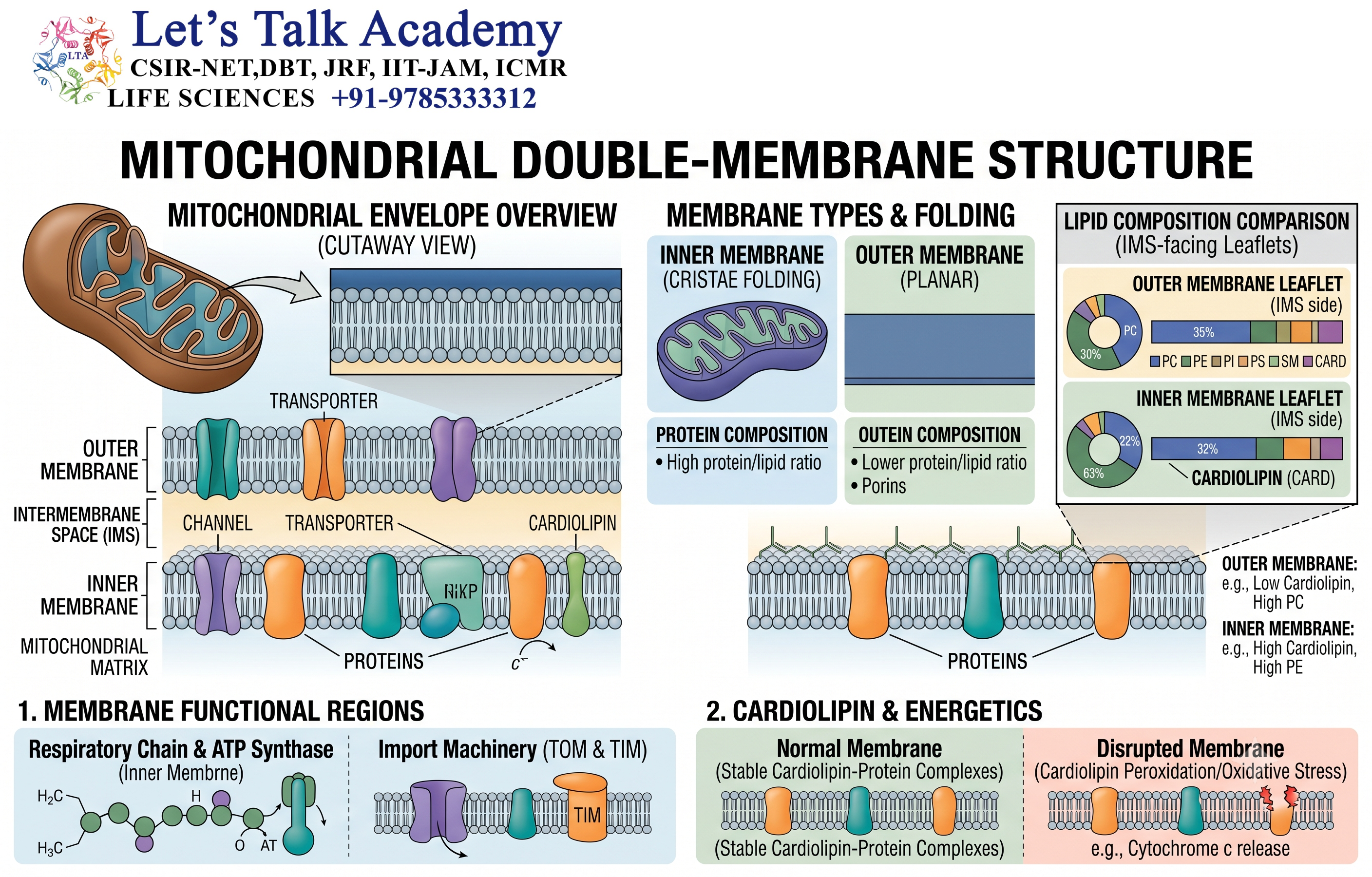
16.1 Double-Membrane Organization
Mitochondria contain:
- Outer membrane
- Intermembrane space
- Inner membrane
- Matrix
This organization creates specialized environments for different biochemical processes.
16.2 Outer Mitochondrial Membrane
The outer membrane contains proteins that regulate communication and transport between the cytosol and the intermembrane space.
It is relatively permeable to many small molecules because of channel-forming proteins such as porins.
16.3 Inner Mitochondrial Membrane
The inner membrane is highly specialized.
It contains:
- Electron transport complexes
- ATP synthase
- Specific transport proteins
It is also highly folded.
These folds are called cristae.
16.4 Cristae
Cristae greatly increase the membrane surface area available for energy-converting processes.
The inner membrane maintains a proton gradient between the intermembrane space and the matrix.
This gradient provides the driving force for ATP production through ATP synthase.
17. Chloroplast Membranes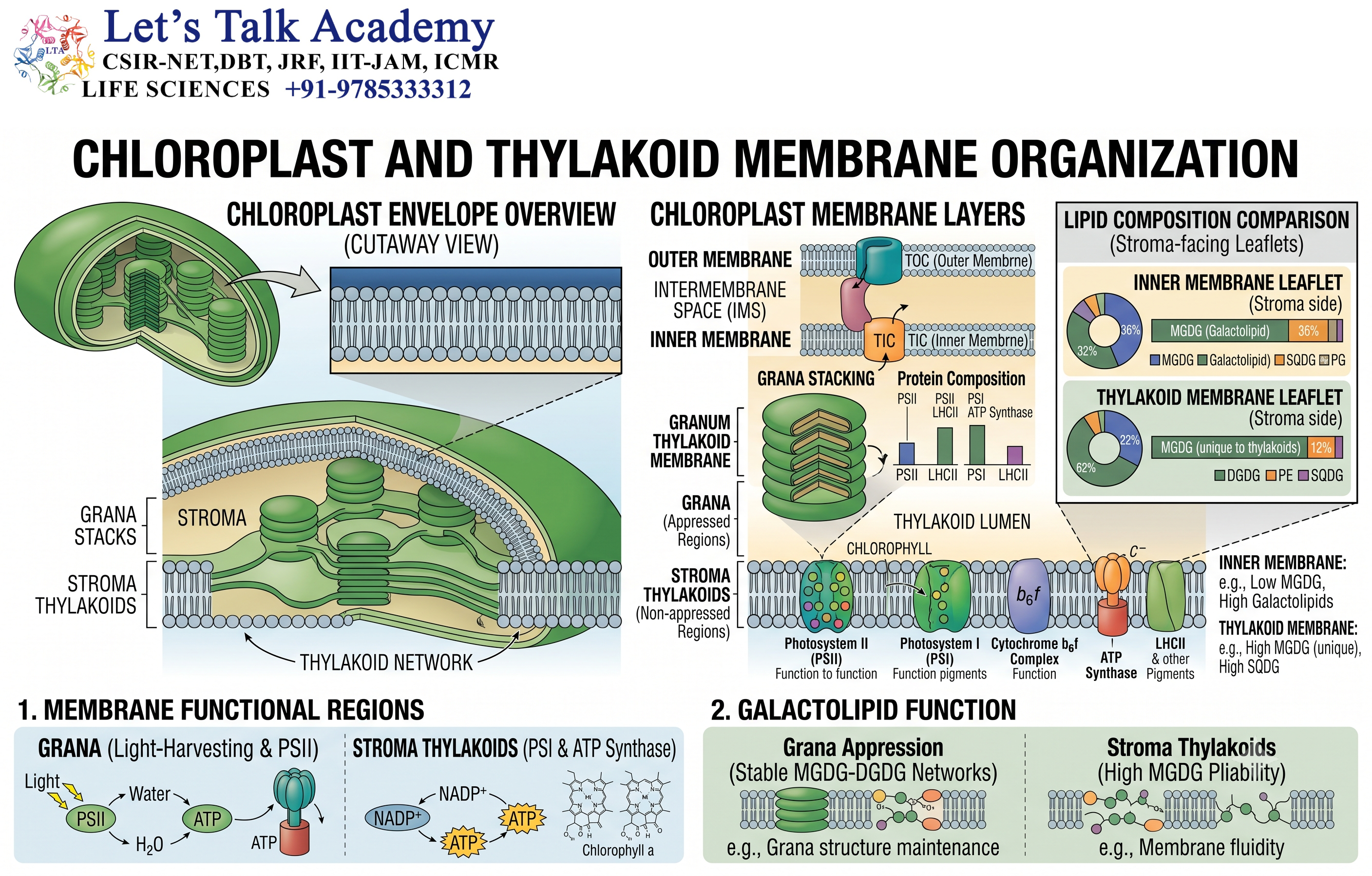
17.1 Chloroplast Envelope
Chloroplasts possess an outer and inner envelope membrane.
These membranes separate the chloroplast from the cytoplasm.
17.2 Thylakoid Membrane
Within the chloroplast is the thylakoid membrane system.
The thylakoid membrane contains:
- Photosystems
- Electron transport components
- ATP synthase
- Photosynthetic pigments
- Associated proteins
17.3 Grana
Thylakoid membranes can form stacks known as grana.
The organization of the thylakoid membrane creates a specialized environment for light-dependent photosynthetic reactions.
Thus, membrane structure directly supports the conversion of light energy into chemical energy.
18. Membrane Domains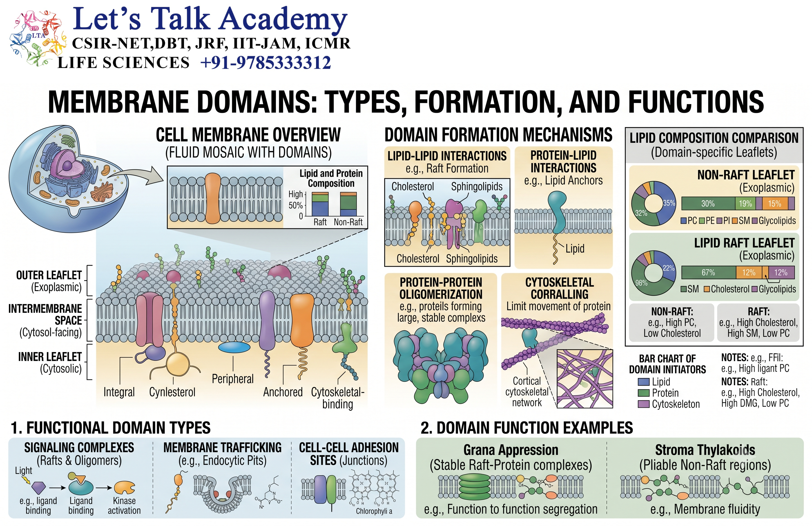
18.1 Concept
Biological membranes are not perfectly homogeneous.
Different lipids and proteins can become concentrated in specific regions.
These regions are often referred to as membrane domains.
18.2 Lipid Rafts
Lipid rafts are proposed membrane domains enriched in particular sterols and sphingolipids.
They can influence the organization of membrane proteins and signaling molecules.
Their exact size, lifetime, and physical properties can vary and remain an active area of research.
18.3 Functional Significance
Membrane domains can contribute to:
- Signal transduction
- Protein sorting
- Membrane trafficking
- Cell adhesion
- Receptor organization
19. Membrane Curvature
19.1 What Is Membrane Curvature?
Although often represented as a flat sheet in diagrams, biological membranes can bend, curve, and form highly complex shapes.
Membrane curvature is essential for:
- Vesicle formation
- Endocytosis
- Exocytosis
- Organelle formation
- Membrane fusion
- Membrane fission
19.2 Lipid Contribution
Different lipid shapes can influence membrane curvature.
Lipids with relatively large head groups and smaller hydrophobic regions may favor different membrane geometries from lipids with more cylindrical shapes.
19.3 Protein Contribution
Certain membrane proteins can induce or stabilize membrane curvature.
Proteins may:
- Insert into the membrane
- Bind curved surfaces
- Form scaffolds
- Interact with cytoskeletal elements
Together, proteins and lipids allow membranes to adopt complex three-dimensional structures.
20. Membrane Permeability
20.1 Selective Permeability
One of the most important properties of the lipid bilayer is selective permeability.
The hydrophobic interior creates a barrier against many charged and polar substances.
Small nonpolar molecules can generally cross relatively easily.
Many ions and large polar molecules require transport proteins.
20.2 Relative Permeability
The ability of a substance to cross the lipid bilayer depends on factors such as:
- Molecular size
- Charge
- Polarity
- Lipid solubility
- Membrane composition
Small hydrophobic molecules generally cross more readily than ions.
20.3 Aquaporins
Water can cross lipid bilayers to some extent, but many cells use specialized membrane proteins called aquaporins.
Aquaporins form selective channels for rapid water movement.
21. Membrane Transport
21.1 Passive Transport
Passive transport does not require direct energy input from ATP.
Substances move down their concentration or electrochemical gradients.
Major forms include:
- Simple diffusion
- Facilitated diffusion
- Osmosis
21.2 Simple Diffusion
Small molecules capable of dissolving in the hydrophobic membrane interior can cross directly.
No transport protein is required.
21.3 Facilitated Diffusion
Facilitated diffusion uses membrane proteins.
The transported substance moves down its electrochemical gradient.
Channels and carriers are important examples.
21.4 Active Transport
Active transport moves substances against their electrochemical gradients.
Energy is required.
Energy may come directly from ATP hydrolysis or indirectly from another ion gradient.
22. Membrane Potential and Electrochemical Gradients
22.1 Membrane Potential
Cells can maintain a difference in electrical potential across their membranes.
This is known as the membrane potential.
It arises from unequal ion distribution and selective permeability.
22.2 Electrochemical Gradient
For ions, movement depends on both:
- Chemical gradient
- Electrical gradient
Together, these form the electrochemical gradient.
Electrochemical gradients are essential for:
- ATP synthesis
- Nutrient transport
- Ion movement
- Electrical signaling
22.3 Proton Motive Force
Many bacteria and mitochondria use proton gradients to store usable energy.
The movement of protons down their electrochemical gradient can drive ATP synthesis.
This principle demonstrates how a physical membrane property can be converted into chemical energy.
23. Membrane Vesicles and Trafficking
23.1 Vesicle Formation
Eukaryotic membranes can bend and form vesicles.
A region of membrane can curve inward or outward, eventually forming a separate membrane-bound compartment.
23.2 Endocytosis
Endocytosis brings material from outside the cell into the cell.
The plasma membrane bends inward and forms a vesicle.
Major forms include:
- Phagocytosis
- Pinocytosis
- Receptor-mediated endocytosis
23.3 Exocytosis
During exocytosis, an intracellular vesicle moves toward the plasma membrane and fuses with it.
Its contents are released outside the cell.
The vesicle membrane becomes part of the plasma membrane.
This process is important for:
- Secretion
- Membrane recycling
- Communication
- Release of signaling molecules
24. Membrane Fusion and Fission
24.1 Membrane Fusion
Fusion occurs when two membrane bilayers merge into one continuous membrane.
It is important during:
- Vesicle trafficking
- Secretion
- Fertilization
- Organelle interactions
Membrane fusion requires overcoming the energetic barrier associated with bringing two hydrophobic bilayers together.
Specialized proteins help control this process.
24.2 Membrane Fission
Fission is the opposite process.
A membrane is divided into separate membrane-bound structures.
Fission is important during:
- Vesicle formation
- Organelle division
- Endocytosis
Together, fusion and fission allow eukaryotic cells to continuously remodel their membrane systems.
25. Comparison of Prokaryotic and Eukaryotic Membranes
25.1 Structural Comparison
| Feature | Prokaryotic Membranes | Eukaryotic Membranes |
|---|---|---|
| Plasma membrane | Present | Present |
| Lipid bilayer | Common | Common |
| Membrane proteins | Present | Present |
| Internal membrane-bound organelles | Generally absent | Extensive |
| Membrane compartmentalization | Relatively limited | Highly developed |
| Energy-converting membrane | Cytoplasmic membrane in many prokaryotes | Mitochondrial inner membrane; thylakoid membrane in chloroplasts |
| Sterols | Variable and lineage-dependent | Important in many eukaryotic membranes |
| Vesicular trafficking | Limited compared with eukaryotes | Extensive |
| Membrane specialization | Present | Extensive |
| Lipid composition | Species- and environment-dependent | Organelle- and tissue-dependent |
25.2 Functional Similarities
Both prokaryotic and eukaryotic membranes:
- Form selective barriers.
- Contain lipids and proteins.
- Regulate molecular transport.
- Maintain ion gradients.
- Participate in signaling.
- Support membrane-associated enzymes.
- Can undergo changes in composition and organization.
25.3 Major Difference in Organization
The most significant organizational difference is the extent of internal compartmentalization.
A typical prokaryotic cell relies heavily on its plasma membrane and specialized membrane-associated structures.
A eukaryotic cell distributes functions among numerous membrane-bound compartments.
This compartmentalization allows different biochemical reactions to occur under different conditions within the same cell.
26. Structure–Function Relationship
26.1 Lipids and Physical Properties
Lipids determine many basic physical characteristics of membranes.
They influence:
- Fluidity
- Thickness
- Curvature
- Permeability
- Stability
26.2 Proteins and Specialized Functions
Proteins provide most of the specialized activities.
They allow membranes to:
- Transport molecules
- Detect signals
- Generate energy
- Catalyze reactions
- Attach to other structures
26.3 Carbohydrates and Recognition
Membrane carbohydrates contribute particularly to:
- Recognition
- Adhesion
- Protection
- Cell–cell interactions
Thus, the membrane functions as an integrated molecular system rather than as a simple lipid barrier.
27. Experimental Study of Model Membranes
27.1 Why Model Membranes Are Used
Natural membranes are chemically complex.
Model membranes simplify this complexity so that individual physical properties can be studied.
Researchers can control:
- Lipid composition
- Temperature
- pH
- Ionic strength
- Protein concentration
- Membrane curvature
27.2 Liposomes
Liposomes are spherical structures consisting of one or more lipid bilayers surrounding an aqueous compartment.
They are useful for studying:
- Membrane permeability
- Drug delivery
- Membrane fusion
- Lipid–protein interactions
27.3 Supported Lipid Bilayers
A supported lipid bilayer is formed on a solid surface.
It provides a convenient experimental platform for studying:
- Membrane protein interactions
- Lipid movement
- Cell adhesion
- Surface signaling
27.4 Giant Unilamellar Vesicles
Giant unilamellar vesicles are large membrane-bound vesicles containing a single lipid bilayer.
Their relatively large size allows researchers to observe membrane shape changes and physical behavior using microscopy.
28. Membrane Organization Under Stress
28.1 Temperature Stress
Changes in temperature can alter membrane fluidity.
Cells respond by modifying lipid composition.
28.2 Osmotic Stress
Changes in external solute concentration can alter water movement across membranes.
Cells must regulate ion and water balance to maintain appropriate volume.
28.3 Oxidative Stress
Reactive oxygen species can damage membrane lipids and proteins.
Cells possess protective mechanisms that help preserve membrane integrity.
28.4 pH and Ionic Stress
Changes in pH and ion concentration can influence membrane proteins, lipid organization, and electrochemical gradients.
Microorganisms have evolved different strategies to maintain membrane function under such conditions.
29. Integrated View of Membrane Structure
29.1 Membrane as a Dynamic System
A biological membrane should be viewed as an integrated system containing:
Lipids + Proteins + Carbohydrates + Ions + Cytoskeletal Interactions
These components continuously interact.
The membrane changes its:
- Shape
- Composition
- Fluidity
- Protein organization
- Curvature
in response to cellular requirements.
29.2 Membrane as a Selective Barrier
The lipid bilayer provides the basic barrier.
Transport proteins determine which substances can cross efficiently.
This combination allows the cell to control its internal environment.
29.3 Membrane as a Communication Platform
Receptors recognize external signals.
These receptors interact with intracellular signaling pathways.
Therefore, the membrane acts as an interface between the external environment and the internal machinery of the cell.


