
1. Introduction to the Lipid Bilayer
Every living cell needs a boundary that separates its internal environment from the surrounding world. This boundary is not simply a rigid wall. It is a dynamic, flexible, selectively permeable structure that allows the cell to maintain its internal conditions while communicating and interacting with its environment.
The fundamental structural component of most biological membranes is the lipid bilayer.
The lipid bilayer forms the basic framework of the plasma membrane and many internal cellular membranes, including the membranes surrounding the nucleus, mitochondria, endoplasmic reticulum, Golgi apparatus, lysosomes, and various vesicles.
Although the membrane appears extremely thin under a microscope, its organization is highly sophisticated. Lipid molecules spontaneously arrange themselves into two opposing layers because of their interaction with water. This simple physical property creates a stable barrier that is essential for life.
The lipid bilayer is therefore much more than a passive boundary. It provides a suitable environment for membrane proteins, contributes to cell signaling, controls molecular transport, supports energy conversion, participates in membrane trafficking, and helps cells maintain their shape and identity.
Understanding the lipid bilayer is fundamental to understanding how cells function.
1.1 What Is a Lipid Bilayer?
A lipid bilayer is a double layer of lipid molecules in which the hydrophilic regions of the lipids face the aqueous environments on either side of the membrane, while their hydrophobic regions face inward toward each other.
The basic arrangement can be represented as:
Water → Hydrophilic heads → Hydrophobic tails → Hydrophobic tails → Hydrophilic heads → Water
This arrangement is primarily driven by the hydrophobic effect.
Most cellular membranes contain large amounts of phospholipids, together with cholesterol, glycolipids, and membrane proteins. The exact composition varies depending on the type of membrane and its cellular function.
The bilayer is typically only a few nanometres thick, yet it provides a remarkably effective barrier to many ions and polar molecules.
The lipid bilayer also possesses an important property known as self-sealing. When a small break occurs in the membrane, lipid molecules can rearrange themselves so that their hydrophobic portions remain protected from water. This allows membranes to maintain their structural integrity despite constant mechanical and biochemical stress.
1.2 Structure of the Lipid Bilayer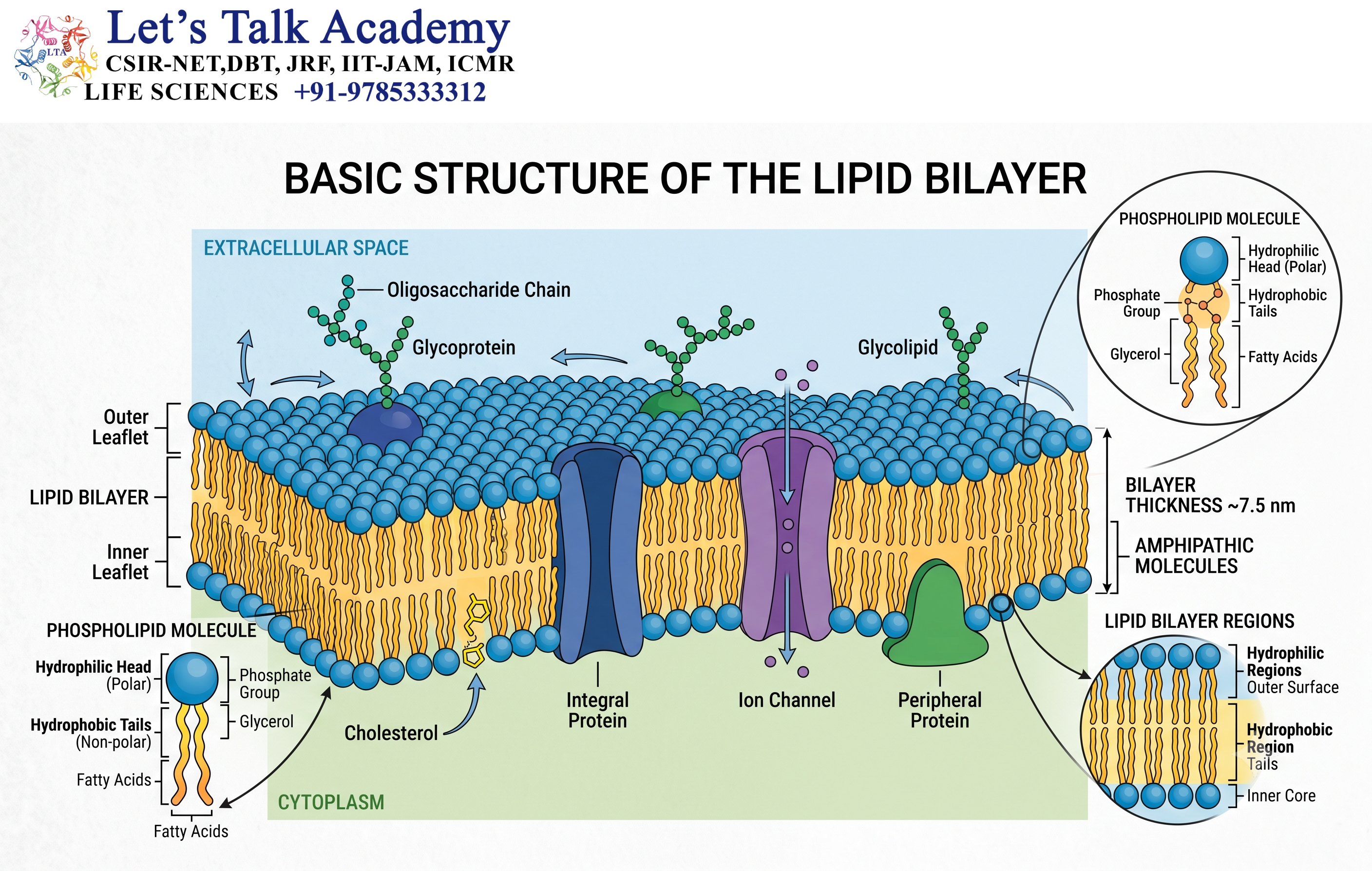
The structure of the lipid bilayer depends largely on the chemical properties of its lipid molecules.
Many membrane lipids are amphipathic, meaning that the same molecule contains both a hydrophilic region and a hydrophobic region.
This dual nature is responsible for the spontaneous formation of biological membranes in an aqueous environment.
1.2.1Amphipathic Nature of Membrane Lipids
An amphipathic molecule contains two chemically different regions.
The hydrophilic region interacts favorably with water, whereas the hydrophobic region avoids contact with water.
Phospholipids are classic examples of amphipathic molecules.
When phospholipids are placed in water, their hydrophilic heads interact with water molecules, while their hydrophobic tails tend to avoid water. As a result, the lipids spontaneously organize into structures such as bilayers, micelles, and vesicles.
This self-assembly generally does not require a direct external energy input.
The formation of the bilayer is therefore an important example of how molecular properties can generate highly organized biological structures.
1.2.2 Hydrophilic Head
The hydrophilic head is the water-attracting portion of a membrane lipid.
In phospholipids, the head group generally contains a phosphate group and may contain additional molecules such as choline, ethanolamine, serine, or inositol.
Because the head region is polar or charged, it interacts strongly with surrounding water.
The heads face the aqueous environment on both sides of the membrane.
One layer faces the cytoplasm, while the other faces the extracellular environment or the lumen of an organelle.
The chemical nature of the head group can influence membrane curvature, interactions with proteins, and signaling processes.
1.2.3 Hydrophobic Tails
The hydrophobic tails are generally composed of hydrocarbon chains derived from fatty acids.
These tails are nonpolar and interact poorly with water.
Inside the membrane, the hydrophobic tails point toward one another, creating a nonpolar interior.
This hydrophobic interior is one of the major reasons that charged particles and many polar molecules cannot freely cross the membrane.
The length and degree of saturation of fatty acid chains also influence membrane thickness, packing, permeability, and fluidity.
2. Major Lipids Present in the Bilayer
2.1 Phospholipids
Phospholipids are among the most abundant membrane lipids.
A typical glycerophospholipid contains:
- A hydrophilic head group
- A phosphate-containing region
- A glycerol backbone
- Two hydrophobic fatty acid tails
Common phospholipids include:
- Phosphatidylcholine
- Phosphatidylethanolamine
- Phosphatidylserine
- Phosphatidylinositol
Different phospholipids have different head groups and physical properties.
Their distribution within the membrane is not random. Specific phospholipids are enriched in particular membrane leaflets and cellular membranes.
For example, phosphatidylinositol and its phosphorylated derivatives are particularly important on the cytosolic side of membranes, where they participate in signaling and membrane recruitment of proteins.
2.2 Cholesterol
Cholesterol is an important component of many animal cell membranes.
It contains a small polar hydroxyl group and a large hydrophobic steroid structure.
Cholesterol is positioned between phospholipid molecules, with its hydroxyl group close to the polar head groups and its hydrophobic region within the membrane interior.
Cholesterol has an important influence on membrane fluidity and permeability.
It can reduce excessive movement of phospholipid molecules at relatively high temperatures and prevent membranes from becoming excessively rigid at lower temperatures.
In this way, cholesterol acts as an important fluidity buffer.
Cholesterol also decreases the permeability of the bilayer to many small water-soluble molecules and ions.
2.3 Glycolipids
Glycolipids are lipids containing carbohydrate groups.
The carbohydrate portion generally faces the extracellular side of the plasma membrane.
Glycolipids contribute to:
- Cell recognition
- Cell-cell interactions
- Membrane stability
- Protection of the cell surface
- Recognition by certain proteins and other molecules
Together with carbohydrate groups attached to membrane proteins, glycolipids contribute to the formation of the glycocalyx on the external surface of many cells.
3. How Does the Lipid Bilayer Form?
3.1 Hydrophobic Effect
The formation of the lipid bilayer is primarily driven by the hydrophobic effect.
Water molecules form organized interactions around nonpolar substances. When amphipathic lipids are present, the system becomes energetically more favorable when the hydrophobic portions are shielded from water.
As a result, phospholipids spontaneously arrange themselves so that:
- Hydrophilic heads remain exposed to water.
- Hydrophobic tails are protected from water.
- The tails interact with one another in the membrane interior.
This self-assembly produces a stable bilayer.
The hydrophobic effect is therefore not simply an attraction between lipid tails. Rather, it is strongly related to the tendency of water to minimize unfavorable interactions with nonpolar surfaces.
3.2 Self-Assembly of Membrane Lipids
When amphipathic lipids are introduced into an aqueous environment, they can spontaneously organize into different structures depending on their molecular shape and chemical properties.
These structures can include:
- Micelles
- Bilayers
- Liposomes
- Multilamellar vesicles
Lipids with two hydrocarbon tails commonly favor bilayer formation because their molecular geometry is well suited to forming two opposing layers.
3.3 Self-Sealing Property
One of the most important characteristics of lipid bilayers is their ability to self-seal.
If a small hole forms, exposing the hydrophobic tails to water, the surrounding lipid molecules rapidly rearrange to minimize this unfavorable exposure.
This property is essential for living cells because membrane damage can occur during movement, mechanical stress, vesicle formation, fusion, and other cellular processes.
4. Fluid mosaic model.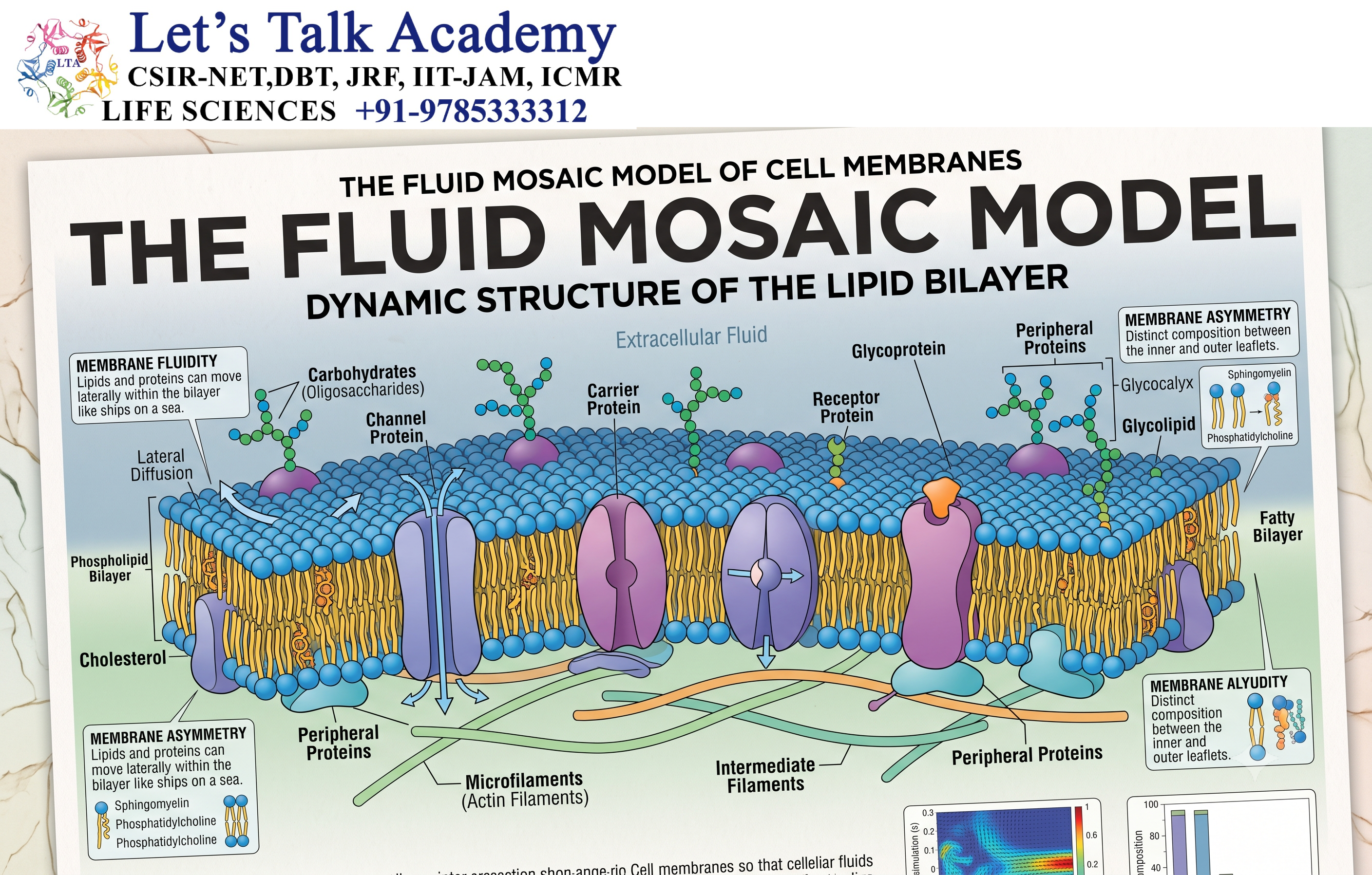
4.1 Basic Concept
The structure of biological membranes is commonly described by the fluid mosaic model.
According to this model, the membrane is a dynamic lipid bilayer containing proteins and other molecules that can move within or associate with the membrane.
The word fluid emphasizes that membrane components are not permanently fixed in one position.
The word mosaic reflects the diverse collection of lipids, proteins, carbohydrates, and other components present in the membrane.
Membrane proteins may be:
- Integral membrane proteins
- Peripheral membrane proteins
- Lipid-anchored proteins
The lipid bilayer therefore provides a flexible environment in which many proteins can function.
4.2 Lateral Movement
Many lipid molecules can move laterally within the same leaflet of the membrane.
This movement contributes to the fluid nature of biological membranes.
However, movement from one leaflet to the opposite leaflet, called flip-flop, is energetically unfavorable for most phospholipids because the polar head group would have to pass through the hydrophobic interior.
Therefore, spontaneous transverse movement is much slower than lateral movement.
Specific enzymes can facilitate lipid movement between leaflets.
5. Membrane Fluidity
5.1 Meaning of Membrane Fluidity
Membrane fluidity refers to the degree to which lipid and protein molecules can move within the membrane.
The membrane is neither a completely rigid structure nor a freely flowing liquid. Its physical state lies between these extremes.
Fluidity is essential because cells require membranes to:
- Change shape
- Fuse with other membranes
- Form vesicles
- Move membrane proteins
- Respond to signals
- Maintain appropriate membrane permeability
Several factors influence membrane fluidity.
5.2 Role of Fatty Acid Saturation
The fatty acid tails of membrane phospholipids can be saturated or unsaturated.
Saturated fatty acids contain no carbon-carbon double bonds. Their relatively straight chains can pack closely together.
This generally increases membrane packing and decreases fluidity.
Unsaturated fatty acids contain one or more carbon-carbon double bonds. Double bonds, especially cis double bonds, introduce bends into the hydrocarbon chains.
These bends prevent tight packing and generally increase membrane fluidity.
Therefore:
More unsaturated fatty acids → Greater fluidity
More saturated fatty acids → Tighter packing and lower fluidity
Cells can regulate membrane composition to maintain appropriate physical properties.
5.3 Effect of Temperature
Temperature has a strong influence on membrane fluidity.
At higher temperatures, lipid molecules generally move more rapidly, increasing membrane fluidity.
At lower temperatures, lipid movement decreases, and membranes tend to become more rigid.
Cells can compensate for temperature changes by modifying their lipid composition.
This ability to adjust membrane lipid composition is often referred to as homeoviscous adaptation.
5.4 Role of Cholesterol
Cholesterol has a complex effect on membrane fluidity.
At high temperatures, cholesterol can restrain phospholipid movement and reduce excessive membrane fluidity.
At low temperatures, cholesterol interferes with close packing of phospholipid tails and helps prevent the membrane from becoming excessively rigid.
Thus, cholesterol helps stabilize membrane physical properties over changing temperatures.
6. Asymmetry of the Lipid Bilayer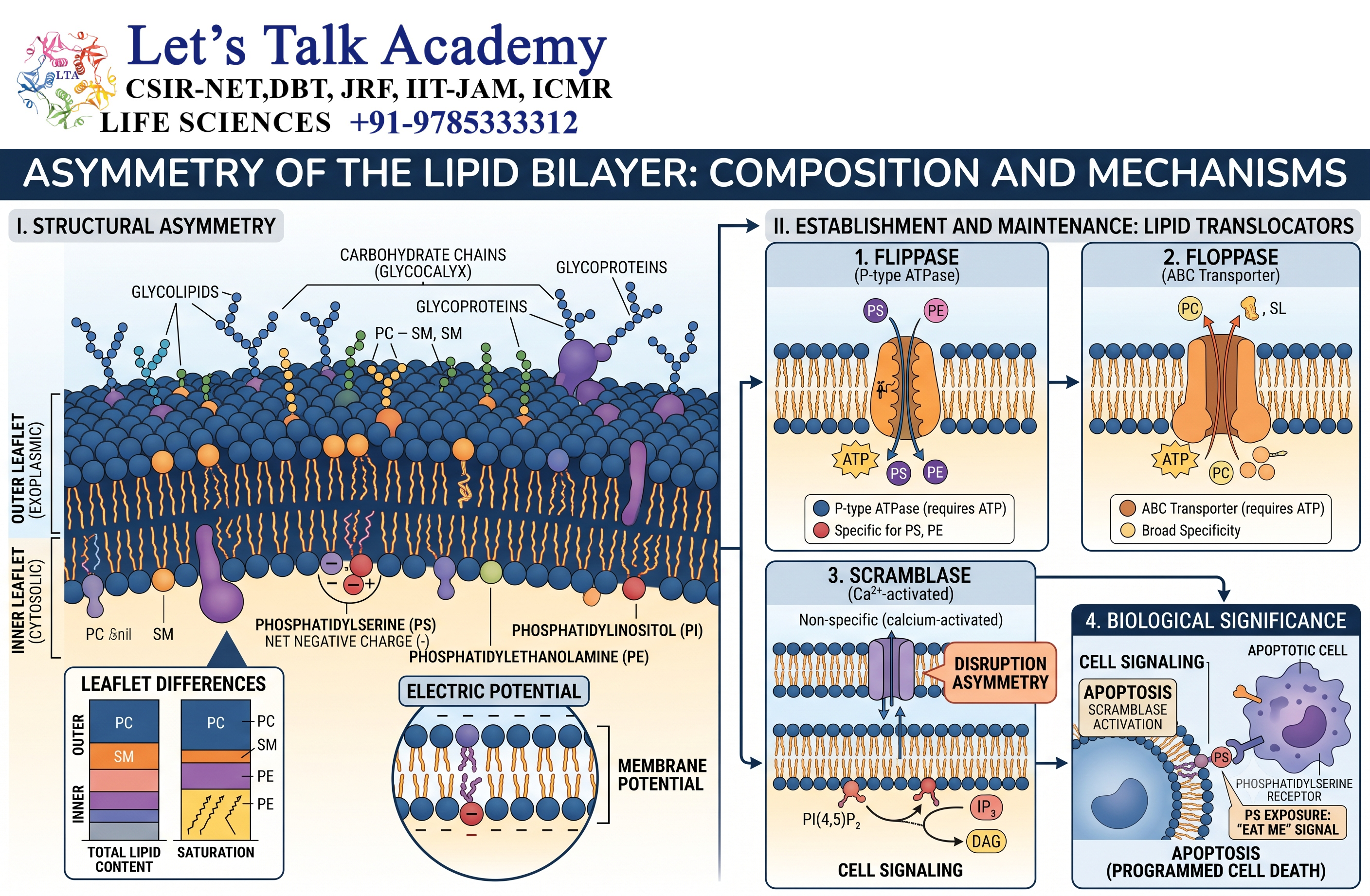
6.1 Meaning of Membrane Asymmetry
The two leaflets of a biological membrane are not identical.
This property is called membrane asymmetry.
Different lipids are preferentially distributed between the inner and outer leaflets.
For example, in the plasma membrane of many animal cells:
- Phosphatidylcholine is abundant in the outer leaflet.
- Sphingomyelin is enriched in the outer leaflet.
- Phosphatidylethanolamine is relatively enriched in the cytosolic leaflet.
- Phosphatidylserine is primarily located in the cytosolic leaflet.
- Phosphatidylinositol and its derivatives are mainly associated with the cytosolic leaflet.
This asymmetric distribution is biologically important.
Phosphatidylserine exposure on the external surface of a cell can act as a signal associated with programmed cell death and recognition by phagocytic cells.
6.2 Maintenance of Membrane Asymmetry
Membrane asymmetry is actively maintained by specialized proteins.
Three important groups are:
- Flippases
- Floppases
- Scramblases
Flippases generally move selected phospholipids from the outer leaflet toward the cytosolic leaflet.
Floppases generally move selected lipids in the opposite direction.
Scramblases promote movement of lipids between the two leaflets with less specificity and can rapidly reduce lipid asymmetry.
The coordinated activity of these proteins allows cells to control membrane composition.
7. Membrane Proteins and Their Association with the Bilayer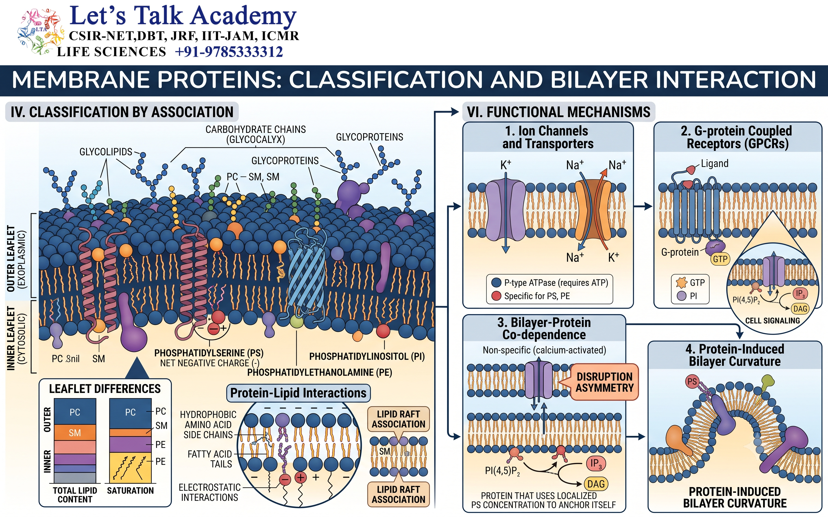
7.1 Integral Membrane Proteins
Integral membrane proteins are closely associated with the lipid bilayer.
Many integral proteins contain hydrophobic regions that interact with the hydrophobic core of the membrane.
Some span the entire membrane and are called transmembrane proteins.
Transmembrane proteins perform numerous functions, including:
- Transport
- Signal reception
- Enzymatic activity
- Cell adhesion
- Cell recognition
7.2 Peripheral Membrane Proteins
Peripheral membrane proteins do not normally penetrate deeply into the hydrophobic core.
They may associate with:
- Polar lipid head groups
- Integral membrane proteins
- Other membrane-associated molecules
Many peripheral proteins participate in signaling, cytoskeletal attachment, and membrane organization.
7.3 Lipid-Anchored Proteins
Some proteins are attached to membranes through covalently linked lipid groups.
These proteins are called lipid-anchored proteins.
The lipid anchor inserts into the membrane, allowing the protein to remain associated with the membrane without necessarily spanning the bilayer.
8. Functions of the Lipid Bilayer
8.1 Formation of the Cellular Boundary
The lipid bilayer forms the basic boundary of cells and many cellular organelles.
It separates different aqueous environments and allows cells to maintain distinct chemical conditions.
8.2 Selective Permeability
The hydrophobic interior of the bilayer prevents many ions and polar molecules from crossing freely.
At the same time, certain small nonpolar molecules can diffuse through the membrane relatively easily.
This property gives the membrane selective permeability.
8.3 Platform for Membrane Proteins
The lipid bilayer provides an environment in which membrane proteins can function.
Transporters, channels, receptors, enzymes, and adhesion proteins depend on the membrane environment for their proper activity.
8.4 Cell Signaling
Membrane lipids can participate directly in signaling.
For example, phosphatidylinositol derivatives can serve as signaling molecules or help recruit signaling proteins to particular membrane regions.
8.5 Energy Conversion
Membranes play essential roles in energy conversion.
The inner mitochondrial membrane is involved in oxidative phosphorylation, while specialized membranes in photosynthetic organisms participate in photosynthetic electron transport.
The lipid bilayer provides the physical barrier necessary for maintaining electrochemical gradients.
8.6 Membrane Trafficking
The ability of membranes to bend, fuse, and undergo fission is essential for vesicle-mediated transport.
Processes such as:
- Endocytosis
- Exocytosis
- Vesicle transport
- Organelle biogenesis
depend on membrane dynamics.
9. Selective Permeability of the Lipid Bilayer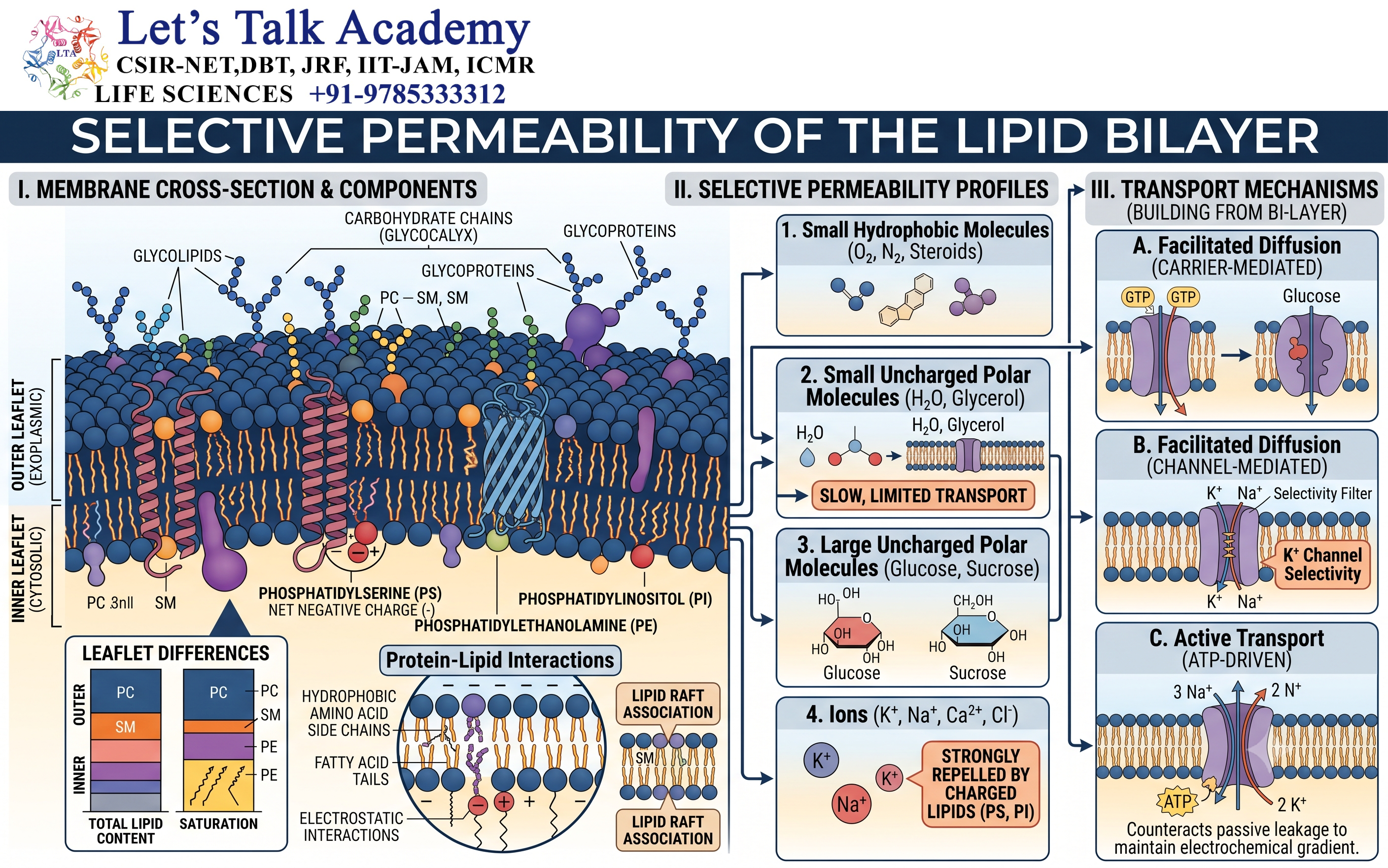
9.1 Permeability of Small Nonpolar Molecules
Small nonpolar molecules generally cross the lipid bilayer relatively easily.
Examples include:
- Oxygen
- Carbon dioxide
- Nitrogen
- Certain lipid-soluble molecules
Their ability to dissolve in the hydrophobic membrane interior facilitates their movement across the bilayer.
9.2 Permeability of Polar Molecules
Small uncharged polar molecules may cross the membrane to some extent, but their permeability is generally lower than that of nonpolar molecules.
Water can cross lipid bilayers slowly, although many cells contain aquaporins, specialized membrane proteins that greatly increase water permeability.
9.3 Permeability of Ions
Ions such as:
- Na⁺
- K⁺
- Ca²⁺
- Cl⁻
- H⁺
cannot readily cross the hydrophobic interior of the lipid bilayer.
They generally require specialized membrane proteins such as ion channels, carriers, or pumps.
This property is fundamental to electrical signaling and the maintenance of ion gradients.
10. Transport Across the Lipid Bilayer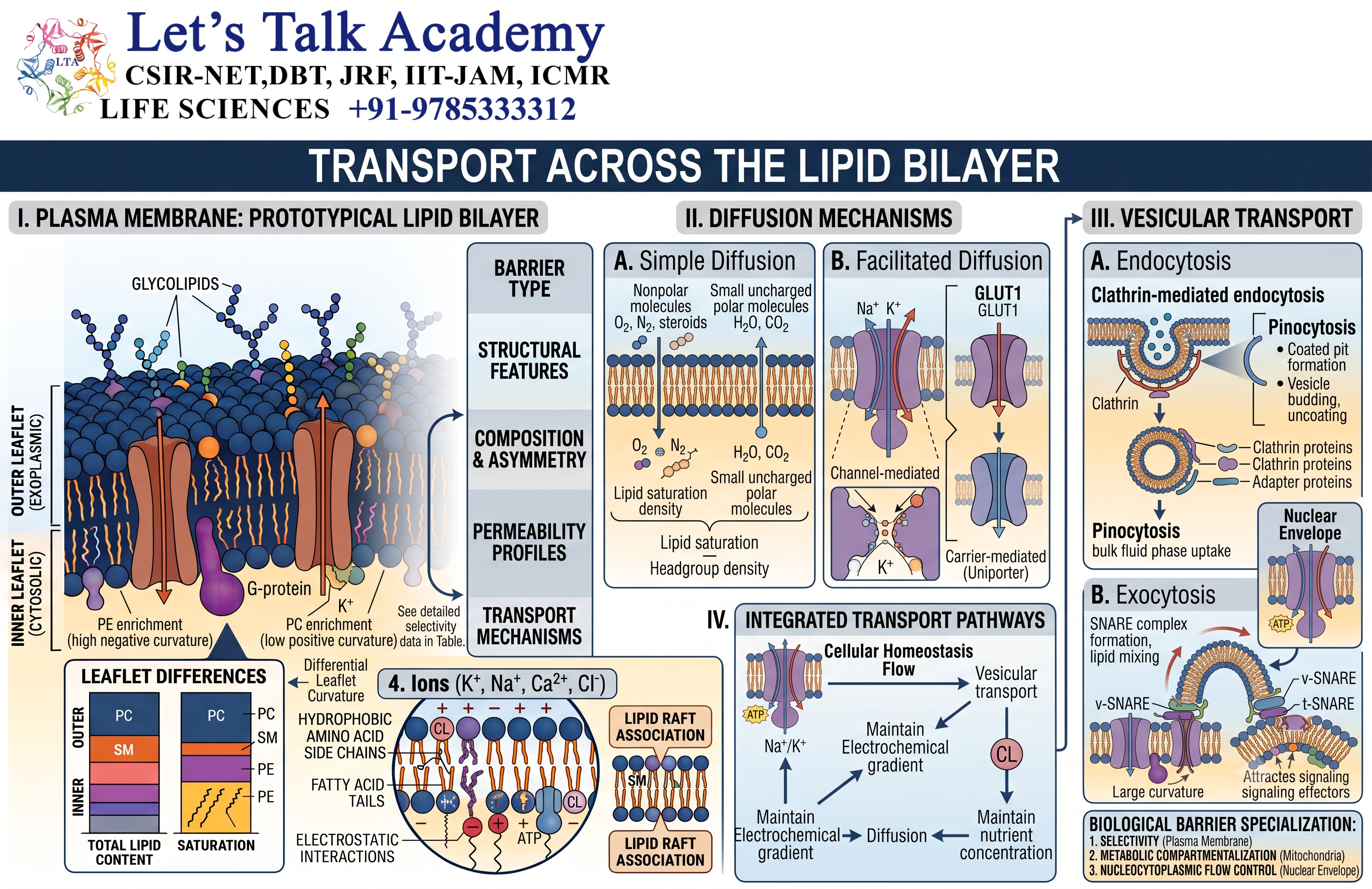
10.1 Simple Diffusion
Simple diffusion is the movement of molecules across the membrane from a region of higher concentration to a region of lower concentration without direct energy expenditure by the cell.
Small hydrophobic molecules are particularly capable of crossing through simple diffusion.
10.2 Facilitated Diffusion
Facilitated diffusion involves membrane proteins that help substances move down their electrochemical gradients.
It does not directly require ATP.
Transport may occur through:
- Ion channels
- Carrier proteins
The movement stops when the relevant electrochemical equilibrium is reached.
10.3 Active Transport
Active transport moves substances against their electrochemical gradients and requires energy.
Energy can be supplied directly by ATP hydrolysis or indirectly through another ion gradient.
Examples include:
- Sodium-potassium pump
- Calcium pumps
- Proton pumps
Active transport allows cells to maintain concentration differences that are essential for cellular function.
10.4 Osmosis
Osmosis is the net movement of water across a selectively permeable membrane in response to differences in water potential or effective solute concentration.
Water movement is particularly important for maintaining:
- Cell volume
- Osmotic balance
- Cellular homeostasis
Aquaporins can facilitate rapid water movement across membranes.
11. Lipid Bilayer and Cell Signaling
11.1 Lipids as Signaling Molecules
Membrane lipids are not merely structural components.
Certain lipids and their derivatives function as signaling molecules.
For example, phosphoinositides can regulate:
- Protein recruitment
- Membrane trafficking
- Cytoskeletal organization
- Cell growth
- Cell survival
- Signal transduction
11.2 Phosphoinositide Signaling
Phosphatidylinositol can be phosphorylated at different positions on its inositol ring.
The resulting phosphoinositides can serve as membrane identity markers and signaling molecules.
For example, different phosphoinositides are enriched in different cellular membranes and can recruit specific proteins containing lipid-binding domains.
This allows cells to organize signaling events spatially.
12.Selective Permeability of the Lipid Bilayer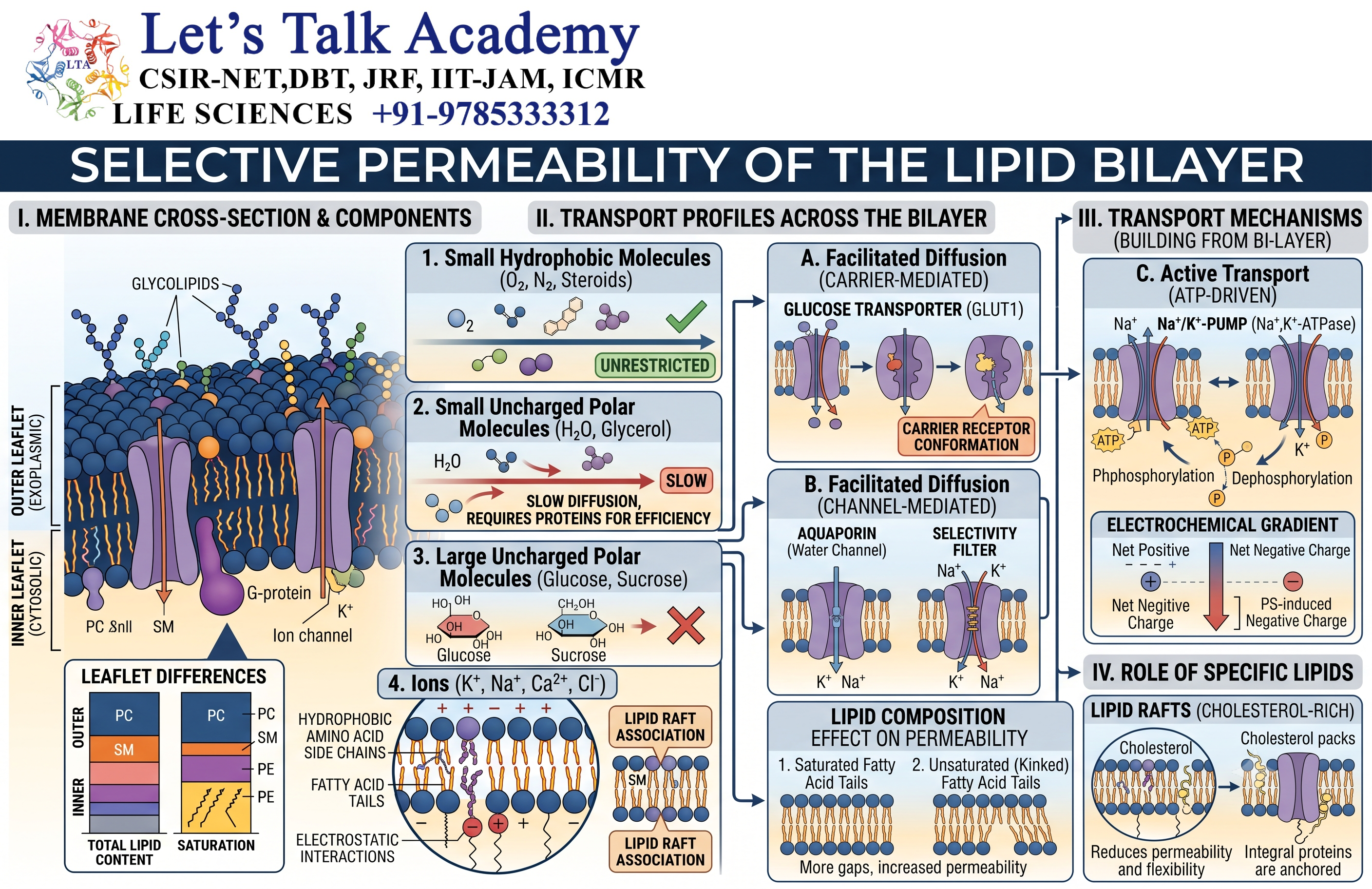
12.1 Membrane Budding
Membranes can bend to form buds that eventually develop into vesicles.
This process is essential for intracellular transport.
Vesicles transport proteins, lipids, and other molecules between cellular compartments.
12.2 Membrane Fusion
During membrane fusion, two lipid bilayers merge to form a continuous membrane.
Fusion is essential during:
- Exocytosis
- Vesicle trafficking
- Endosomal transport
- Synaptic vesicle release
Specialized proteins control the fusion process and ensure that it occurs at the correct location.
12.3 Endocytosis
Endocytosis allows cells to take material from the extracellular environment into the cell.
The plasma membrane bends inward and forms a vesicle containing extracellular material.
Different forms include:
- Phagocytosis
- Pinocytosis
- Receptor-mediated endocytosis
13. Lipid Rafts and Membrane Organization
13.1 Concept of Lipid Rafts
Biological membranes are not necessarily completely uniform.
Certain regions can become enriched in particular lipids and proteins.
These regions are commonly discussed as lipid rafts.
They are often enriched in cholesterol and sphingolipids and can influence the organization of signaling proteins and membrane trafficking machinery.
13.2 Functional Importance
Membrane organization can help cells concentrate specific molecules in particular regions.
This spatial organization can improve the efficiency of:
- Signal transduction
- Protein sorting
- Membrane trafficking
- Cell adhesion
The exact physical nature and lifetime of lipid rafts can vary, and their organization is an active area of research.
14. Experimental Evidence for the Lipid Bilayer
14.1 Early Studies of Membrane Lipids
Early studies of biological membranes demonstrated that lipids are major structural components of membranes.
The amount of lipid relative to membrane surface area provided important evidence that membranes contain a lipid-based structure.
14.2 Monolayer Experiments
Studies involving spreading membrane lipids as monolayers at air-water interfaces provided evidence that membrane lipids could occupy an area consistent with a bilayer arrangement.
These experiments contributed to the development of early membrane models.
14.3 Freeze-Fracture Electron Microscopy
Freeze-fracture electron microscopy provided important structural evidence about membrane organization.
When a membrane is fractured at low temperature, the fracture plane often passes through the hydrophobic interior of the bilayer.
This technique revealed structures associated with membrane proteins and helped establish the concept that proteins are embedded within the membrane.
14.4 Membrane Protein Mobility
Experiments involving fluorescently labeled membrane proteins demonstrated that many membrane proteins can move laterally within the membrane.
Such observations provided important experimental support for the dynamic nature of biological membranes.
15. Factors Affecting Lipid Bilayer Stability
15.1 Lipid Composition
Different lipid compositions produce membranes with different physical properties.
The relative amounts of phospholipids, cholesterol, sphingolipids, and other components can affect:
- Fluidity
- Thickness
- Curvature
- Permeability
- Mechanical stability
15.2 Fatty Acid Chain Length
Longer hydrocarbon chains generally produce stronger hydrophobic interactions and can increase membrane thickness and order.
Shorter chains generally increase fluidity and can reduce membrane thickness.
15.3 Degree of Unsaturation
Increasing the number of cis double bonds generally reduces tight packing and increases fluidity.
15.4 Cholesterol Concentration
Changes in cholesterol concentration can alter membrane packing, permeability, and fluidity.
15.5 Temperature
Temperature changes influence lipid movement and membrane physical state.
Cells therefore regulate lipid composition to maintain suitable membrane properties.
16. Biological Importance of the Lipid Bilayer
16.1 Maintenance of Cellular Homeostasis
The lipid bilayer allows cells to control what enters and leaves the cell.
This is essential for maintaining appropriate concentrations of:
- Ions
- Nutrients
- Metabolites
- Water
- Signaling molecules
16.2 Maintenance of Electrochemical Gradients
Because the bilayer is relatively impermeable to ions, cells can maintain differences in ion concentration across membranes.
These gradients are important for:
- ATP production
- Nerve impulses
- Muscle contraction
- Secondary active transport
- Cellular signaling
16.3 Cell Recognition
Membrane carbohydrates attached to lipids and proteins contribute to cell recognition.
This is important in processes such as immune recognition and cell-cell interactions.
16.4 Cellular Communication
Membrane receptors detect extracellular signals and transmit information into the cell.
The lipid bilayer provides the physical environment required for many of these receptors and signaling complexes.
17. Lipid Bilayer in Different Cellular Membranes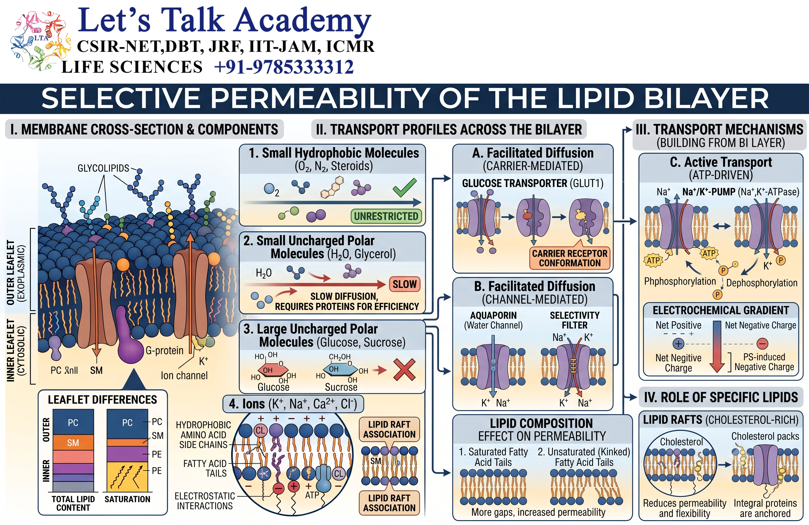
17.1 Plasma Membrane
The plasma membrane surrounds the cell and separates the cytoplasm from the extracellular environment.
It is involved in:
- Transport
- Signaling
- Cell adhesion
- Recognition
- Communication
17.2 Mitochondrial Membranes
Mitochondria contain an outer membrane and an inner membrane.
The inner mitochondrial membrane is highly specialized and contains components of the electron transport chain and ATP synthase.
Its relatively low permeability to ions is essential for establishing the proton gradient required for ATP synthesis.
17.3 Endoplasmic Reticulum
The endoplasmic reticulum is surrounded by a membrane system involved in:
- Protein synthesis
- Lipid synthesis
- Protein processing
- Calcium storage
The endoplasmic reticulum is also a major site of membrane lipid production.
17.4 Golgi Apparatus
The Golgi apparatus contains membrane-bound compartments that modify, sort, and distribute proteins and lipids.
Its membrane composition changes across different Golgi regions, contributing to the specialized functions of these compartments.
17.5 Lysosomal Membranes
Lysosomal membranes separate hydrolytic enzymes from the cytoplasm.
They contain proteins that help maintain the acidic environment required for lysosomal digestion.
18. Lipid Bilayer and Membrane Curvature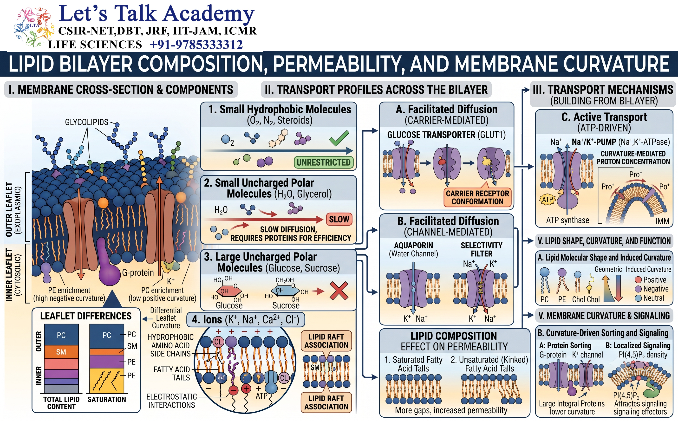
18.1 Importance of Membrane Curvature
Biological membranes are not always flat.
They can bend, curve, and form highly complex structures.
Membrane curvature is important during:
- Vesicle formation
- Endocytosis
- Exocytosis
- Organelle formation
- Membrane fusion
- Membrane fission
18.2 Factors Influencing Curvature
Membrane curvature can be influenced by:
- Lipid shape
- Lipid composition
- Protein binding
- Protein insertion
- Cytoskeletal forces
- Membrane tension
Lipids with different molecular geometries can favor different membrane curvatures.
19. Lipid Bilayer and Electrochemical Gradients
19.1 Ion Gradients
The lipid bilayer acts as an electrical barrier because ions cannot readily pass through its hydrophobic interior.
Membrane proteins can selectively transport ions across the membrane.
This allows cells to establish differences in ion concentration and electrical potential.
19.2 Membrane Potential
The unequal distribution of charged ions across a membrane can generate a membrane potential.
Membrane potential is particularly important in:
- Neurons
- Muscle cells
- Secretory cells
- Mitochondria
- Chloroplasts
19.3 Proton Gradients
Proton gradients across membranes are particularly important for energy conversion.
In mitochondria, electron transport establishes a proton gradient across the inner mitochondrial membrane.
ATP synthase uses this electrochemical gradient to produce ATP.
A similar principle operates in chloroplasts during photosynthesis.
20. Lipid Bilayer and Cell Death
20.1 Phosphatidylserine Exposure
Under normal conditions, phosphatidylserine is predominantly located on the cytosolic side of the plasma membrane.
During apoptosis, this asymmetry can be disrupted, causing phosphatidylserine to become exposed on the extracellular surface.
This exposure acts as an important recognition signal for phagocytic cells.
20.2 Membrane Integrity During Cell Death
Different forms of cell death can involve different patterns of membrane disruption.
In apoptosis, the plasma membrane can remain relatively intact during early stages, whereas uncontrolled membrane damage is a prominent feature of necrotic processes.
Thus, membrane integrity is closely connected to the biological outcome of cellular stress and death.
21. Lipid Bilayer Compared with Other Biological Barriers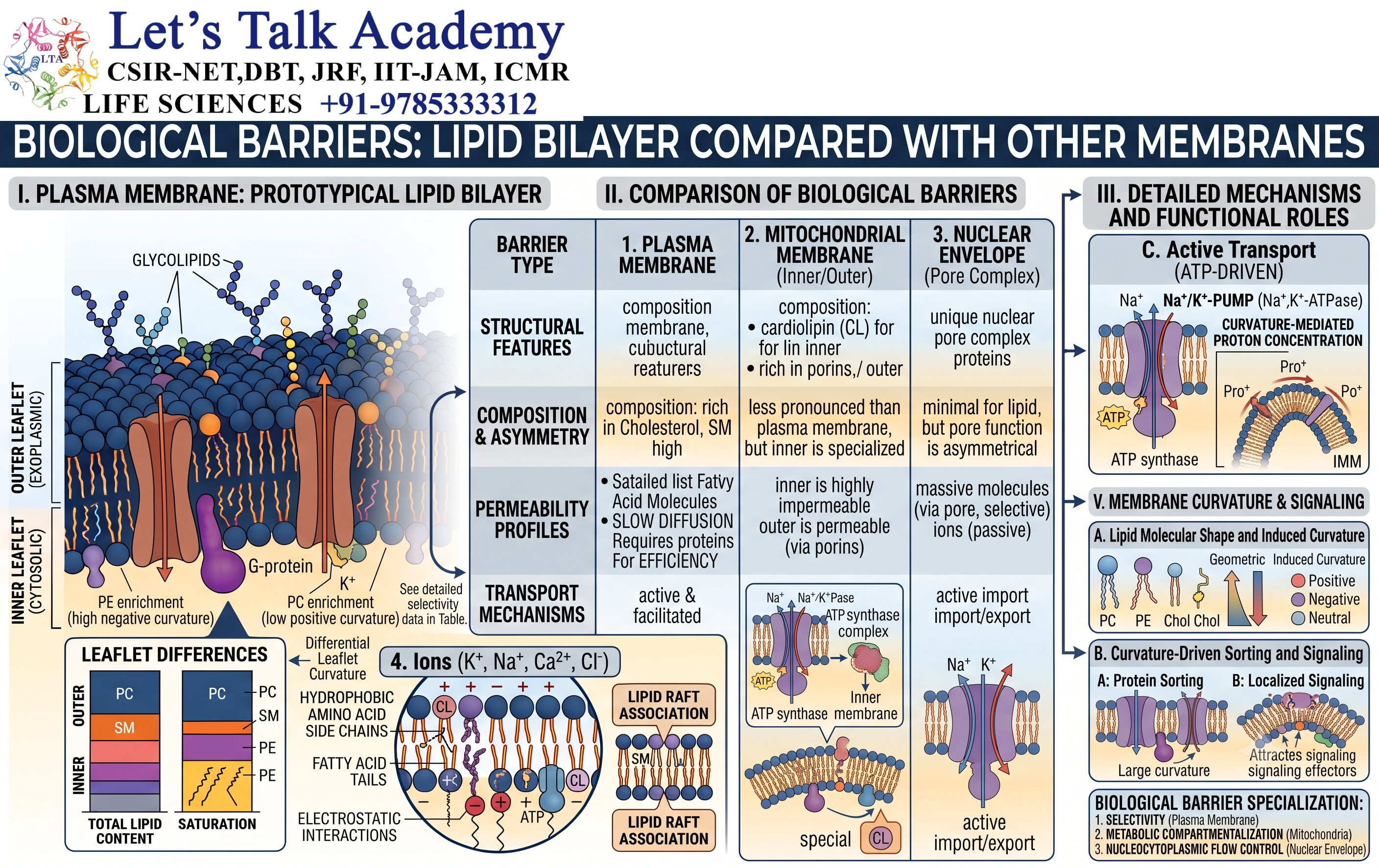
The lipid bilayer should not be confused with the cell wall.
A cell membrane is primarily a lipid-protein structure, whereas a cell wall is an external structural layer found in organisms such as plants, fungi, and bacteria.
For example:
Cell membrane: Flexible, lipid-based, selectively permeable.
Cell wall: More rigid, provides structural support and protection.
In plants, the plasma membrane is located inside the cell wall.


