
1. Cellular Organization
1.1 Cell Wall and Cell Membrane: Structure and Function
1.1.1 Introduction
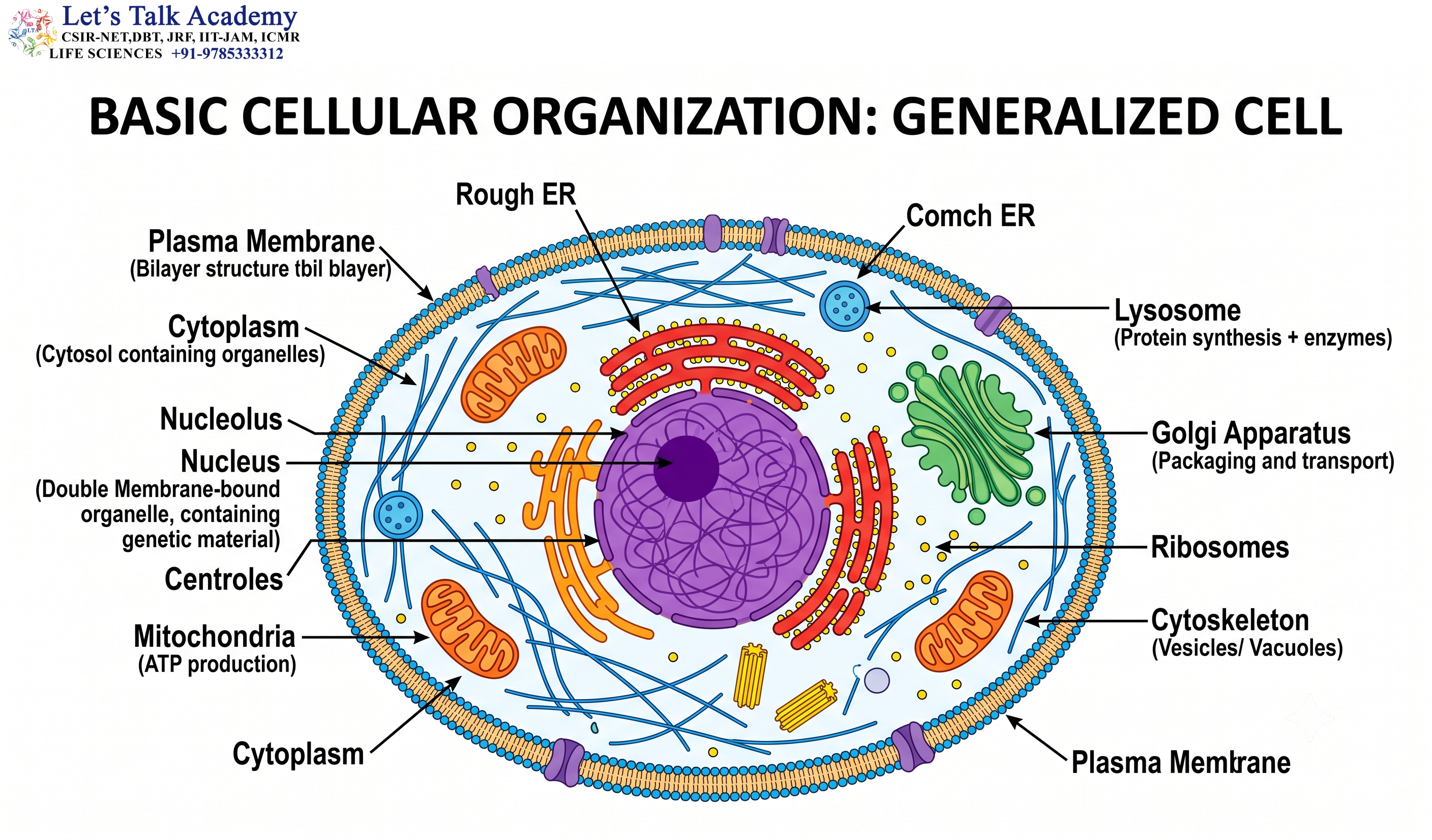
The cell is the fundamental structural and functional unit of life. Every living organism, from a single-celled bacterium to a highly complex multicellular organism such as a human being or a flowering plant, is made up of cells.
Although cells differ greatly in their size, shape, internal organization, and functions, they share several fundamental characteristics. One of the most important characteristics of a cell is its ability to maintain a distinct internal environment while continuously interacting with its surroundings.
This ability depends largely on specialized structures that separate the cell from its surroundings and provide mechanical support, protection, selective transport, and communication.
Two important structures associated with the outer boundary of many cells are the cell wall and the cell membrane.
The cell membrane, also known as the plasma membrane, is a thin, flexible, selectively permeable membrane that surrounds the cytoplasm of every living cell. It regulates the movement of substances into and out of the cell and also participates in cell signaling, recognition, adhesion, and communication.
The cell wall, in contrast, is a relatively rigid extracellular structure found outside the plasma membrane in plants, fungi, bacteria, archaea, and several other organisms. It provides mechanical support, protects the cell, maintains cellular shape, and helps prevent excessive expansion caused by water uptake.
Although the cell wall and cell membrane are closely associated, they are structurally and functionally different. Their properties allow cells to maintain their internal organization while interacting effectively with their external environment.
Understanding the structure and function of these cellular boundaries provides an important foundation for studying cell biology, molecular biology, microbiology, plant biology, physiology, and biochemistry.
1.2 Cellular Boundaries
A cell must maintain a controlled internal environment in order to survive. At the same time, it must continuously exchange materials and information with its surroundings.
For example, a cell may need to:
- Take up nutrients
- Absorb ions
- Obtain oxygen
- Remove carbon dioxide
- Eliminate metabolic waste
- Receive signals from other cells
- Respond to changes in temperature, pH, nutrients, and other environmental conditions
If the cell were completely open to its surroundings, essential molecules could diffuse away and harmful substances could enter freely. Therefore, cells require specialized boundaries that separate the internal environment from the external environment while still allowing controlled exchange.
The plasma membrane performs this essential function in all living cells.
In many organisms, an additional cell wall provides mechanical support and protection outside the plasma membrane.
A simplified arrangement in a plant cell can therefore be represented as:
External environment → Cell wall → Plasma membrane → Cytoplasm
However, this arrangement is not identical in every organism. Animal cells lack a cell wall, bacterial and archaeal cell envelopes have different compositions, and fungal cells possess a wall with a composition distinct from that of plants.
The diversity of cell boundaries reflects the different structural and environmental requirements of living organisms.
2. Cell Wall
2.1 Definition of the Cell Wall
The cell wall is a relatively rigid or semi-rigid extracellular layer located outside the plasma membrane in many organisms.
It provides structural support and protection and helps cells resist mechanical and osmotic stress.
The chemical composition of the cell wall differs considerably among different groups of organisms. This variation is an important example of how biological structures are adapted to particular cellular requirements.
In plants, the major structural component is cellulose.
In fungi, chitin is an important structural component.
In most bacteria, peptidoglycan forms the major structural framework of the cell wall.
Archaea have diverse cell-envelope structures and generally do not possess the bacterial type of peptidoglycan.
Thus, the term cell wall describes a general structural feature rather than a chemically identical structure shared by all organisms.
2.2 General Characteristics of the Cell Wall
The cell wall has several important characteristics.
First, it is located outside the plasma membrane.
Second, it is generally more rigid than the plasma membrane.
Third, its composition varies among organisms.
Fourth, although it provides structural resistance, it is not simply an impermeable barrier. Many cell walls contain pores and spaces through which water, ions, and small molecules can move.
The physical properties of the cell wall are closely related to its molecular composition.
For example, cellulose microfibrils provide tensile strength in plants, while the cross-linked structure of peptidoglycan provides mechanical strength in bacteria.
2.3 Functions of the Cell Wall
2.3.1 Mechanical Support
One of the primary functions of the cell wall is to provide mechanical strength.
Cells contain water and dissolved substances that exert pressure against their surrounding structures. The cell wall resists this pressure and helps maintain the structural integrity of the cell.
This function is especially important in plants, where water uptake can generate considerable internal pressure.
2.3.2 Maintenance of Cell Shape
The cell wall helps determine and maintain the characteristic shape of cells.
Different types of cells may have different wall thicknesses and compositions depending on their functions.
Cells involved in mechanical support may have thick and highly strengthened walls, whereas actively growing cells generally have more flexible walls.
2.3.3 Protection
The cell wall provides protection against mechanical damage and environmental stresses.
It forms an external structural layer that can help protect the plasma membrane and the cellular contents.
In microorganisms, the cell wall can also be important for survival under changing environmental conditions.
2.3.4 Prevention of Excessive Cell Expansion
Water can enter cells through osmosis when the external environment has a higher water potential than the cell.
Without sufficient mechanical resistance, excessive water entry could cause the cell to swell and rupture.
The cell wall provides resistance to expansion and helps prevent this from occurring in many walled cells.
2.3.5 Regulation of Cell Growth
The cell wall is strong but not completely static.
Growing plant cells must modify their cell walls in a controlled manner to permit expansion.
This involves enzymatic modification of wall components and changes in interactions between structural polymers.
Therefore, the cell wall should be regarded as a dynamic structure rather than a passive protective shell.
2.3.6 Interaction with the Environment
The cell wall can also participate in interactions between the cell and its environment.
In plants, the wall contains molecules involved in recognition, defense, signaling, and interactions with neighboring cells and microorganisms.
3. Plant Cell Wall
3.1 General Organization of the Plant Cell Wall
The plant cell wall is a highly organized extracellular matrix surrounding the plasma membrane.
It is composed primarily of polysaccharides along with structural and functional proteins.
The major components include:
- Cellulose
- Hemicellulose
- Pectin
- Structural proteins
- Lignin, particularly in secondary cell walls
The relative proportions of these components vary depending on cell type, developmental stage, tissue, and physiological condition.
For example, a growing cell generally possesses a relatively flexible primary wall, whereas cells specialized for mechanical support or water conduction may develop thick and highly lignified secondary walls.
3.2 Layers of the Plant Cell Wall
The plant cell wall can be broadly described in terms of three major regions:
- Middle lamella
- Primary cell wall
- Secondary cell wall
These regions are not simply separate layers with completely independent compositions. Instead, they form an integrated extracellular structure.
3.2.1 Middle Lamella
The middle lamella is the region located between the primary cell walls of adjacent plant cells.
It is particularly rich in pectic substances.
The middle lamella acts as an adhesive layer that helps neighboring cells remain attached to one another.
Cell adhesion is essential for maintaining the structural organization of plant tissues.
Changes in pectin during fruit ripening can contribute to the softening of tissues because the adhesion between neighboring cells becomes weaker.
3.2.2 Primary Cell Wall
The primary cell wall develops while a plant cell is growing.
It is relatively thin and flexible compared with the secondary wall.
Its major components include:
- Cellulose
- Hemicellulose
- Pectin
- Proteins
Cellulose microfibrils provide tensile strength, while hemicellulose and pectin contribute to the surrounding matrix.
The primary cell wall must maintain a balance between strength and flexibility.
It needs sufficient strength to resist internal pressure but must remain flexible enough to allow controlled cell expansion.
3.2.3 Secondary Cell Wall
Some plant cells develop a secondary cell wall after the major phase of cell expansion has been completed.
The secondary wall is generally thicker and more rigid than the primary wall.
It can contain substantial amounts of:
- Cellulose
- Hemicellulose
- Lignin
The secondary wall provides additional mechanical strength and is especially important in specialized tissues.
Cells associated with xylem and mechanical tissues often possess strongly thickened secondary walls.
4. Components of the Plant Cell Wall
4.1 Cellulose
4.1.1 Structure of Cellulose
Cellulose is one of the most abundant biological polymers on Earth and is a major structural component of plant cell walls.
It is a linear polysaccharide composed of glucose molecules connected through β(1→4) glycosidic bonds.
The linear arrangement of glucose residues allows cellulose chains to associate closely with one another through hydrogen bonding.
Groups of cellulose molecules form microfibrils, which provide considerable tensile strength to the cell wall.
4.1.2 Structural Importance of Cellulose
The strength of cellulose is closely related to its molecular structure.
The glucose chains are:
- Linear
- Long
- Closely associated
- Stabilized by hydrogen bonding
The resulting microfibrils form an important load-bearing component of the plant cell wall.
A useful comparison is that cellulose contains β-linked glucose units, whereas starch contains mainly α-linked glucose units. This difference in linkage contributes to major differences in their three-dimensional structures and biological functions.
4.2 Hemicellulose
Hemicellulose refers to a diverse group of polysaccharides associated with cellulose in plant cell walls.
Unlike cellulose, which has a relatively uniform structure, hemicelluloses include several different types of polymers.
Hemicellulose interacts with cellulose microfibrils and helps organize the wall matrix.
It contributes to:
- Mechanical strength
- Flexibility
- Cell-wall architecture
- Regulation of cell expansion
The composition of hemicellulose can vary between different tissues and developmental stages.
4.3 Pectin
Pectin is another major component of the plant cell wall.
It is particularly abundant in the middle lamella and contributes significantly to the adhesion of neighboring cells.
Pectin also influences:
- Water retention
- Cell-wall porosity
- Wall flexibility
- Cell adhesion
- Tissue structure
Because pectins contain many charged groups and interact strongly with water, they contribute to the physical properties of the cell-wall matrix.
4.4 Lignin
Lignin is a complex phenolic polymer associated mainly with the secondary walls of certain plant cells.
It contributes to:
- Rigidity
- Mechanical strength
- Resistance to compression
- Hydrophobicity
- Resistance to degradation
Lignification is particularly important in vascular plants.
In water-conducting tissues, lignified walls help prevent cells from collapsing under the mechanical stresses associated with water transport.
Thus, lignin is not simply a strengthening substance. It also contributes to the functional specialization of plant tissues.
4.5 Cell Wall Proteins
Although carbohydrates constitute much of the structural framework of the plant cell wall, proteins are also important.
Wall-associated proteins and enzymes participate in:
- Cell-wall remodeling
- Cell growth
- Defense
- Cell signaling
- Modification of extracellular polymers
Certain enzymes modify polysaccharides and change the mechanical properties of the wall.
This demonstrates that the plant cell wall is a biologically active and continuously remodeled structure.
5. Plasmodesmata
5.1 Structure and Function
Plant cells are not completely isolated from one another.
Specialized microscopic channels called plasmodesmata connect adjacent plant cells.
These channels pass through the cell wall and establish cytoplasmic continuity between neighboring cells.
Through plasmodesmata, cells can exchange certain:
- Ions
- Metabolites
- Small molecules
- Signaling molecules
- Proteins
- RNA molecules
The plasma membrane lining the plasmodesma is continuous with the plasma membranes of the connected cells.
Many plasmodesmata also contain a narrow structure derived from the endoplasmic reticulum called the desmotubule.
5.2 Biological Importance of Plasmodesmata
Plasmodesmata allow neighboring plant cells to coordinate their activities.
They are important in:
- Cell-to-cell communication
- Distribution of metabolites
- Developmental signaling
- Coordination of tissue responses
- Movement of certain macromolecules
The permeability of plasmodesmata can also be regulated.
Plants can modify the effective size of the channel, thereby controlling the movement of substances between cells.
6. Bacterial Cell Wall
6.1 General Structure
Bacteria possess a cell envelope that provides protection and helps maintain cellular shape.
For most bacteria, the principal structural component of the cell wall is peptidoglycan.
Peptidoglycan forms a mesh-like structure surrounding the cell.
It consists of alternating sugar residues:
- N-acetylglucosamine (NAG)
- N-acetylmuramic acid (NAM)
These glycan chains are interconnected by peptide cross-links.
The resulting network provides mechanical strength and helps bacteria withstand osmotic pressure.
6.2 Peptidoglycan
Peptidoglycan is a characteristic structural polymer of bacterial cell walls.
Its strength results from the combination of:
- Long glycan chains
- Peptide side chains
- Cross-linking between neighboring chains
This creates a strong network surrounding the bacterial plasma membrane.
Because the bacterial cytoplasm may have a higher solute concentration than the external environment, bacteria can experience substantial osmotic pressure.
The peptidoglycan layer provides resistance against this pressure.
6.3 Cell Wall Synthesis
Bacterial cell-wall synthesis is a highly coordinated process.
New peptidoglycan must be synthesized and incorporated into the existing wall as the bacterial cell grows and divides.
The process involves enzymes responsible for:
- Synthesis of precursor molecules
- Polymerization of glycan chains
- Cross-linking of peptide chains
- Remodeling of existing peptidoglycan
This continuous remodeling allows the bacterial wall to expand while maintaining structural integrity.
7. Gram-Positive and Gram-Negative Bacteria
7.1 Gram-Positive Cell Wall
Gram-positive bacteria generally possess a thick peptidoglycan layer.
Their cell envelope commonly contains:
- Peptidoglycan
- Teichoic acids
- Lipoteichoic acids
They do not possess the outer membrane characteristic of Gram-negative bacteria.
The thick peptidoglycan layer contributes significantly to the mechanical strength of the cell envelope.
7.2 Gram-Negative Cell Envelope
Gram-negative bacteria generally possess:
- A relatively thin peptidoglycan layer
- A periplasmic space
- An outer membrane
The outer membrane contains lipopolysaccharide (LPS).
LPS can be divided into three broad regions:
- Lipid A
- Core polysaccharide
- O-antigen
Lipid A is embedded in the outer membrane and is responsible for much of the endotoxin activity associated with LPS.
The outer membrane also contains porins, which form channels through which certain small hydrophilic molecules can pass.
7.3 Comparison of Gram-Positive and Gram-Negative Bacteria
| Feature | Gram-Positive | Gram-Negative |
|---|---|---|
| Peptidoglycan | Thick | Thin |
| Outer membrane | Absent | Present |
| Teichoic acids | Present | Generally absent |
| Lipopolysaccharide | Absent | Present |
| Periplasmic region | Less prominent | Prominent |
| Porins in outer membrane | Absent | Present |
The differences between these two types of bacterial envelopes influence their staining properties, permeability, interactions with the environment, and responses to antimicrobial compounds.
8. Fungal Cell Wall
8.1 General Structure
Fungi possess a cell wall outside their plasma membrane.
The fungal cell wall contains several important components, including:
- Chitin
- β-glucans
- Mannoproteins
The relative amounts of these components vary among fungal species and developmental stages.
8.2 Chitin
Chitin is a structural polysaccharide composed of N-acetylglucosamine residues.
It provides mechanical strength to the fungal cell wall.
The chemical distinction between major structural polymers can be summarized as:
Plants → Cellulose
Fungi → Chitin
Bacteria → Peptidoglycan
These differences are useful for understanding the structural diversity of cell walls.
9. Archaeal Cell Envelopes
9.1 General Characteristics
Archaea are prokaryotic organisms, but their cell envelopes differ significantly from those of bacteria.
Many archaea do not contain peptidoglycan.
Depending on the organism, their cell surface may contain:
- Proteinaceous S-layers
- Polysaccharides
- Pseudomurein in certain groups
Therefore, peptidoglycan should not be considered a universal structural component of all prokaryotic cell envelopes.
The diversity of archaeal cell envelopes reflects their adaptation to a wide range of environmental conditions.
10. Cell Membrane
10.1 Definition
The cell membrane, also called the plasma membrane, is a thin, flexible, selectively permeable membrane surrounding the cytoplasm.
It is present in all living cells.
The plasma membrane separates the intracellular environment from the extracellular environment while allowing controlled exchange of materials.
However, the plasma membrane is much more than a simple physical barrier.
It is involved in:
- Transport
- Cell signaling
- Cell recognition
- Cell adhesion
- Energy conversion
- Maintenance of ion gradients
- Communication with the external environment
10.2 Major Functions of the Plasma Membrane
The plasma membrane performs several essential functions.
10.2.1 Selective Barrier
The membrane controls which substances can enter or leave the cell.
10.2.2 Transport
Specific proteins allow ions, nutrients, and other substances to cross the membrane.
10.2.3 Communication
Membrane receptors detect signals from outside the cell and transmit information to intracellular signaling pathways.
10.2.4 Recognition
Carbohydrates and proteins on the cell surface help cells recognize one another.
10.2.5 Adhesion
Membrane proteins participate in interactions between neighboring cells and between cells and the extracellular matrix.
10.2.6 Energy Conversion
Specialized membranes are involved in energy transduction.
For example, mitochondrial membranes are central to oxidative phosphorylation, while photosynthetic membranes participate in light-dependent reactions.
11. Chemical Composition of the Cell Membrane
11.1 Major Components
The plasma membrane is primarily composed of:
- Lipids
- Proteins
- Carbohydrates
The exact composition varies among organisms, cell types, and cellular membranes.
Lipids form the structural framework, proteins perform many specialized functions, and carbohydrates are particularly important for recognition and interactions at the cell surface.
12. Phospholipid Bilayer
12.1 Phospholipids
Phospholipids are major structural components of biological membranes.
A typical phospholipid contains:
- A hydrophilic head
- Hydrophobic hydrocarbon tails
Because phospholipids contain both hydrophilic and hydrophobic regions, they are called amphipathic molecules.
When phospholipids are placed in an aqueous environment, they spontaneously arrange themselves so that the hydrophobic portions are shielded from water while the hydrophilic portions interact with water.
This property is fundamental to the formation of biological membranes.
12.2 Organization of the Bilayer
The plasma membrane consists primarily of a phospholipid bilayer.
The two layers are called leaflets.
The hydrophilic heads face the aqueous environments on either side of the membrane.
The hydrophobic tails face inward toward one another.
The general arrangement can be represented as:
Aqueous environment → Hydrophilic heads → Hydrophobic tails → Hydrophilic heads → Aqueous environment
The hydrophobic core forms a major permeability barrier against many charged and highly polar molecules.
13. Fluid Mosaic Model
13.1 Concept of the Fluid Mosaic Model
The organization of the plasma membrane is commonly explained using the Fluid Mosaic Model, proposed by S. J. Singer and G. L. Nicolson in 1972.
According to this model, the membrane consists of a dynamic lipid bilayer containing proteins and other components.
The word fluid refers to the ability of many membrane components to move within the plane of the membrane.
The word mosaic reflects the heterogeneous distribution of different proteins, lipids, and associated molecules.
The membrane is therefore not a rigid sheet. It is a dynamic structure in which its components continuously interact and, in many cases, move laterally.
13.2 Importance of Membrane Fluidity
Membrane fluidity is essential for:
- Membrane transport
- Vesicle formation
- Membrane fusion
- Cell signaling
- Cell movement
- Cell division
- Distribution and function of membrane proteins
If a membrane becomes excessively rigid or excessively fluid, normal cellular processes can be disrupted.
14. Factors Affecting Membrane Fluidity
14.1 Temperature
Temperature strongly influences membrane fluidity.
At higher temperatures, lipid molecules generally move more rapidly, increasing membrane fluidity.
At lower temperatures, lipid molecules tend to pack more tightly, decreasing fluidity.
Cells can adjust their membrane composition to maintain an appropriate degree of fluidity.
14.2 Fatty-Acid Unsaturation
Unsaturated fatty acids contain one or more carbon-carbon double bonds.
These double bonds introduce bends or kinks into the hydrocarbon chains.
As a result, phospholipids cannot pack as tightly.
Therefore:
Greater unsaturation → Generally greater membrane fluidity
In contrast:
Greater saturation → Tighter packing and generally lower membrane fluidity
14.3 Fatty-Acid Chain Length
Longer hydrocarbon chains generally interact more strongly with neighboring chains and tend to reduce membrane fluidity.
Shorter chains generally increase fluidity.
Thus, both chain length and degree of unsaturation contribute to membrane properties.
15. Cholesterol
15.1 Structure and Location
Cholesterol is an important sterol found in many animal cell membranes.
It contains a small polar hydroxyl group and a larger hydrophobic region.
Its structure allows it to position itself between phospholipid molecules.
15.2 Functions of Cholesterol
Cholesterol helps regulate membrane properties.
At relatively high temperatures, cholesterol restrains excessive movement of phospholipids.
At low temperatures, it interferes with the close packing of phospholipid tails and helps prevent the membrane from becoming excessively rigid.
Therefore, cholesterol acts as an important fluidity buffer.
It can also reduce the permeability of the membrane to certain small water-soluble molecules.
16. Membrane Proteins
16.1 Classification
Membrane proteins perform many specialized functions.
They can broadly be divided into:
- Integral membrane proteins
- Peripheral membrane proteins
16.2 Integral Membrane Proteins
Integral membrane proteins are closely associated with the lipid bilayer.
Many extend across the entire membrane and are called transmembrane proteins.
They perform functions such as:
- Transport
- Signal reception
- Enzymatic activity
- Cell adhesion
- Cell recognition
Ion channels, receptors, transporters, and many pumps are examples of integral membrane proteins.
16.3 Peripheral Membrane Proteins
Peripheral membrane proteins are associated mainly with the surface of the membrane.
They may interact with:
- Membrane proteins
- Lipid head groups
- Cytoskeletal components
- Signaling molecules
They can therefore serve as links between the plasma membrane and the cytoskeleton or participate in signaling pathways.
17. Membrane Carbohydrates and Glycocalyx
17.1 Membrane Carbohydrates
Carbohydrates associated with the plasma membrane are generally located on the extracellular surface.
They may be attached to proteins, forming glycoproteins, or to lipids, forming glycolipids.
These carbohydrate-containing structures are important for interactions between cells and their surroundings.
17.2 Glycocalyx
The glycocalyx is a carbohydrate-rich layer associated with the external surface of many cells.
It participates in:
- Cell recognition
- Cell adhesion
- Protection
- Cell signaling
- Interaction with extracellular molecules
The glycocalyx is particularly important in multicellular organisms, where cells must distinguish between different types of cells and interact with their surrounding tissues.
18. Membrane Asymmetry
18.1 Distribution of Membrane Lipids
The two leaflets of a biological membrane are not chemically identical.
Different lipids are preferentially distributed in different leaflets.
For example, in many eukaryotic plasma membranes:
- Phosphatidylcholine is enriched in the outer leaflet.
- Sphingomyelin is enriched in the outer leaflet.
- Phosphatidylethanolamine is enriched in the inner leaflet.
- Phosphatidylserine is normally concentrated in the inner leaflet.
This asymmetric distribution is actively maintained by cellular mechanisms.
18.2 Biological Importance of Membrane Asymmetry
Membrane asymmetry is important for:
- Cell signaling
- Membrane curvature
- Cell recognition
- Vesicle trafficking
- Programmed cell death
Thus, the two sides of the membrane are functionally different.
18.3 Phosphatidylserine and Apoptosis
Under normal conditions, phosphatidylserine is mainly located on the cytoplasmic side of the plasma membrane.
During apoptosis, phosphatidylserine can become exposed on the extracellular surface.
This exposure acts as an important recognition signal for phagocytic cells.
Therefore, changes in lipid distribution can serve as signals that influence cellular behavior.
19. Selective Permeability
19.1 Nature of Selective Permeability
The plasma membrane is described as selectively permeable because it does not allow every substance to cross freely.
The ability of a molecule to cross the membrane depends on factors such as:
- Molecular size
- Electrical charge
- Polarity
- Lipid solubility
- Presence of specific transport proteins
Small nonpolar molecules generally cross the lipid bilayer more easily than ions and large polar molecules.
This selective permeability is essential for maintaining cellular homeostasis.
20. Transport Across the Cell Membrane
20.1 Major Transport Mechanisms
Substances cross the plasma membrane through several mechanisms:
- Simple diffusion
- Facilitated diffusion
- Osmosis
- Active transport
- Endocytosis
- Exocytosis
Each mechanism has a different molecular basis and is suited to particular types of substances.
21. Simple Diffusion
21.1 Definition
Simple diffusion is the passive movement of molecules from a region of higher concentration to a region of lower concentration.
The process does not require direct expenditure of ATP.
Small nonpolar molecules such as oxygen and carbon dioxide can cross biological membranes relatively easily.
21.2 Factors Affecting Diffusion
The rate of diffusion is influenced by:
- Concentration gradient
- Temperature
- Surface area
- Diffusion distance
- Molecular size
- Membrane permeability
A greater concentration difference generally provides a stronger driving force for diffusion.
22. Facilitated Diffusion
22.1 General Mechanism
Facilitated diffusion is a passive transport process in which molecules move across the membrane with the assistance of membrane proteins.
The transported substance moves down its concentration or electrochemical gradient.
No direct ATP expenditure is required.
Two major types of membrane proteins involved are:
- Channel proteins
- Carrier proteins
22.2 Channel Proteins
Channel proteins form hydrophilic pathways through the membrane.
They are particularly important for the movement of:
- Ions
- Water
- Certain small molecules
Many channels are selective and can open or close in response to specific signals.
Channels may be regulated by:
- Voltage
- Ligand binding
- Mechanical forces
22.3 Carrier Proteins
Carrier proteins bind specific molecules and undergo conformational changes that move those molecules across the membrane.
Examples include transport proteins involved in the movement of:
- Glucose
- Amino acids
- Ions
Carrier-mediated transport differs from channel-mediated transport because the transported substance interacts directly with the carrier during the transport cycle.
23. Active Transport
23.1 General Mechanism
Active transport moves substances against their concentration or electrochemical gradients.
Because movement against a gradient requires energy, active transport depends on an energy source.
Active transport is essential because cells must often maintain ion concentrations that are very different from those present outside the cell.
There are two broad categories:
- Primary active transport
- Secondary active transport
23.2 Primary Active Transport
Primary active transport directly uses energy, commonly obtained from ATP hydrolysis.
A well-known example is the Na⁺/K⁺-ATPase found in animal cells.
For each ATP molecule hydrolyzed, the pump typically transports:
3 Na⁺ out of the cell
and
2 K⁺ into the cell
This contributes to:
- Maintenance of ion gradients
- Membrane potential
- Osmotic balance
- Secondary active transport
23.3 Secondary Active Transport
Secondary active transport does not directly use ATP at the transporter itself.
Instead, it uses the energy stored in an electrochemical gradient.
That gradient is generally established by primary active transport.
Secondary transport can occur through two major mechanisms.
23.3.1 Symport
In symport, two substances move in the same direction across the membrane.
23.3.2 Antiport
In antiport, two substances move in opposite directions.
Secondary active transport demonstrates how energy stored in an ion gradient can be converted into useful cellular work.
24. Electrochemical Gradient
24.1 Chemical Gradient
A chemical gradient results from a difference in the concentration of a substance between two sides of a membrane.
A substance tends to move down its concentration gradient when an appropriate pathway is available.
24.2 Electrical Gradient
For charged particles, the electrical difference across the membrane also influences movement.
A difference in electrical charge between the two sides of the membrane produces an electrical gradient.
24.3 Electrochemical Gradient
The combination of the chemical and electrical components is called the electrochemical gradient.
This concept is particularly important for ion movement and membrane potential.
25. Osmosis and Water Transport
25.1 Osmosis
Osmosis is the movement of water across a selectively permeable membrane in response to differences in water potential or effective solute concentration.
Water can cross membranes through the lipid bilayer to some extent, but specialized membrane proteins called aquaporins greatly facilitate water movement.
Osmosis is important for:
- Cell volume regulation
- Plant water relations
- Kidney function
- Tissue fluid balance
- Maintenance of cellular homeostasis
25.2 Tonicity
The effect of an external solution on cell volume is commonly described using the terms:
- Hypotonic
- Isotonic
- Hypertonic
25.2.1 Hypotonic Solution
A hypotonic environment generally promotes net water entry into a cell.
25.2.2 Isotonic Solution
An isotonic environment does not produce a sustained net change in cell volume due to water movement.
25.2.3 Hypertonic Solution
A hypertonic environment generally promotes net water loss from a cell.
The actual response depends on the permeability properties of the solutes involved.
26. Turgor Pressure and Plasmolysis
26.1 Turgor Pressure
When water enters a plant cell, the cell contents press against the cell wall.
The resulting pressure is called turgor pressure.
Turgor is important for:
- Maintaining plant tissue rigidity
- Supporting leaves and young stems
- Cell expansion
- Maintaining cellular structure
When plant cells lose water and turgor pressure decreases, plant tissues can become limp or wilted.
26.2 Plasmolysis
Plasmolysis is a characteristic response of plant cells to a sufficiently hypertonic environment.
Water leaves the cell, causing the plasma membrane and cytoplasm to shrink away from the cell wall.
The cell wall remains relatively fixed while the living contents contract inward.
Plasmolysis demonstrates the different physical properties of the cell wall and plasma membrane.
27. Endocytosis
27.1 General Mechanism
Endocytosis is the process by which cells internalize extracellular material through the formation of membrane-bound vesicles.
During endocytosis, a region of the plasma membrane bends inward and eventually pinches off to form an intracellular vesicle.
Major forms include:
- Phagocytosis
- Pinocytosis
- Receptor-mediated endocytosis
28. Phagocytosis
28.1 Mechanism
Phagocytosis involves the uptake of relatively large particles.
Specialized cells can engulf:
- Microorganisms
- Dead cells
- Cellular debris
- Large particles
The engulfed material becomes enclosed within a vesicle called a phagosome.
The phagosome can subsequently interact with lysosomal compartments, where the contents may be degraded.
Phagocytosis is important in cellular defense and tissue maintenance.
29. Pinocytosis
29.1 General Mechanism
Pinocytosis refers broadly to the uptake of extracellular fluid and dissolved substances through small vesicles.
It allows cells to continuously sample and internalize components of their surrounding environment.
Unlike phagocytosis, which involves large particles, pinocytosis generally involves fluid and relatively small dissolved substances.
30. Receptor-Mediated Endocytosis
30.1 Selective Uptake
Receptor-mediated endocytosis provides a selective mechanism for internalizing specific extracellular molecules.
A molecule first binds to a specific receptor on the cell surface.
The receptor and its bound cargo become concentrated in specialized regions of the plasma membrane.
The membrane then bends inward and forms a vesicle containing the selected material.
This mechanism allows cells to efficiently internalize specific molecules even when their extracellular concentration is relatively low.
31. Exocytosis
31.1 General Mechanism
Exocytosis is the process by which intracellular vesicles fuse with the plasma membrane and release their contents outside the cell.
It is important for:
- Hormone secretion
- Neurotransmitter release
- Enzyme secretion
- Extracellular protein release
- Delivery of membrane components
During exocytosis, the vesicle membrane becomes continuous with the plasma membrane.
The process therefore contributes not only to secretion but also to the maintenance and expansion of the plasma membrane.
32. Cell Membrane in Cell Signaling
32.1 Role in Cellular Communication
The plasma membrane is a major site of cellular communication.
Cells continuously receive information about their surroundings.
Signals may include:
- Hormones
- Neurotransmitters
- Growth factors
- Cytokines
- Extracellular metabolites
Many signaling molecules bind to specific receptors located in the plasma membrane.
The receptor then undergoes a conformational or biochemical change that initiates intracellular signaling pathways.
The plasma membrane therefore acts as both a physical boundary and an information-processing interface.
33. Membrane Receptors
33.1 Major Classes
Membrane receptors are proteins that recognize specific extracellular signals.
Important classes include:
- G-protein-coupled receptors
- Receptor tyrosine kinases
- Ligand-gated ion channels
Binding of a signaling molecule to its receptor can initiate a cascade of intracellular events.
These signaling pathways can influence:
- Gene expression
- Metabolism
- Cell growth
- Cell division
- Cell movement
- Cell survival
34. Cell Membrane and Cell Adhesion
34.1 Cell Adhesion
Cells frequently need to attach to neighboring cells or to components of the extracellular matrix.
Membrane proteins called cell adhesion molecules participate in these interactions.
Cell adhesion is essential for:
- Tissue formation
- Development
- Wound repair
- Cell migration
- Cellular communication
Thus, the plasma membrane contributes directly to the organization and stability of multicellular tissues.
35. Biological Membranes Beyond the Plasma Membrane
35.1 Membrane-Bound Organelles
Membranes are not restricted to the outer surface of the cell.
Eukaryotic cells contain numerous membrane-bound organelles, including:
- Endoplasmic reticulum
- Golgi apparatus
- Lysosomes
- Endosomes
- Mitochondria
- Chloroplasts
- Peroxisomes
- Nuclear envelope
Each membrane has a specialized composition and performs particular functions.
Although the basic principles of lipid-bilayer organization apply broadly, the exact lipid and protein composition varies between organelles.
35.2 Functional Compartmentalization
Membrane-bound compartments allow different biochemical processes to occur in controlled environments.
For example:
- The nucleus provides an environment for DNA storage and many nuclear processes.
- The endoplasmic reticulum is involved in protein and lipid synthesis.
- The Golgi apparatus modifies and sorts many proteins and lipids.
- Lysosomes provide an acidic environment for degradation.
- Mitochondria contain specialized membranes involved in ATP production.
- Chloroplasts contain membranes required for photosynthesis.
This compartmentalization increases the functional efficiency of eukaryotic cells.
36. Membrane Dynamics
36.1 Dynamic Nature of Biological Membranes
The plasma membrane is continuously remodeled.
Membrane components can:
- Move laterally
- Associate with proteins
- Be internalized
- Be recycled
- Be transported through vesicles
- Fuse with other membranes
This dynamic behavior is essential for:
- Endocytosis
- Exocytosis
- Cell migration
- Cell division
- Signal transduction
- Membrane repair
The membrane should therefore be viewed as a dynamic biological system rather than a static covering.
37. Membrane Repair
37.1 Maintenance of Membrane Integrity
Because the plasma membrane is thin and dynamic, cells can experience physical damage.
Cells possess mechanisms that detect and repair disruptions in membrane integrity.
Small membrane lesions can sometimes be repaired through rapid changes in membrane organization and vesicle-mediated processes.
Maintaining membrane integrity is essential because uncontrolled leakage of ions and metabolites can rapidly disrupt cellular homeostasis.
38. Cell Wall Remodeling
38.1 Dynamic Nature of the Plant Cell Wall
Plant cell walls undergo continuous modification throughout growth and development.
During cell expansion, enzymes can modify:
- Cellulose-associated structures
- Pectins
- Hemicelluloses
- Wall proteins
These changes alter the mechanical properties of the wall.
The cell can therefore coordinate wall loosening with internal pressure to achieve controlled expansion.
Cell-wall remodeling is particularly important during:
- Root growth
- Stem growth
- Leaf development
- Fruit development
- Cell differentiation
39. Structural Integration of the Cell Surface
39.1 Cell Surface as an Integrated System
The cell surface is not simply composed of isolated layers.
It represents an integrated system involving:
- Cell wall
- Plasma membrane
- Cytoskeleton
- Extracellular matrix
- Cell adhesion proteins
- Signaling molecules
The plasma membrane interacts with the cytoskeleton on its cytoplasmic side.
In plants, the plasma membrane is closely associated with the cell wall.
In animals, membrane proteins connect cells to the extracellular matrix.
This structural integration allows mechanical forces and biochemical signals to be transmitted across the cell boundary.
40. Structure–Function Relationships
40.1 Structure Determines Function
The study of cell walls and membranes illustrates one of the central principles of biology: structure and function are closely related.
40.1.1 Cellulose Structure and Mechanical Strength
The linear structure and extensive hydrogen bonding of cellulose allow plant cell walls to resist mechanical stress.
40.1.2 Amphipathic Lipids and Bilayer Formation
The amphipathic nature of phospholipids allows them to spontaneously organize into bilayers in aqueous environments.
40.1.3 Lipid Composition and Membrane Fluidity
The proportions of saturated and unsaturated lipids influence the physical state of the membrane.
40.1.4 Membrane Proteins and Selective Transport
Specific membrane proteins allow cells to control the movement of ions and molecules.
40.1.5 Ion Gradients and Cellular Function
Ion gradients provide energy for secondary transport and contribute to electrical signaling.
40.1.6 Cell Wall and Osmotic Resistance
The rigid cell wall helps prevent excessive expansion when water enters a plant cell.
41. Comparative Overview of Cell Envelopes
41.1 Major Differences Among Organisms
| Organism | Major Outer Structural Feature |
|---|---|
| Plants | Cellulose-rich cell wall |
| Fungi | Chitin- and glucan-rich cell wall |
| Bacteria | Peptidoglycan-containing cell wall |
| Archaea | Variable surface layers; generally no bacterial-type peptidoglycan |
| Animals | No cell wall; plasma membrane and extracellular matrix |
These differences demonstrate that organisms have evolved different structural solutions for maintaining cellular integrity.
42. Cell Wall and Cell Membrane: Comparative Analysis
42.1 Major Differences
| Feature | Cell Wall | Cell Membrane |
|---|---|---|
| Nature | Rigid or semi-rigid extracellular structure | Flexible lipid bilayer |
| Location | Outside plasma membrane | Boundary of cytoplasm |
| Presence | Found in plants, fungi, bacteria, archaea, and some other organisms | Present in all living cells |
| Major plant component | Cellulose | Phospholipids and proteins |
| Major bacterial component | Peptidoglycan | Lipids and proteins |
| Major fungal component | Chitin and glucans | Lipids and proteins |
| Main function | Support and protection | Selective transport and communication |
| Permeability | Generally porous | Selectively permeable |
| Fluidity | Not a fluid lipid bilayer | Dynamic and fluid |
| Signaling role | Supportive and indirect | Major role |
| Transport role | Provides extracellular framework | Directly regulates membrane transport |
43. Relationship Between Cell Wall and Cell Membrane
43.1 Coordinated Functions
The cell wall and plasma membrane should not be considered completely independent structures.
They work together to maintain cellular integrity.
In a plant cell, for example:
Cell wall → Provides mechanical resistance
Plasma membrane → Controls molecular exchange
Cytoplasm → Contains the machinery for metabolism and cellular activities
When water enters the cell:
- Water crosses the plasma membrane.
- The cellular contents expand.
- Pressure develops against the cell wall.
- The cell wall resists further expansion.
- Turgor pressure develops.
This example demonstrates how the physical properties of different cellular structures combine to produce a functional response.
44. Biological Membranes and Cellular Compartmentalization
44.1 Importance of Membrane-Bound Compartments
Eukaryotic cells are highly compartmentalized.
Different organelles maintain distinct chemical environments that allow specialized biochemical reactions to occur.
For example, lysosomes maintain an acidic environment that supports hydrolytic enzymes.
Mitochondria maintain proton gradients across their inner membrane that are essential for ATP synthesis.
Chloroplasts establish proton gradients across thylakoid membranes during photosynthesis.
Thus, membrane structure is directly linked to cellular energy metabolism and biochemical organization.


