
Oxidative Phosphorylation
1. Introduction
1.1 Meaning of Oxidative Phosphorylation
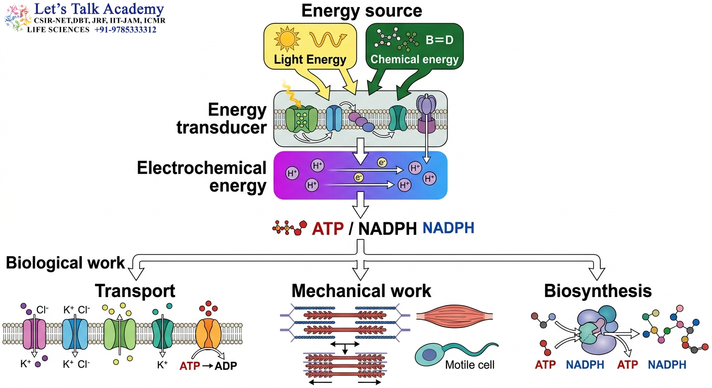
Oxidative phosphorylation is the major ATP-producing process of aerobic respiration. It is the process in which the energy released during the oxidation of reduced electron carriers such as NADH and FADH₂ is ultimately used to synthesize ATP.
The term oxidative phosphorylation contains two important ideas:
- Oxidative refers to the oxidation of NADH and FADH₂ and the transfer of their electrons through the electron transport chain.
- Phosphorylation refers to the addition of inorganic phosphate (Pi) to ADP to form ATP.
The overall reaction can be represented as:
ADP + Pi → ATP
However, the energy required for this reaction does not come directly from NADH or FADH₂. Instead, it is transferred through several intermediate energy-conversion steps.
The complete sequence is:
NADH/FADH₂ oxidation → Electron transport → Proton pumping → Proton-motive force → ATP synthase → ATP
This sequence is one of the most important concepts in cellular bioenergetics.
1.2 Site of Oxidative Phosphorylation
In eukaryotic cells, oxidative phosphorylation occurs primarily at the:
Inner mitochondrial membrane
The inner mitochondrial membrane contains:
- Electron transport chain complexes
- Coenzyme Q
- Cytochrome c-associated electron-transfer system
- ATP synthase
- Transport proteins required for oxidative phosphorylation
The mitochondrial matrix is on one side of this membrane, while the intermembrane space is on the other.
1.3 Why Oxidative Phosphorylation Is Important
ATP is required for almost every energy-dependent process in a living cell.
ATP generated by oxidative phosphorylation supports:
- Active transport
- Muscle contraction
- Protein synthesis
- DNA replication
- RNA synthesis
- Biosynthetic reactions
- Ion homeostasis
- Cell movement
- Signal transduction
- Maintenance of membrane potentials
Therefore, oxidative phosphorylation provides the major energy supply for many aerobic cells.
1.4 Central Principle
The most important principle to remember is:
The electron transport chain does not directly synthesize ATP. It generates a proton-motive force, which is subsequently used by ATP synthase to produce ATP.
Thus:
Electron transport → Proton gradient → ATP synthesis
1.5 Oxidative Phosphorylation as Energy Transduction
Oxidative phosphorylation is an example of biological energy transduction, in which energy changes from one form to another.
The energy conversion can be represented as:
Chemical energy of nutrients
↓
Redox energy of NADH/FADH₂
↓
Electrochemical energy of proton gradient
↓
Rotational energy of ATP synthase
↓
Chemical energy stored in ATP
This makes oxidative phosphorylation a highly organized energy-conversion system.
2. Oxidation-Reduction Reactions
2.1 Meaning of Redox Reactions
Oxidation-reduction reactions, commonly called redox reactions, are reactions involving the transfer of electrons between molecules.
Whenever one molecule loses electrons, another molecule must gain those electrons.
Therefore:
Oxidation and reduction always occur together.
2.2 Oxidation
Oxidation is defined as the:
Loss of electrons
A useful mnemonic is:
OIL = Oxidation Is Loss
In biological systems, oxidation can also involve:
- Loss of hydrogen
- Gain of oxygen
- Increase in oxidation state
However, electron loss is the fundamental definition.
2.3 Reduction
Reduction is defined as the:
Gain of electrons
A useful mnemonic is:
RIG = Reduction Is Gain
Reduction may also involve:
- Gain of hydrogen
- Loss of oxygen
- Decrease in oxidation state
2.4 Electron Donors
A molecule that donates electrons is called an electron donor.
In mitochondrial oxidative phosphorylation, important electron donors include:
- NADH
- FADH₂
These molecules are generated during oxidation of metabolic substrates.
2.5 Electron Acceptors
A molecule that accepts electrons is called an electron acceptor.
In the mitochondrial ETC, electrons are transferred through a series of acceptors before reaching oxygen.
The final electron acceptor is:
Oxygen (O₂)
Therefore, oxygen is called the:
Terminal electron acceptor
2.6 Oxidizing and Reducing Agents
The molecule that accepts electrons acts as an oxidizing agent, whereas the molecule that donates electrons acts as a reducing agent.
Therefore:
Electron donor = reducing agent
Electron acceptor = oxidizing agent
2.7 Redox Potential
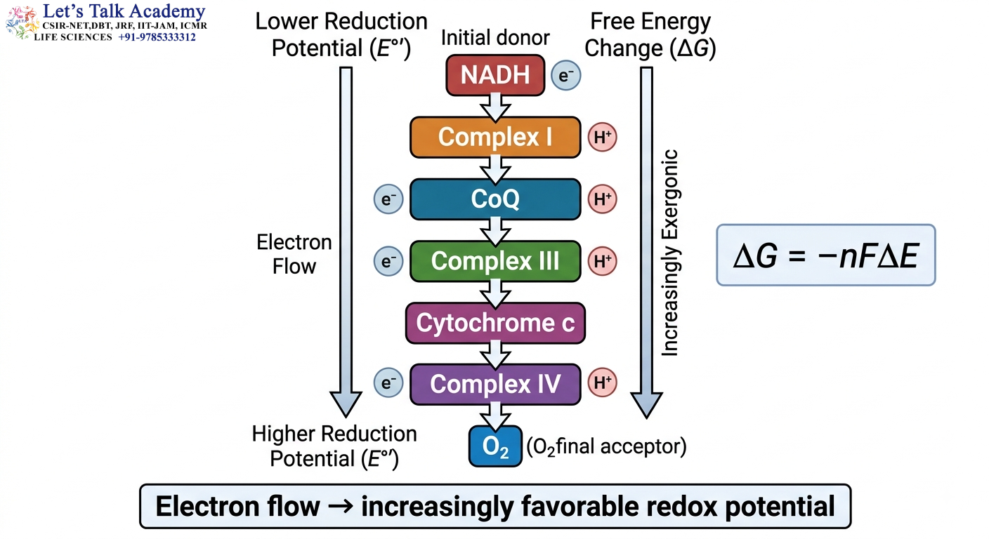
The tendency of a molecule to accept electrons is described by its reduction potential.
Electrons tend to move spontaneously from carriers with lower reduction potential toward carriers with higher reduction potential.
The free-energy change associated with electron transfer is:
ΔG = −nFΔE
Where:
- ΔG = change in Gibbs free energy
- n = number of electrons transferred
- F = Faraday constant
- ΔE = difference in reduction potential
When ΔE is positive:
ΔG becomes negative
and electron transfer is thermodynamically favorable.
2.8 Importance in the ETC
The ETC is organized so that electrons move through a series of carriers with progressively favorable redox potentials.
Ultimately, electrons are transferred to oxygen.
Therefore:
NADH/FADH₂ → ETC → O₂
3. Mitochondrial Organization
3.1 General Structure of Mitochondria

Mitochondria are double-membrane-bound organelles found in most eukaryotic cells.
A typical mitochondrion contains four major regions:
- Outer mitochondrial membrane
- Intermembrane space
- Inner mitochondrial membrane
- Mitochondrial matrix
Each region has a specialized role.
3.2 Outer Mitochondrial Membrane
The outer mitochondrial membrane surrounds the organelle.
It contains channel proteins called porins, particularly voltage-dependent anion channels (VDACs).
These channels make the outer membrane relatively permeable to many small molecules and ions.
3.3 Intermembrane Space
The intermembrane space lies between the outer and inner mitochondrial membranes.
During electron transport, protons are pumped into this compartment.
Therefore:
H⁺ concentration increases in the intermembrane space.
This proton accumulation is essential for generating the proton-motive force.
3.4 Inner Mitochondrial Membrane
The inner mitochondrial membrane is the major site of oxidative phosphorylation.
It contains:
- Complex I
- Complex II
- Complex III
- Complex IV
- Coenzyme Q
- ATP synthase
- Numerous transport proteins
The inner membrane is highly selective and relatively impermeable to protons.
This property is essential for maintaining the proton gradient.
3.5 Mitochondrial Matrix
The matrix contains enzymes involved in several major metabolic pathways.
Important pathways associated with the matrix include:
- Pyruvate oxidation
- Citric acid cycle
- Fatty acid β-oxidation
- Several amino acid metabolic pathways
The matrix also contains:
- Mitochondrial DNA
- Mitochondrial ribosomes
- Enzymes involved in mitochondrial gene expression
3.6 Cristae
The inner mitochondrial membrane forms numerous folds called:
Cristae
Cristae increase the surface area of the inner mitochondrial membrane.
This provides more space for:
- Respiratory complexes
- ATP synthase
- Electron transport
- Proton translocation
3.7 Functional Compartmentalization
Mitochondrial organization is essential for oxidative phosphorylation because the ETC and ATP synthase are separated from the cytosol and positioned within a membrane capable of maintaining an electrochemical gradient.
Thus:
Mitochondrial structure is directly related to mitochondrial function.
4. Inner Mitochondrial Membrane
4.1 Importance of the Inner Membrane
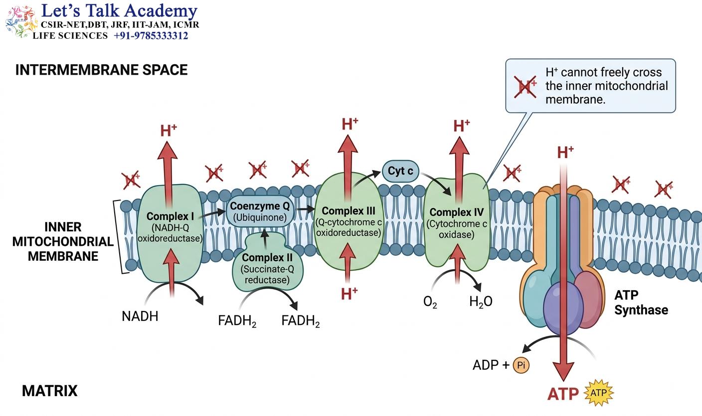
The inner mitochondrial membrane is the central structural platform for oxidative phosphorylation.
It separates:
Mitochondrial matrix ↔ Intermembrane space
and allows the cell to maintain a difference in:
- Proton concentration
- Electrical charge
- Chemical potential
4.2 Electron Transport Components
The inner membrane contains the major respiratory complexes:
Complex I → Complex II → Complex III → Complex IV
It also contains ATP synthase.
4.3 Proton Impermeability
The inner mitochondrial membrane is highly impermeable to H⁺.
This is essential because the ETC pumps protons from the matrix into the intermembrane space.
If H⁺ could freely cross the membrane, the gradient would rapidly disappear.
Therefore:
Low proton permeability → Proton gradient maintained → ATP synthesis possible
4.4 Membrane Potential
Because protons are pumped out of the matrix, the matrix becomes relatively negative.
The intermembrane space becomes relatively positive.
Therefore, an electrical gradient develops across the inner membrane.
4.5 Cardiolipin
The inner mitochondrial membrane is rich in the distinctive phospholipid:
Cardiolipin
Cardiolipin contributes to:
- Membrane organization
- Stability of respiratory complexes
- Mitochondrial membrane function
- Formation and organization of cristae
4.6 Transport Proteins
The inner mitochondrial membrane also contains transport systems required for movement of:
- ADP
- ATP
- Phosphate
- Pyruvate
- Metabolic intermediates
- Other ions and metabolites
Therefore, ATP production depends not only on ETC and ATP synthase but also on proper mitochondrial transport.
5. Electron Transport Chain
5.1 Definition
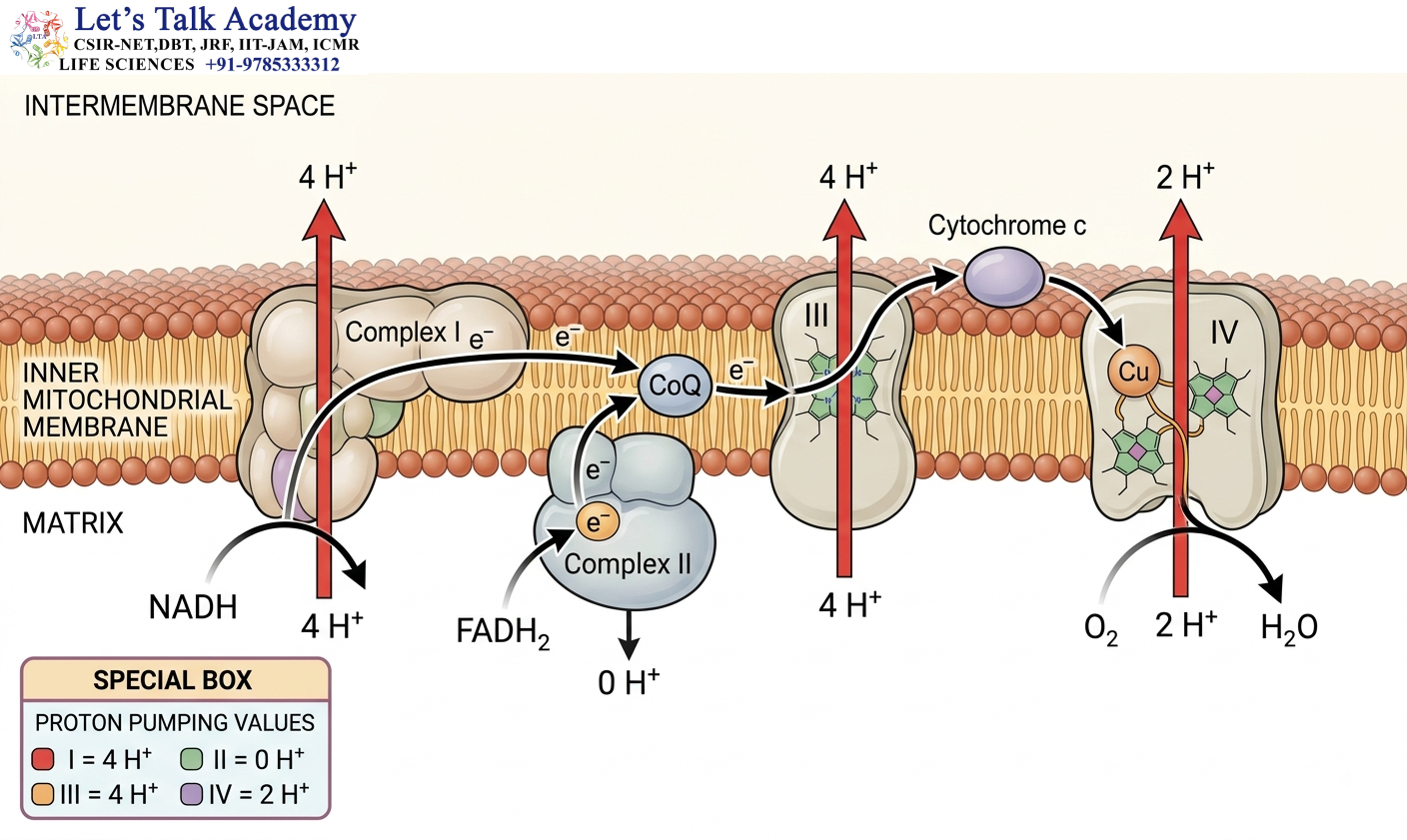
The electron transport chain (ETC) is a series of protein complexes and electron carriers that transfer electrons from reduced metabolic cofactors to oxygen.
In mitochondria, the ETC is located in the:
Inner mitochondrial membrane
5.2 Major ETC Complexes
The mitochondrial ETC contains four classical complexes:
- Complex I — NADH:ubiquinone oxidoreductase
- Complex II — Succinate dehydrogenase
- Complex III — Cytochrome bc₁ complex
- Complex IV — Cytochrome c oxidase
5.3 Mobile Electron Carriers
Two important mobile carriers connect the complexes:
- Coenzyme Q
- Cytochrome c
5.4 Electron Flow from NADH
The simplified pathway is:
NADH → Complex I → CoQ → Complex III → Cytochrome c → Complex IV → O₂
5.5 Electron Flow from FADH₂
The simplified pathway is:
FADH₂-linked electrons → Complex II → CoQ → Complex III → Cytochrome c → Complex IV → O₂
5.6 Proton-Pumping Pattern
The major proton-pumping complexes are:
Complex I → Pumps H⁺
Complex II → Does not pump H⁺
Complex III → Pumps H⁺
Complex IV → Pumps H⁺
A useful numerical pattern is:
4 – 0 – 4 – 2
for:
Complex I – II – III – IV
5.7 Total Proton Translocation
For one pair of electrons entering through Complex I:
Complex I = 4 H⁺
Complex III = 4 H⁺
Complex IV = 2 H⁺
Total:
Approximately 10 H⁺
For electrons entering through Complex II:
Complex II = 0 H⁺
Complex III = 4 H⁺
Complex IV = 2 H⁺
Total:
Approximately 6 H⁺
5.8 Central Principle of the ETC
The ETC performs two major functions:
1. Electron transfer
2. Energy conservation through proton pumping
Therefore:
Electron transfer → Energy release → Proton pumping
6. Complex I
6.1 Name of Complex I
Complex I is known as:
NADH:Ubiquinone Oxidoreductase
It is also called:
NADH dehydrogenase
6.2 Function
Complex I accepts electrons from:
NADH
and transfers them to:
Coenzyme Q
The overall reaction can be represented as:
NADH + H⁺ + CoQ → NAD⁺ + CoQH₂
6.3 Electron Entry
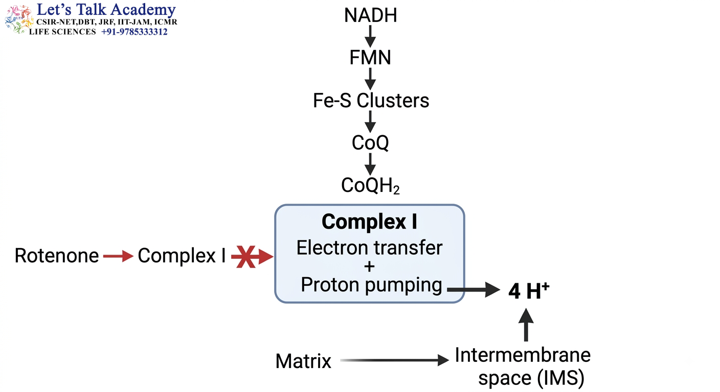
NADH transfers electrons to the flavin cofactor:
FMN — Flavin Mononucleotide
Electrons are then transferred through a series of:
Iron-sulfur (Fe-S) centers
Finally, they reach CoQ.
6.4 Proton Pumping
Complex I uses energy released during electron transfer to pump approximately:
4 H⁺ per pair of electrons
from:
Matrix → Intermembrane space
6.5 Important Components
Complex I contains:
- FMN
- Fe-S clusters
- Membrane-associated subunits
6.6 Functional Significance
Complex I is an important entry point for NADH-derived electrons.
Because it pumps protons, NADH-derived electrons contribute strongly to the proton gradient.
NOTE:- Complex I accepts electrons from NADH, transfers them to CoQ through FMN and Fe-S centers, and pumps approximately four protons per pair of electrons.
6.7 Complex I Inhibitor
An important inhibitor of Complex I is: Rotenone
7. Complex II
7.1 Name
Complex II is known as: Succinate Dehydrogenase
7.2 Dual Role
Complex II is unique because it participates in both: Citric acid cycle and Electron transport chain
7.3 Reaction in the Citric Acid Cycle
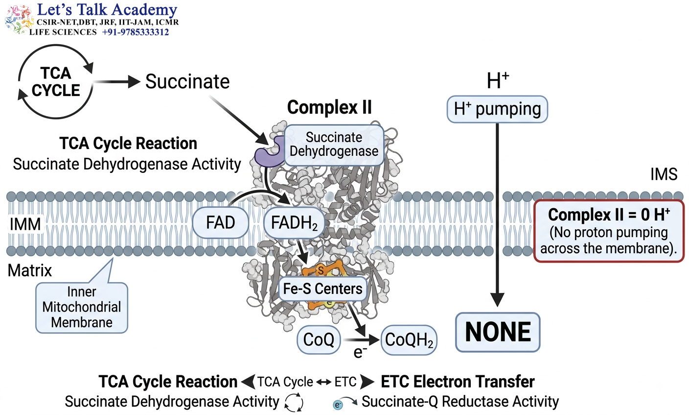
Complex II catalyzes:
Succinate → Fumarate
During this reaction:
FAD → FADH₂
The electrons are subsequently transferred through Fe-S centers toward CoQ.
7.4 Electron Flow
The simplified pathway is:
Succinate → FAD → Fe-S centers → CoQ
7.5 Proton Pumping
Complex II:
Does not pump protons.
Therefore:
Complex II = 0 H⁺ pumped
7.6 Why Is Complex II Important?
Although Complex II does not pump protons, it provides an important route for electrons to enter the ETC.
It connects the citric acid cycle directly with the respiratory chain.
NOTE:- Complex II is both a citric acid cycle enzyme and an ETC complex, but it does not contribute directly to proton pumping.
7.7 Comparison with Complex I
Complex I:
NADH → CoQ + proton pumping
Complex II:
Succinate/FAD-linked electrons → CoQ + no proton pumping
8. Coenzyme Q
8.1 Other Names
Coenzyme Q is also called:
- Ubiquinone
- CoQ
- UQ
8.2 Nature of CoQ
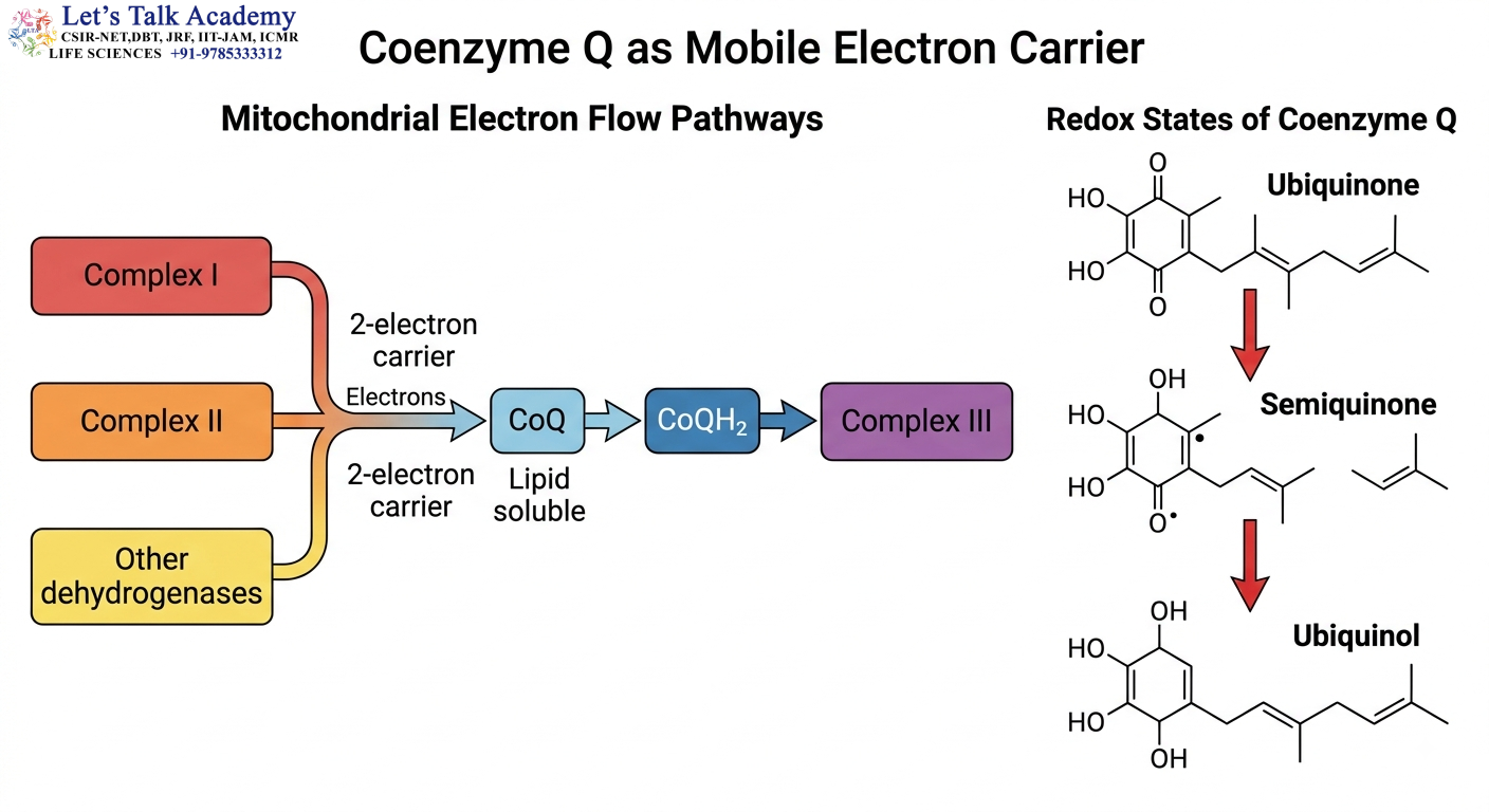
CoQ is a:
Lipid-soluble electron carrier
Because of its hydrophobic nature, it can move within the lipid environment of the inner mitochondrial membrane.
8.3 Function
CoQ receives electrons from:
- Complex I
- Complex II
- Other mitochondrial dehydrogenases
It transfers them toward:
Complex III
8.4 Redox States
CoQ can exist in several redox states:
Ubiquinone (Q)
↓
Semiquinone
↓
Ubiquinol (QH₂)
8.5 Electron-Carrying Capacity
Reduced CoQ, or QH₂, can carry:
2 electrons
and associated protons.
This is important because cytochrome c carries only one electron.
8.6 CoQ as a Mobile Electron Pool
CoQ acts as a central collection point for electrons from several pathways.
Therefore:
Complex I + Complex II + other dehydrogenases → CoQ → Complex III
9. Complex III
9.1 Name
Complex III is called:
Cytochrome bc₁ Complex
It is also known as:
Ubiquinol:cytochrome c oxidoreductase
9.2 Function
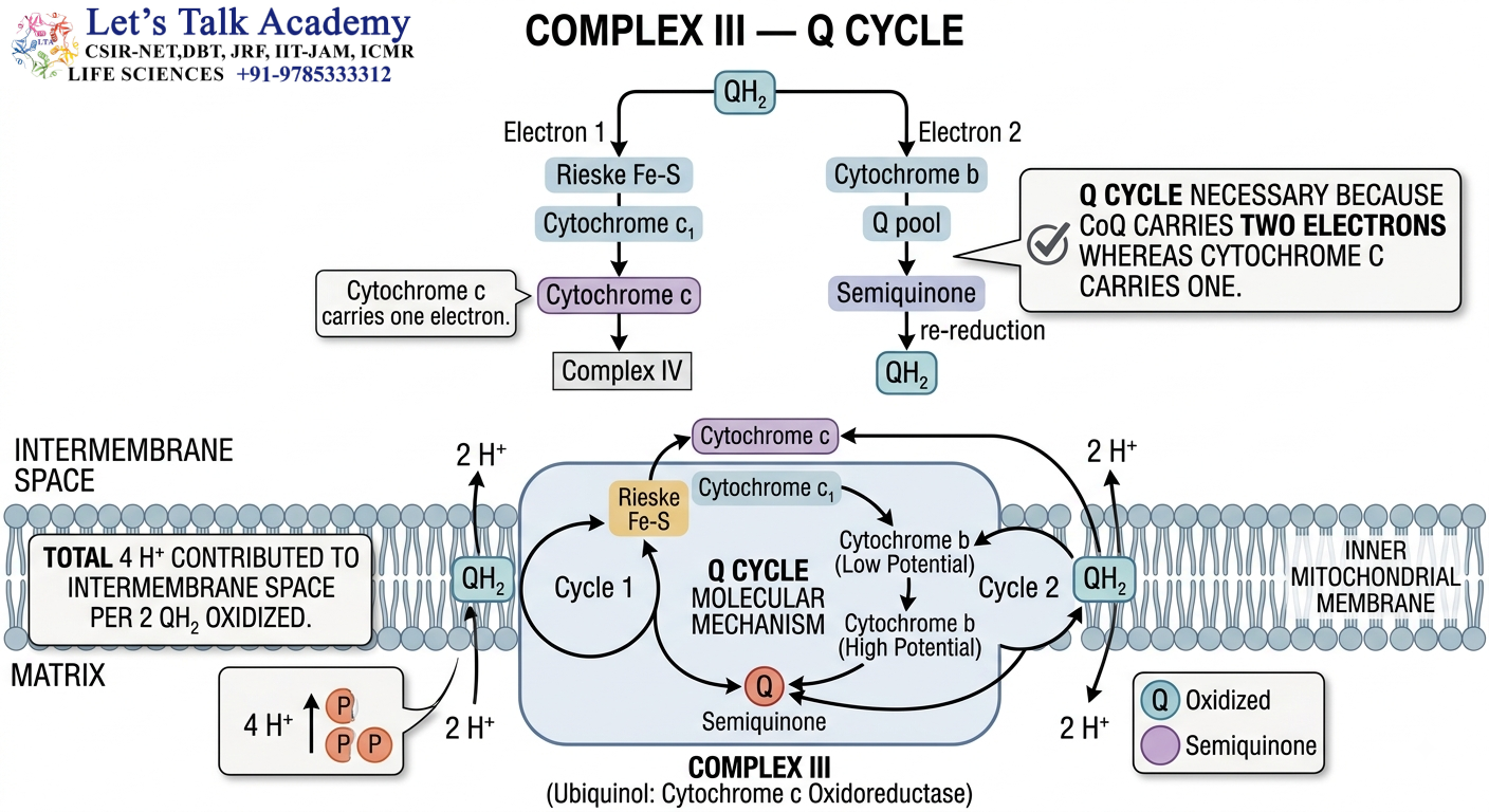
Complex III transfers electrons from:
QH₂ → Cytochrome c
9.3 Major Components
Important components include:
- Cytochrome b
- Cytochrome c₁
- Rieske Fe-S protein
9.4 The Q Cycle
Complex III functions through the:
Q cycle
The Q cycle is important because:
CoQ carries 2 electrons
whereas:
Cytochrome c carries 1 electron
The Q cycle solves this electron-transfer problem by allowing the two electrons from QH₂ to follow different routes.
9.5 Semiquinone Intermediate
The Q cycle involves the formation of a:
Semiquinone intermediate
This allows stepwise electron transfer.
9.6 Proton Translocation
The overall operation of Complex III contributes approximately:
4 H⁺ per pair of electrons
to the proton gradient.
10. Cytochrome c
10.1 Nature of Cytochrome c
Cytochrome c is a small:
Heme-containing protein
associated with the outer surface of the inner mitochondrial membrane.
It is located on the:
Intermembrane-space side
10.2 Function in ETC
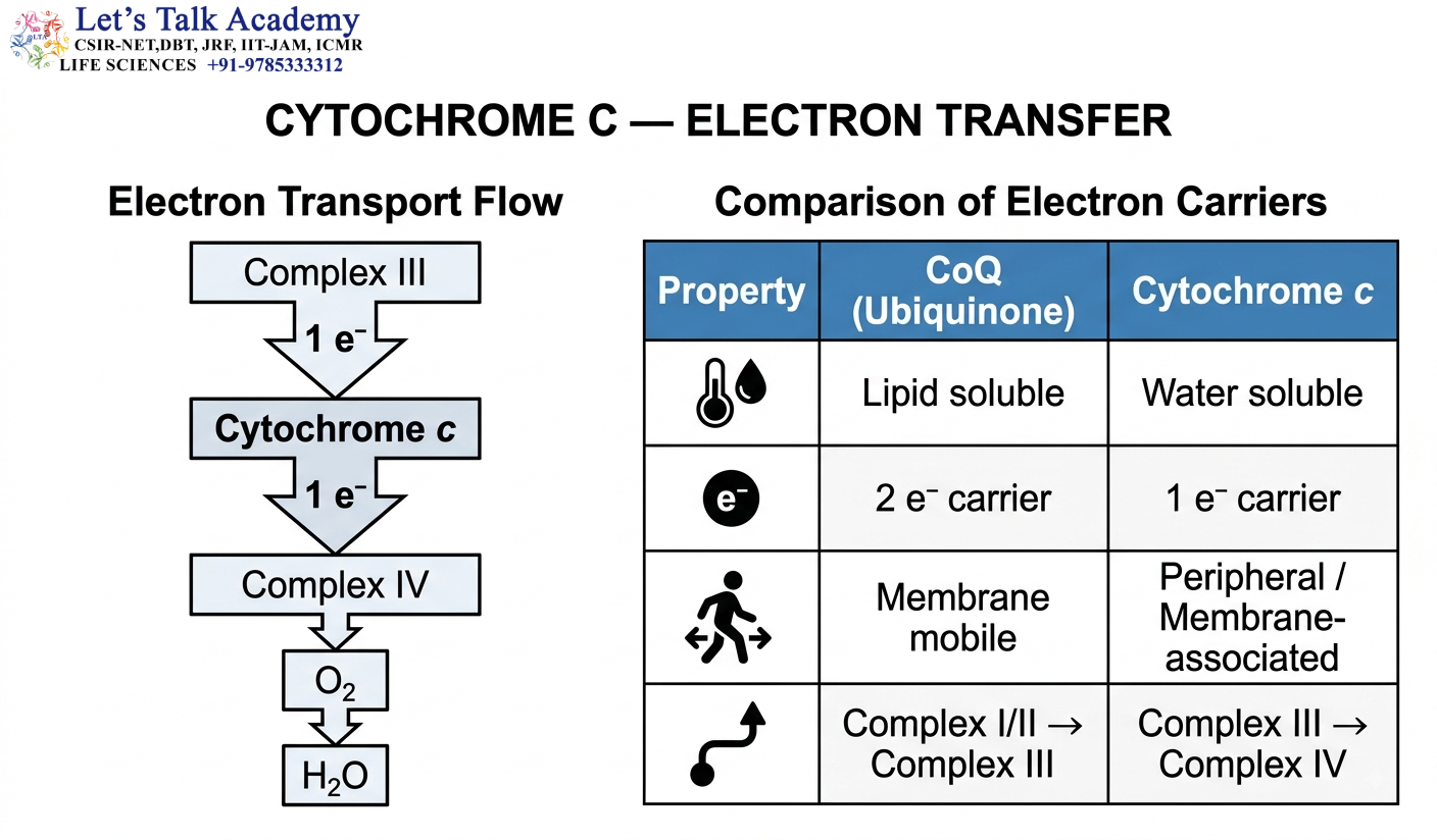
Cytochrome c transfers electrons from:
Complex III → Complex IV
10.3 Number of Electrons
Cytochrome c transfers:
One electron at a time
This is an important distinction from CoQ.
10.4 CoQ versus Cytochrome c
CoQ:
- Lipid soluble
- Membrane mobile
- Two-electron carrier
Cytochrome c:
- Water soluble
- Peripheral membrane-associated
- One-electron carrier
10.5 Role in Apoptosis
Cytochrome c has another major biological function.
During intrinsic apoptosis, mitochondrial outer membrane permeabilization can cause cytochrome c release into the cytosol.
Cytochrome c then contributes to:
Apoptosome formation
which promotes activation of:
Caspase-9
and subsequent apoptotic signaling.
11. Complex IV
11.1 Name
Complex IV is called:
Cytochrome c Oxidase
It is the terminal complex of the mitochondrial electron transport chain.
11.2 Function
Complex IV accepts electrons from:
Cytochrome c
and transfers them to:
Oxygen
11.3 Oxygen Reduction
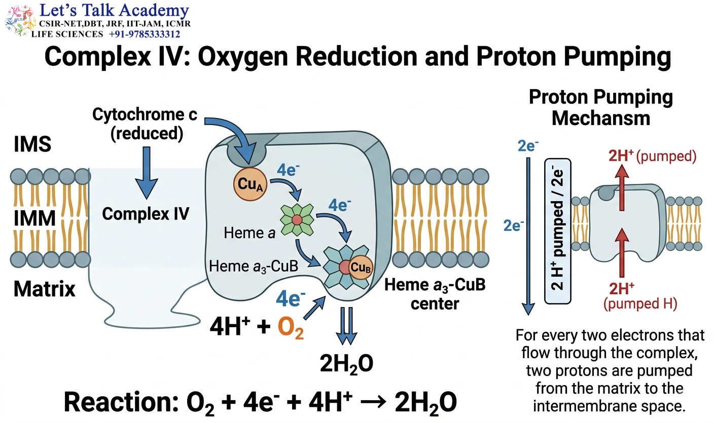
The overall reaction is:
O₂ + 4e⁻ + 4H⁺ → 2H₂O
Therefore, oxygen is reduced to water.
11.4 Major Metal Centers
Complex IV contains:
- CuA
- Heme a
- Heme a₃
- CuB
The heme a₃-CuB center is particularly important in oxygen reduction.
11.5 Proton Pumping
Complex IV pumps approximately:
2 H⁺ per pair of electrons
into the intermembrane space.
Additional matrix protons are chemically consumed during water formation.
11.6 Importance
Complex IV is essential because it provides the final route for electrons to leave the ETC.
If Complex IV is blocked:
Electron accumulation occurs
↓
Electron transport decreases
↓
Proton pumping decreases
↓
ATP synthesis decreases
12. Oxygen as Terminal Electron Acceptor
12.1 Why Oxygen Is Required
Oxygen is the final electron acceptor of aerobic mitochondrial respiration.
The final reaction is:
O₂ + 4e⁻ + 4H⁺ → 2H₂O
12.2 Oxygen Does Not Directly Produce ATP

Oxygen does not directly phosphorylate ADP.
Instead:
O₂ accepts electrons
↓
Electron transport continues
↓
Proton pumping continues
↓
Proton-motive force is maintained
↓
ATP synthase produces ATP
12.3 Consequences of Oxygen Absence
If oxygen is unavailable:
- Complex IV cannot transfer electrons to oxygen.
- Electron flow through the ETC becomes blocked.
- NADH oxidation decreases.
- NAD⁺ regeneration decreases.
- Proton pumping decreases.
- Oxidative phosphorylation decreases.
12.4 Important Statement
Oxygen is essential for oxidative phosphorylation because it acts as the terminal electron acceptor, not because it directly supplies energy to ATP synthase.
13. Proton Pumping
13.1 Meaning
Proton pumping refers to the movement of H⁺ from the mitochondrial matrix into the intermembrane space.
This movement is driven by energy released during electron transfer.
13.2 Direction
Matrix → Intermembrane space
13.3 Complexes Responsible
Only three classical ETC complexes pump protons:
Complex I
Complex III
Complex IV
Complex II does not.
13.4 Proton-Pumping Values
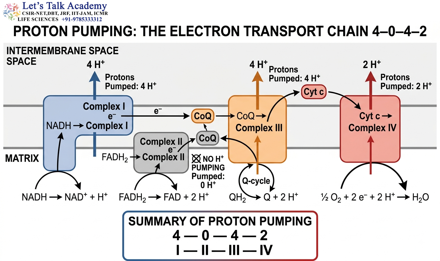
Complex |
Approximate H⁺/2e⁻ |
|---|---|
| Complex I | 4 |
| Complex II | 0 |
| Complex III | 4 |
| Complex IV | 2 |
13.5 Total for NADH
4 + 4 + 2 = 10 H⁺
13.6 Total for FADH₂-Linked Electrons
0 + 4 + 2 = 6 H⁺
13.7 Importance
Proton pumping converts:
Redox energy → Electrochemical energy
The resulting gradient is then used by ATP synthase.
14. Proton-Motive Force
14.1 Definition
The proton-motive force (PMF) is the electrochemical potential difference for protons across the inner mitochondrial membrane.
It is generated by proton pumping.
14.2 Components
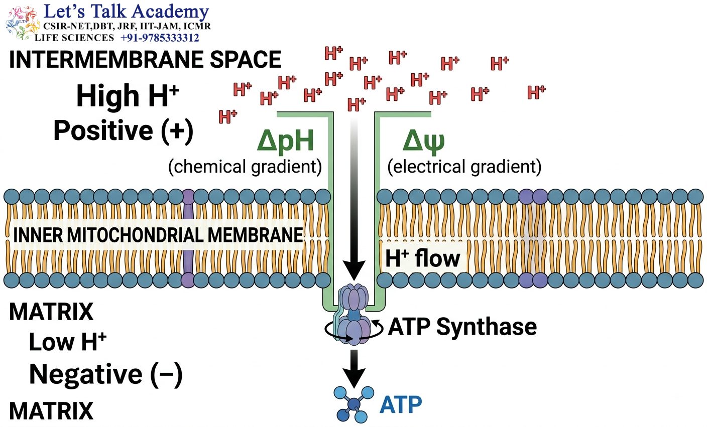
The PMF has two major components:
- Electrical gradient (Δψ)
- Chemical gradient (ΔpH)
14.3 Electrical Gradient
As positive protons are pumped out of the matrix:
Matrix becomes relatively negative
Intermembrane space becomes relatively positive
This creates a membrane potential.
14.4 Chemical Gradient
Because H⁺ accumulates in the intermembrane space:
Intermembrane space → Higher H⁺ concentration
Matrix → Lower H⁺ concentration
Therefore, a pH gradient develops.
14.5 Proton Movement
The electrochemical gradient favors movement of H⁺:
Intermembrane space → Matrix
ATP synthase provides the major controlled pathway for this movement.
14.6 Energy Stored in PMF
The proton-motive force represents stored electrochemical energy.
This energy is converted into ATP through ATP synthase.
15. Chemiosmotic Theory
15.1 Definition
The chemiosmotic theory explains how electron transport is coupled to ATP synthesis through an electrochemical proton gradient.
15.2 Proposed By
The theory was proposed by:
Peter Mitchell
15.3 Basic Mechanism
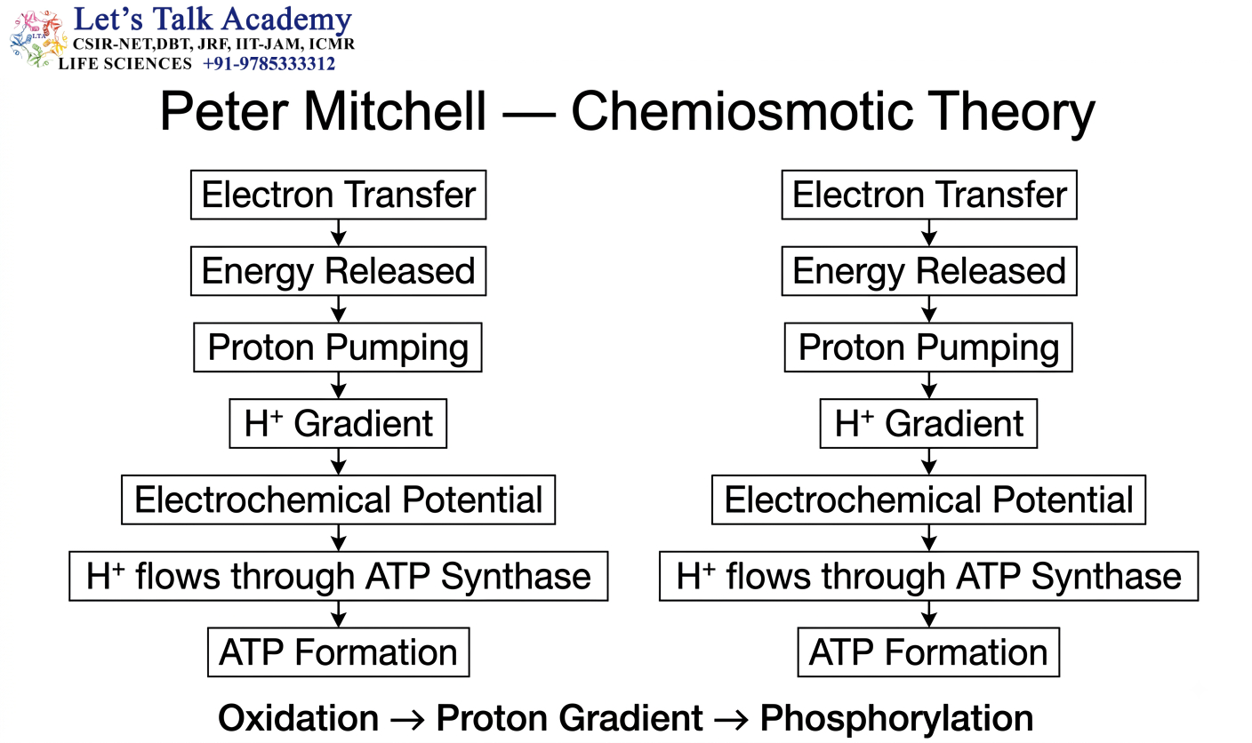
According to the chemiosmotic theory:
- Electrons move through the ETC.
- Energy released during electron transfer is used to pump protons.
- A proton gradient develops.
- The inner membrane stores this electrochemical energy.
- Protons flow back through ATP synthase.
- ATP synthase uses this energy to produce ATP.
15.4 Complete Sequence
Electron transport
↓
Proton pumping
↓
Proton gradient
↓
Proton-motive force
↓
Proton flow through ATP synthase
↓
ATP synthesis
15.5 Importance of Membrane Integrity
Chemiosmosis requires an intact membrane.
If the membrane becomes highly permeable to protons:
Proton gradient collapses and: ATP synthesis decreases
15.6 Importance in Bioenergetics
Chemiosmosis provides the mechanistic link between:
Oxidation and Phosphorylation
Therefore:
Oxidation → Proton gradient → Phosphorylation
16. Peter Mitchell
16.1 Contribution
Peter Mitchell proposed the chemiosmotic theory of oxidative phosphorylation.
His work established the concept that biological membranes can conserve energy through electrochemical gradients.
16.2 Nobel Prize
Peter Mitchell received the:
Nobel Prize in Chemistry in 1978
for his contribution to the understanding of biological energy transfer.
Peter Mitchell → Chemiosmotic theory
This should be directly remembered for examination purposes.
17. ATP Synthase
17.1 Definition
ATP synthase is a membrane-associated molecular enzyme complex that synthesizes ATP using the energy of the proton-motive force.
It is also called:
F₀F₁-ATP synthase

17.2 Location
ATP synthase is located in the:
Inner mitochondrial membrane
Its catalytic portion faces the:
Mitochondrial matrix
17.3 Overall Reaction
ADP + Pi → ATP
17.4 Energy Source
ATP synthesis is powered by:
Proton movement down the electrochemical gradient
Thus:
PMF → ATP synthase → ATP
17.5 ATP Synthase as a Rotary Machine
ATP synthase is a remarkable molecular motor.
It converts:
Electrochemical energy
into:
Rotational energy
which is then converted into:
Chemical energy stored in ATP
17.6 Major Components
ATP synthase consists of:
- F₀ component
- F₁ component
- Central stalk
- Peripheral stalk
- Rotary ring components
18. F₀ and F₁ Components
18.1 F₀ Component
F₀ is the membrane-embedded portion of ATP synthase.
Its major role is:
Proton translocation and rotation
It contains membrane-associated components including the:
c-ring
18.2 Proton Movement Through F₀
Protons move through the membrane-associated pathway.
This causes:
c-ring rotation
The rotational movement is transmitted to the central stalk.
18.3 F₁ Component
F₁ projects toward the mitochondrial matrix.
It contains the major catalytic sites responsible for ATP synthesis.
Its principal catalytic arrangement includes:
α₃β₃
The β subunits contain the principal catalytic sites.
18.4 Functional Difference
F₀ → Proton-conducting and rotary component
F₁ → Catalytic ATP-forming component
19. Binding-Change Mechanism
19.1 Concept
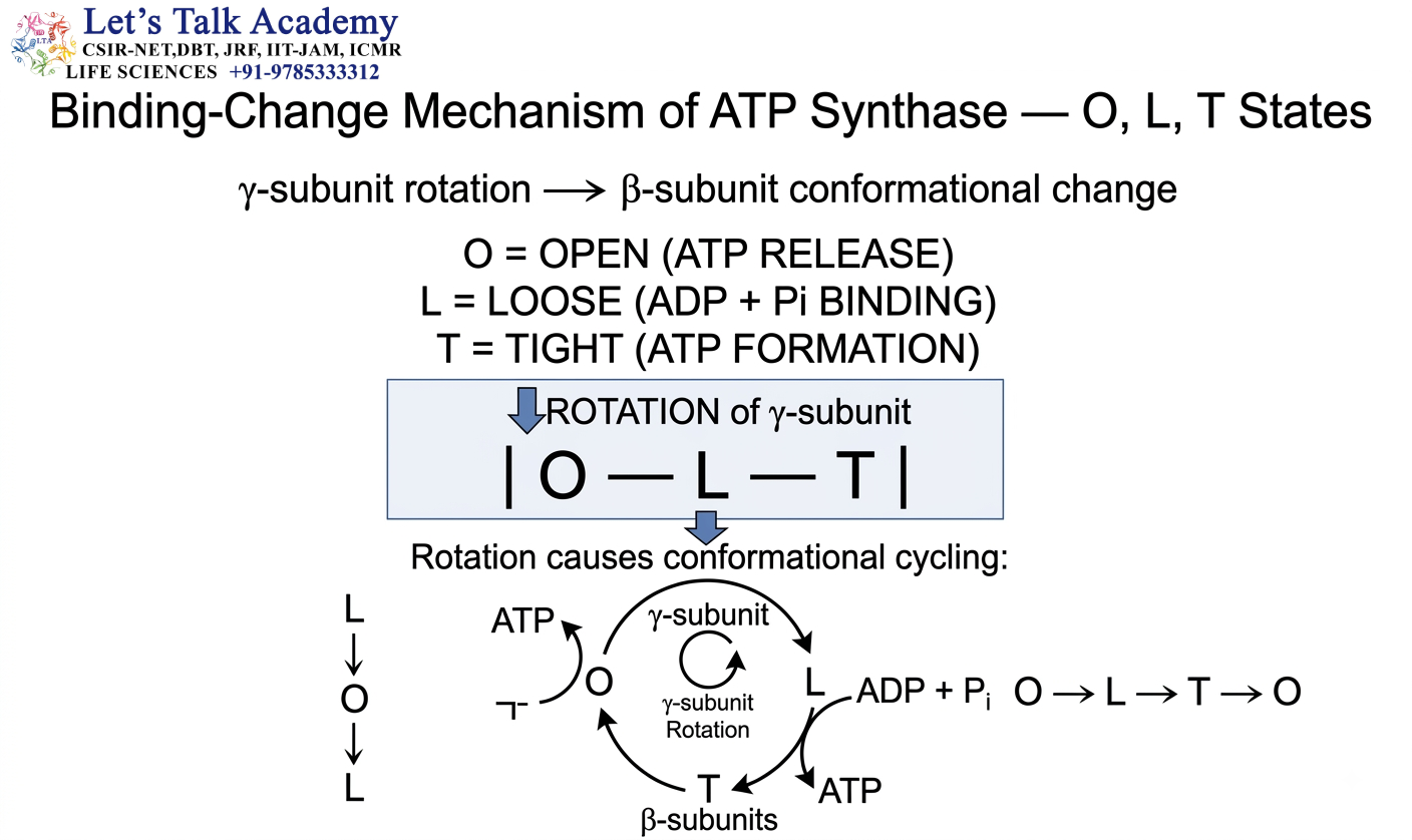
The binding-change mechanism explains how ATP synthase produces ATP through conformational changes in its catalytic β subunits.
The mechanism is strongly associated with:
Paul Boyer
19.2 Three Conformational States
The β subunits cycle through three major states:
- O = Open
- L = Loose
- T = Tight
19.3 Open State
The O state has low affinity for nucleotides and facilitates ATP release.
19.4 Loose State
The L state binds:
ADP + Pi
19.5 Tight State
The T state binds substrates tightly and favors ATP formation.
19.6 Role of Rotation
Rotation of the central γ subunit causes conformational changes in the β subunits.
These changes alter their affinity for:
- ADP
- Pi
- ATP
19.7 Simplified Cycle
Loose → Tight → Open → Loose
The process can be summarized as:
ADP + Pi binding
↓
ATP formation
↓
ATP release
The proton gradient does not directly provide a phosphate group to ADP. It drives ATP synthase rotation, which produces conformational changes required for ATP synthesis and release.
20. NADH vs FADH₂

20.1 NADH
NADH transfers electrons to:
Complex I
Therefore:
NADH → Complex I → CoQ → III → Cytochrome c → IV
Approximately: 10 H⁺
are translocated per pair of electrons.
Modern approximate ATP yield:
1 NADH ≈ 2.5 ATP
20.2 FADH₂
FADH₂-linked electrons enter through:
Complex II
Therefore:
FADH₂ → Complex II → CoQ → III → Cytochrome c → IV
Approximately: 6 H⁺
are translocated per pair of electrons.
Modern approximate ATP yield:
1 FADH₂ ≈ 1.5 ATP
20.3 Reason for Difference
The major reason for the lower ATP yield from FADH₂ is:
FADH₂-linked electrons bypass Complex I.
Therefore, the proton-pumping contribution of Complex I is lost.
20.4 Comparison
Feature |
NADH |
FADH₂ |
|---|---|---|
| Entry | Complex I | Complex II |
| Complex I used | Yes | No |
| Approx. H⁺ | 10 | 6 |
| ATP yield | ~2.5 | ~1.5 |
21. ATP Yield
21.1 Modern ATP Values
Modern biochemical estimates are:
1 NADH ≈ 2.5 ATP
1 FADH₂ ≈ 1.5 ATP
21.2 Older ATP Values
Older textbooks commonly used:
NADH = 3 ATP
FADH₂ = 2 ATP
These values were based on simplified estimates.
Modern values are generally preferred because they account more realistically for proton translocation and ATP synthesis.
21.3 Why ATP Yield Is Not an Exact Whole Number
The ATP yield is an approximation because ATP synthesis depends on:
- Proton-pumping stoichiometry
- ATP synthase proton requirements
- ADP/ATP transport
- Phosphate transport
- Shuttle systems
- Membrane leak
- Cellular conditions
22. P/O Ratio
22.1 Definition
The P/O ratio describes the relationship between:
Phosphorylation and Oxygen reduction
during oxidative phosphorylation.
It is commonly interpreted as the approximate number of ATP molecules generated per oxygen atom reduced under specified conditions.
22.2 Approximate Values
Modern approximate values are:
NADH → P/O ≈ 2.5
FADH₂ → P/O ≈ 1.5
22.3 Significance
The P/O ratio provides an estimate of the efficiency with which respiratory electron transfer is coupled to ATP synthesis.
22.4 Important Statement
A decrease in coupling efficiency, such as during uncoupling, can reduce ATP production relative to electron transport and oxygen consumption.
23. Shuttle Systems

23.1 Why Shuttle Systems Are Required
Glycolysis occurs in the: Cytosol and generates: NADH
However, the inner mitochondrial membrane is not freely permeable to NADH itself.
Therefore, the reducing equivalents of cytosolic NADH must be transferred into mitochondria through shuttle systems.
The major systems are:
- Malate-aspartate shuttle
- Glycerol-3-phosphate shuttle
24. Malate-Aspartate Shuttle
24.1 Basic Principle
The malate-aspartate shuttle transfers the reducing equivalents of cytosolic NADH into the mitochondrial matrix.
The reducing equivalents are ultimately transferred to mitochondrial NAD⁺ to produce:
Mitochondrial NADH
24.2 ETC Entry
The mitochondrial NADH generated by this shuttle transfers electrons to:
Complex I
Therefore, the reducing equivalents yield approximately:
2.5 ATP
24.3 Important Tissues
This shuttle is particularly important in:
- Liver
- Heart
- Kidney
24.4 Significance
The malate-aspartate shuttle is relatively efficient because the reducing equivalents enter the ETC at the NADH/Complex I level.
25. Glycerol-3-Phosphate Shuttle
25.1 Basic Principle
The glycerol-3-phosphate shuttle transfers reducing equivalents from cytosolic NADH to a mitochondrial FAD-linked pathway.
The electrons are subsequently transferred to:
CoQ
25.2 ETC Entry
Because these electrons enter at the level of the CoQ pool, they bypass Complex I.
Therefore, their approximate yield is:
1.5 ATP per cytosolic NADH equivalent
25.3 Comparison with Malate-Aspartate Shuttle
Malate-aspartate → Complex I level → ~2.5 ATP
Glycerol-3-phosphate → CoQ level → ~1.5 ATP
26. Respiratory Control
26.1 Definition
Respiratory control refers to the regulation of mitochondrial respiration according to the cellular demand for ATP.
A major controlling factor is:
ADP availability
26.2 High ADP Condition
When ATP is being consumed rapidly:
ATP ↓
↓
ADP ↑
↓
ATP synthase activity ↑
↓
Proton gradient is utilized
↓
Electron transport ↑
↓
O₂ consumption ↑
26.3 Low ADP Condition
When ATP demand is low:
ADP ↓
↓
ATP synthesis ↓
↓
Proton gradient builds up
↓
Electron transport slows
↓
O₂ consumption decreases
26.4 Acceptor Control
Because ADP is an acceptor of phosphate during ATP synthesis, this form of regulation is also called:
Acceptor control
26.5 Other Factors
Respiratory activity can also be influenced by:
- Oxygen concentration
- ADP concentration
- ATP/ADP ratio
- NADH/NAD⁺ ratio
- Substrate availability
- Pi availability
- Membrane potential
- Proton gradient
- Mitochondrial integrity
27. Uncoupling
27.1 Definition
Uncoupling occurs when electron transport becomes separated from ATP synthesis.
Under normal conditions:
Electron transport → Proton gradient → ATP synthesis
During uncoupling: Electron transport continues but ATP synthesis is reduced
because the proton gradient is dissipated without efficiently passing through ATP synthase.
27.2 Mechanism
An uncoupler provides an alternative pathway for protons to return to the matrix.
Normally:
Intermembrane space → ATP synthase → Matrix
During uncoupling:
Intermembrane space → Uncoupling pathway → Matrix
27.3 Consequences
Uncoupling generally causes:
- Proton gradient ↓
- ATP synthesis ↓
- Heat production ↑
- Substrate oxidation ↑
- Oxygen consumption may increase
28. UCP1
28.1 Full Name
UCP1 = Uncoupling Protein 1
28.2 Location
UCP1 is predominantly found in:
Brown adipose tissue
28.3 Function
UCP1 provides a pathway for proton re-entry into the mitochondrial matrix.
Instead of converting the proton gradient primarily into ATP, the energy is released as:
Heat
28.4 Non-Shivering Thermogenesis
UCP1 is therefore important for:
Non-shivering thermogenesis
This is particularly important in:
- Newborn mammals
- Cold exposure
- Thermoregulation
28.5 Mechanism
Fatty acid oxidation
↓
NADH/FADH₂ production
↓
ETC
↓
Proton gradient
↓
UCP1-mediated proton leak
↓
Heat production
28.6 ATP Synthase versus UCP1
ATP synthase:
PMF → ATP
UCP1:
PMF → Heat
29. ETC Inhibitors
29.1 Importance of ETC Inhibitors
ETC inhibitors are useful for understanding the organization and function of the respiratory chain.
By blocking a specific component, researchers can determine:
- Where electrons normally flow
- Which complexes pump protons
- How oxygen consumption changes
- How ATP synthesis depends on electron transport
Important inhibitors include:
- Rotenone
- Antimycin A
- Cyanide
- Carbon monoxide
- Oligomycin
30. Rotenone
30.1 Site of Action
Rotenone inhibits:
Complex I
30.2 Effect
It interferes with electron transfer from Complex I toward CoQ.
Therefore:
NADH-derived electron transport ↓
Electron pathways entering the ETC downstream of Complex I may remain functional if they are not otherwise inhibited.
31. Antimycin A
31.1 Site of Action
Antimycin A inhibits:
Complex III
31.2 Effect
It interferes with electron transfer through the cytochrome bc₁ complex.
Therefore:
CoQ → Cytochrome c electron transfer is inhibited.
31.3 ROS
Complex III inhibition can promote electron leakage and increase ROS formation under appropriate conditions.
32. Cyanide
32.1 Site of Action
Cyanide inhibits:
Complex IV
32.2 Mechanism
It interferes with the oxygen-reducing function of cytochrome c oxidase.
Therefore:
Electron transfer to O₂ is blocked.
32.3 Consequences
- Oxygen utilization decreases.
- Electron transport stops.
- Proton pumping decreases.
- Oxidative phosphorylation is severely inhibited.
Cyanide inhibits cellular oxygen utilization by blocking cytochrome c oxidase rather than by removing oxygen from the environment.
33. Carbon Monoxide
33.1 Site of Action
Carbon monoxide can inhibit:
Complex IV
33.2 Mechanism
It can interact with the oxygen-binding region of cytochrome c oxidase and interfere with oxygen utilization.
33.3 Importance
Because Complex IV is responsible for terminal electron transfer to oxygen, its inhibition severely affects oxidative phosphorylation.
34. Oligomycin
34.1 Site of Action
Oligomycin inhibits:
ATP synthase
Specifically, it interferes with the proton-conducting:
F₀ component
34.2 Effect on ATP
Because proton flow through ATP synthase decreases:
ATP synthesis decreases
34.3 Effect on Electron Transport
The proton gradient becomes increasingly high because protons cannot efficiently return through ATP synthase.
This creates increased resistance to further proton pumping.
Consequently:
Electron transport decreases secondarily.
34.4 Important Comparison
Oligomycin → blocks ATP synthase
Uncoupler → bypasses ATP synthase and dissipates proton gradient
35. ETC Inhibitor
Inhibitor |
Primary Target |
Major Effect |
|---|---|---|
| Rotenone | Complex I | Blocks NADH-linked electron transfer |
| Antimycin A | Complex III | Blocks electron transfer through Complex III |
| Cyanide | Complex IV | Blocks oxygen reduction |
| Carbon monoxide | Complex IV | Interferes with oxygen utilization |
| Oligomycin | ATP synthase/F₀ | Blocks proton flow through ATP synthase |
36. ROS Generation
36.1 Meaning of ROS
ROS stands for:
Reactive Oxygen Species
ROS are oxygen-containing molecules capable of participating in highly reactive chemical reactions.
Important mitochondrial ROS include:
- Superoxide
- Hydrogen peroxide
- Hydroxyl radical
36.2 Why ROS Are Generated
Electron transport is normally tightly controlled.
However, a small fraction of electrons can escape from the normal pathway and prematurely reduce oxygen.
This can generate:
Superoxide (O₂•⁻)
36.3 Superoxide Formation
The reaction can be represented as:
O₂ + e⁻ → O₂•⁻
36.4 Major ETC Sites
Important mitochondrial sites associated with ROS generation include:
Complex I and Complex III
36.5 Hydrogen Peroxide
Superoxide can undergo dismutation to form: H₂O₂
Hydrogen peroxide is less reactive than the hydroxyl radical but can participate in further reactions.
36.6 Hydroxyl Radical
The: Hydroxyl radical (•OH) is extremely reactive and can damage biomolecules.
37. Effects of Excessive ROS
37.1 Lipid Damage
ROS can cause:
Lipid peroxidation
This can alter membrane:
- Fluidity
- Permeability
- Protein function
37.2 Protein Damage
ROS can oxidize amino acid residues and alter:
- Protein structure
- Enzyme activity
- Protein stability
37.3 DNA Damage
ROS can damage mitochondrial and nuclear DNA.
Potential effects include:
- Base oxidation
- Strand damage
- Mutations
37.4 Mitochondrial Dysfunction
Excessive ROS can cause:
- ETC dysfunction
- Membrane damage
- Loss of membrane potential
- Reduced ATP production
- Increased mitochondrial stress
37.5 Beneficial ROS
ROS are not always harmful.
Controlled ROS levels can participate in:
- Cell signaling
- Stress responses
- Immune defense
- Adaptation
- Regulation of gene expression
Therefore, the biological effect of ROS depends strongly on:
Concentration + cellular context + antioxidant capacity
38. Antioxidant Defense
38.1 Superoxide Dismutase
Superoxide dismutase converts superoxide into hydrogen peroxide.
Reaction:
2O₂•⁻ + 2H⁺ → H₂O₂ + O₂
38.2 Catalase
Catalase converts hydrogen peroxide into water and oxygen.
Reaction:
2H₂O₂ → 2H₂O + O₂
38.3 Glutathione Peroxidase
Glutathione peroxidase reduces:
- Hydrogen peroxide
- Lipid hydroperoxides
using reduced glutathione.
38.4 Redox Balance
The cell must maintain a balance between:
ROS production and Antioxidant defense
Excessive ROS relative to antioxidant capacity can result in: Oxidative stress
39. Mitochondrial Energy Transduction
39.1 Definition
Energy transduction is the conversion of energy from one form into another.
Mitochondrial oxidative phosphorylation is one of the clearest examples of energy transduction in biology.
39.2 Step 1 — Substrate Oxidation
Nutrients such as carbohydrates and fatty acids are oxidized.
This generates:
NADH + FADH₂
39.3 Step 2 — Electron Transfer
NADH and FADH₂ donate electrons to the ETC.
The electrons move toward oxygen.
39.4 Step 3 — Proton Pumping
The energy released during electron transfer is used by:
Complex I + III + IV to pump H⁺.
39.5 Step 4 — Proton-Motive Force
The proton gradient stores electrochemical energy.
39.6 Step 5 — ATP Synthase
Protons flow through ATP synthase.
This causes rotation and conformational changes.
39.7 Step 6 — ATP Formation
The energy is ultimately used to synthesize: ATP
Thus:
Nutrient energy → NADH/FADH₂ → ETC → PMF → ATP
40. Integrated Oxidative Phosphorylation Pathway
40.1 From Nutrients to ATP
The complete process begins with oxidation of metabolic substrates.
Glucose
↓
Glycolysis
↓
Pyruvate
↓
Acetyl-CoA
↓
Citric Acid Cycle
↓
NADH + FADH₂
↓
Electron Transport Chain
↓
Proton Gradient
↓
ATP Synthase
↓
ATP
40.2 NADH Electron Pathway
NADH
↓
Complex I
↓
CoQ
↓
Complex III
↓
Cytochrome c
↓
Complex IV
↓
O₂
↓
H₂O
At the same time:
Complex I + III + IV
↓
H⁺ pumping
↓
Proton-motive force
↓
ATP synthase
↓
ATP
40.3 FADH₂ Electron Pathway
FADH₂-linked electrons
↓
Complex II
↓
CoQ
↓
Complex III
↓
Cytochrome c
↓
Complex IV
↓
O₂
↓
H₂O
At the same time:
Complex III + IV
↓
H⁺ pumping
↓
Proton-motive force
↓
ATP synthase
↓
ATP
41. Integrated Proton Movement
41.1 During Electron Transport
Protons are pumped:
Matrix → Intermembrane space
This produces:
- High H⁺ concentration outside the matrix
- Positive charge outside
- Negative matrix
41.2 During ATP Synthesis
Protons move:
Intermembrane space → Matrix
through:
ATP synthase
41.3 Importance
The proton gradient therefore acts as an energy reservoir.
The ETC charges the reservoir.
ATP synthase allows controlled discharge of this stored energy.
This is a useful conceptual model for understanding chemiosmosis.
42. Integrated Relationship Between ETC and ATP Synthase
42.1 ETC Function
The ETC:
Transfers electrons
and:
Pumps protons
42.2 ATP Synthase Function
ATP synthase:
Uses proton flow
to:
Synthesize ATP
42.3 Coupling
The two processes are coupled through:
Proton-motive force
Therefore:
ETC ↔ Proton gradient ↔ ATP synthase
42.4 Important Statement
The proton-motive force is the energetic link between electron transport and ATP synthesis.
43. Oxidative Phosphorylation and Cellular Energy Demand
43.1 High Energy Demand
When the cell requires more ATP:
ATP consumption increases
↓
ADP increases
↓
ATP synthase activity increases
↓
Proton gradient is utilized
↓
Electron transport increases
↓
O₂ consumption increases
Thus, mitochondria increase respiration according to cellular energy demand.
43.2 Low Energy Demand
When ATP demand is low:
ADP decreases
↓
ATP synthesis decreases
↓
Proton gradient increases
↓
Electron transport slows
This demonstrates the principle of respiratory control.
44. Oxidative Phosphorylation and Uncoupling
44.1 Normal Condition
Under normal conditions:
Electron transport
↓
Proton gradient
↓
ATP synthase
↓
ATP
44.2 Uncoupled Condition
During uncoupling:
Electron transport
↓
Proton gradient
↓
Proton leak
↓
Heat
ATP production decreases because the proton gradient is dissipated without productive passage through ATP synthase.
44.3 Important Comparison
Feature |
Normal Oxidative Phosphorylation |
Uncoupling |
|---|---|---|
| Electron transport | Active | Active/often increased |
| Proton gradient | Maintained | Dissipated |
| ATP synthesis | High | Decreased |
| Oxygen consumption | Regulated | Often increased |
| Heat production | Lower | Increased |
45. ETC Inhibitor vs Uncoupler
45.1 ETC Inhibitor
An ETC inhibitor blocks electron transfer.
Example:
Rotenone
Result:
Electron transport ↓
Proton pumping ↓
ATP production ↓
45.2 Uncoupler
An uncoupler dissipates the proton gradient.
Result:
Proton gradient ↓
ATP synthesis ↓
but:
Electron transport may increase
45.3 Key Difference
ETC inhibitor blocks electron flow, whereas an uncoupler separates electron transport from ATP synthesis.
46. Oligomycin vs Uncoupling
46.1 Oligomycin
Oligomycin blocks:
ATP synthase/F₀
Therefore:
Proton flow through ATP synthase ↓
ATP synthesis ↓
Proton gradient ↑
Electron transport eventually ↓
46.2 Uncoupler
An uncoupler allows:
Proton flow without ATP synthesis
Therefore:
Proton gradient ↓
ATP synthesis ↓
Electron transport often ↑
47. Oxidative vs Substrate-Level Phosphorylation
47.1 Oxidative Phosphorylation
ATP formation depends on:
ETC + Proton gradient + ATP synthase
47.2 Substrate-Level Phosphorylation
ATP/GTP is formed directly by transfer of a phosphate group from a high-energy substrate.
Examples: Glycolysis
Succinyl-CoA synthetase reaction
47.3 Major Difference
Feature |
Oxidative Phosphorylation |
Substrate-Level Phosphorylation |
|---|---|---|
| ETC required | Yes | No |
| Proton gradient required | Yes | No |
| ATP synthase required | Yes | No |
| Direct phosphate transfer from substrate | No | Yes |
| Major location | Inner mitochondrial membrane | Glycolysis/TCA reactions |


