7. In mitochondria the enzymes of electron transport chain are located at (1) Outer membrane (2) Inter membrane space (3) Inner membrane (4) Matrix The electron transport chain (ETC) […]

No posts found.
7. In mitochondria the enzymes of electron transport chain are located at (1) Outer membrane (2) Inter membrane space (3) Inner membrane (4) Matrix The electron transport chain (ETC) […]
6. The site for oxidative phosphorylation in eukaryotes is (1) Inner membrane of mitochondria (2) Vacuole membrane (3) Plasma membrane (4) Thylakoid membrane of chloroplast Oxidative phosphorylation is a […]
5.In bacteria site of respiration is- (1) Mesosome only (2) Plasma membrane (3) Endoplasmic reticulum (4) Microsomes Respiration is a vital process in all living organisms, including bacteria, where […]
4. The process of photosynthesis which leads to formation of glucose from C02 and H20 is an example of (1) Oxidation (2) Reduction (3) Condensation (4) Fixation Photosynthesis is a […]
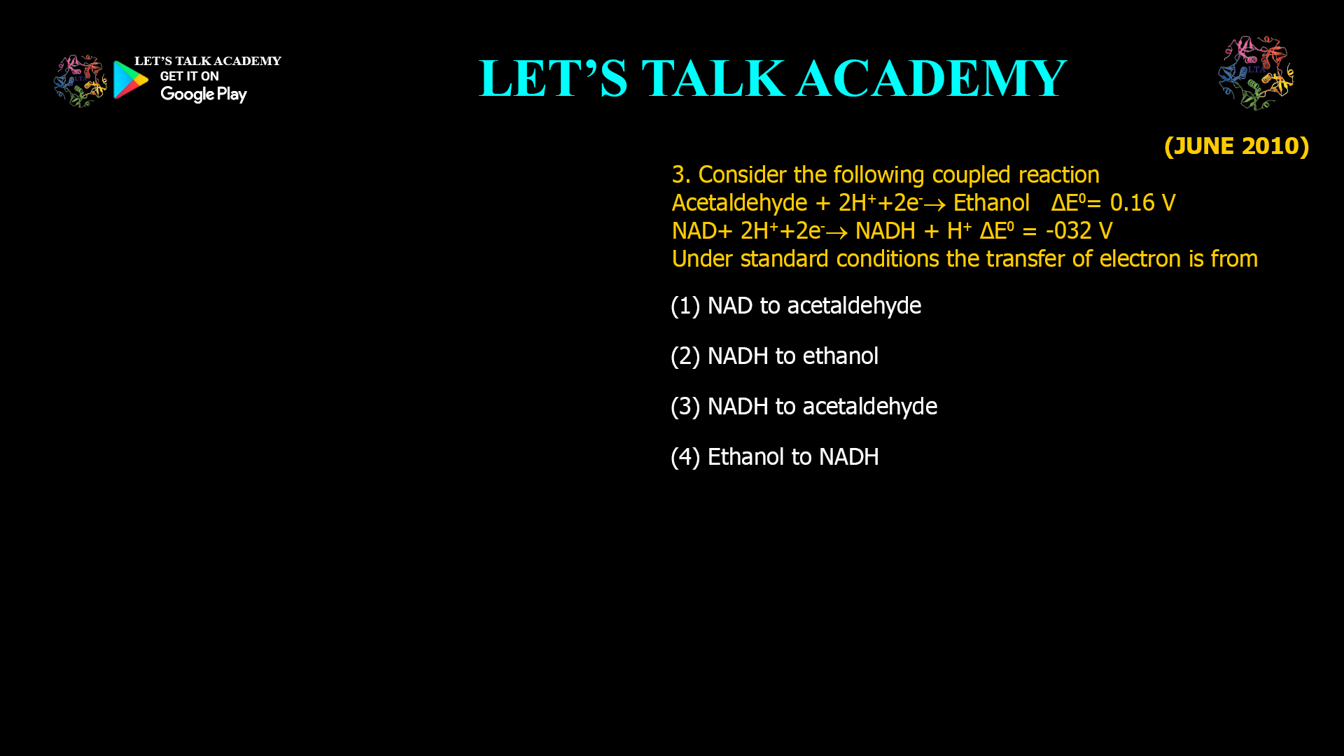
Consider the following coupled reaction Acetaldehyde + 2H++2e– à Ethanol ΔE0= 0.16 V NAD+ 2H++2e– à NADH + H+ ΔE0 = -032 V Under standard conditions the transfer of electron […]
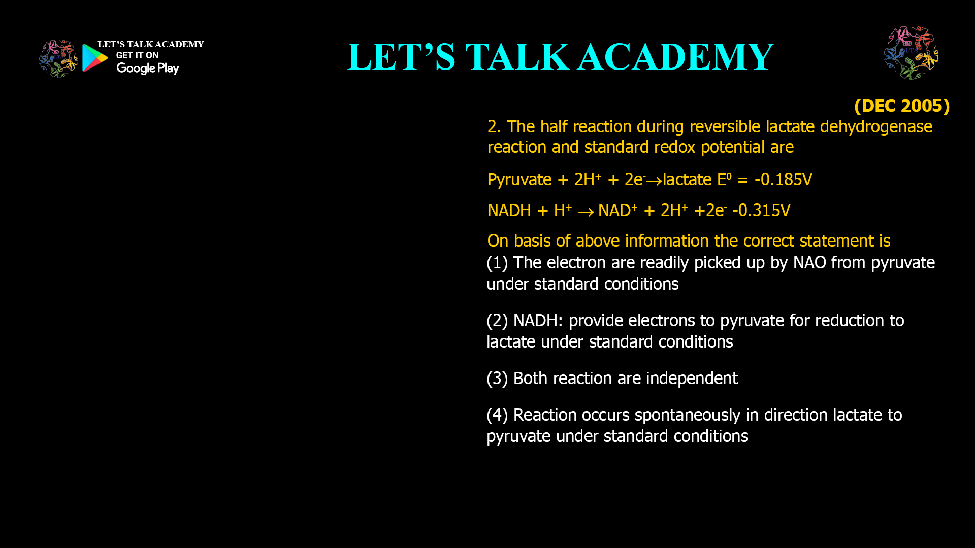
The half reaction during reversible lactate dehydrogenase reaction and standard redox potential are Pyruvate + 2H+ + 2e– à lactate E0 = -0.185V NADH + H+ à NAD+ + 2H+ […]
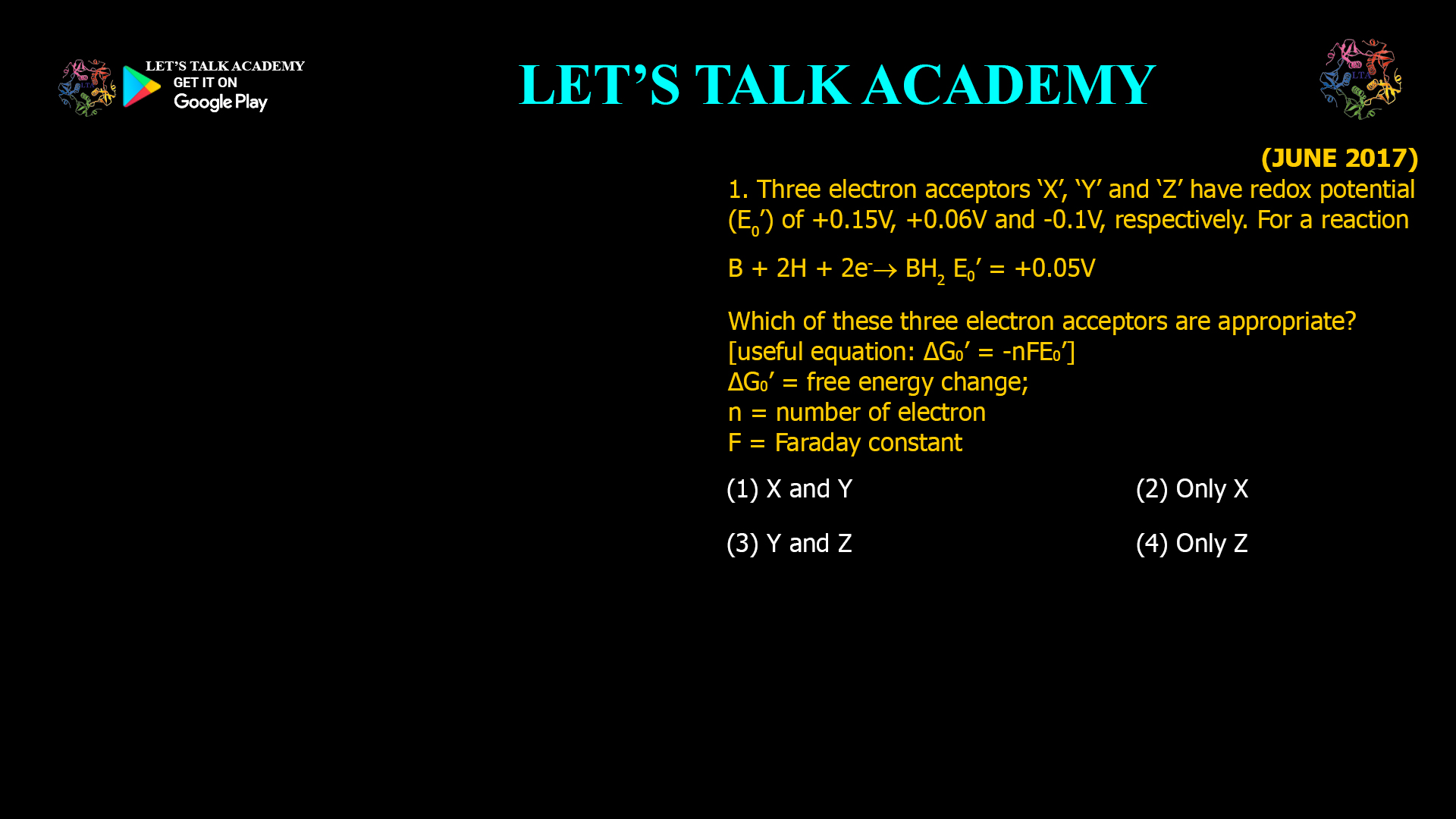
1. Three electron acceptors ‘X’, ‘Y’ and ‘Z’ have redox potential (E0’) of +0.15V, +0.06V and -0.1V, respectively. For a reaction B + 2H + 2e– à BH2 E0’ = […]

Heating of some nucleic acids shows an increase in the absorbance at 260 nm (A260) typified by the plot shown above. The sharp transition midpoint is defined as melting temperature […]

Double stranded DNA can be destabilized and denatured by (1) Formamide (2) Polyglycine (3) Sucrose (4) PEG Introduction DNA denaturation involves the separation of the two complementary strands of the […]

Denaturation profiles of DNA are shown below The differences in the profiles arise because (1) the DNA is single stranded but of different sizes (2) A + T content of […]


