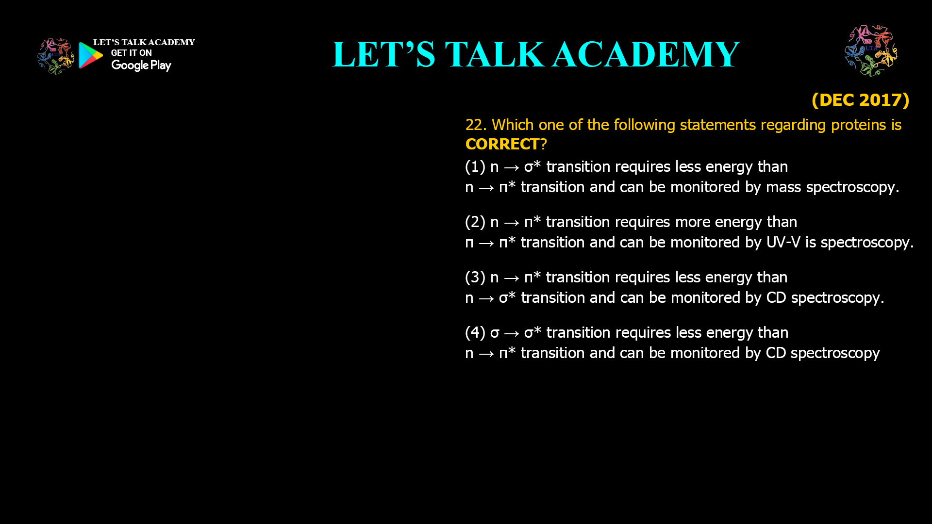
22. Which one of the following statements regarding proteins is CORRECT?
(1) n → σ* transition requires less energy than n → π* transition and can be monitored by mass spectroscopy.
(2) n → π* transition requires more energy than π → π* transition and can be monitored by UV-VIS spectroscopy.
(3) n → π* transition requires less energy than n → σ* transition and can be monitored by CD spectroscopy.
(4) σ → σ* transition requires less energy than n → π* transition and can be monitored by CD spectroscopy.
Which Statement Regarding Electronic Transitions in Proteins is Correct?
Correct Answer
Option (3): n → π* Transition Requires Less Energy than n → σ* Transition and Can Be Monitored by Circular Dichroism (CD) Spectroscopy
The correct answer is Option (3). The energy required for an electronic transition depends on the energy difference between the occupied orbital and the corresponding antibonding orbital. The n → π* transition involves excitation of a non-bonding electron to a π* antibonding orbital and therefore requires less energy than the n → σ* transition, which involves excitation into the higher-energy σ* antibonding orbital.
Proteins possess peptide bonds that exhibit both π → π* and n → π* transitions. These transitions are responsible for the characteristic spectra observed in the far-ultraviolet region. Circular Dichroism spectroscopy measures the differential absorption of left and right circularly polarized light arising from these electronic transitions in optically active proteins, making it highly suitable for studying protein secondary structure.
Understanding Electronic Transitions
Electrons occupy molecular orbitals of different energies. When ultraviolet radiation is absorbed, electrons may be promoted from bonding or non-bonding orbitals into antibonding orbitals. The most common electronic transitions observed in biological molecules include σ → σ*, n → σ*, π → π*, and n → π* transitions.
Among these, the energy requirement generally follows the order:
σ → σ* > n → σ* > π → π* > n → π*
Therefore, the n → π* transition requires the least energy among the transitions relevant to peptide bonds and occurs at relatively longer ultraviolet wavelengths.
Role of Circular Dichroism Spectroscopy
Circular Dichroism spectroscopy detects differences in the absorption of left- and right-circularly polarized light by chiral molecules such as proteins. The technique primarily monitors the π → π* and n → π* transitions of peptide bonds. These transitions generate characteristic CD spectra that reveal the presence of α-helices, β-sheets, and random coil structures.
Because protein secondary structure directly influences these electronic transitions, CD spectroscopy has become one of the most widely used methods for studying protein folding, unfolding, thermal stability, and conformational changes.
Why Option (1) is Incorrect
This option incorrectly states that the n → σ* transition requires less energy than the n → π* transition. In reality, the σ* antibonding orbital lies at a much higher energy than the π* antibonding orbital, making the n → σ* transition significantly more energetic. Furthermore, mass spectrometry measures the mass-to-charge ratio of ions and does not monitor electronic transitions.
Why Option (2) is Incorrect
Although UV-Visible spectroscopy is capable of detecting electronic transitions, the statement incorrectly claims that the n → π* transition requires more energy than the n → σ* transition. The opposite is true. The n → π* transition occurs at lower energy and longer wavelength because excitation into a π* orbital requires less energy than excitation into a σ* orbital.
Why Option (3) is Correct
The n → π* transition is a lower-energy transition than the n → σ* transition because the π* antibonding orbital lies at lower energy than the σ* antibonding orbital. These transitions contribute to the characteristic Circular Dichroism spectra of proteins, allowing researchers to determine secondary structure and monitor conformational changes. Therefore, this statement is scientifically accurate.
Why Option (4) is Incorrect
Although Circular Dichroism spectroscopy is indeed used to study proteins, the statement incorrectly reverses the relative energies of the electronic transitions. The n → σ* transition does not require less energy than the n → π* transition. Consequently, the statement is incorrect despite identifying the appropriate spectroscopic technique.
Applications in Protein Spectroscopy
Electronic transitions of peptide bonds are fundamental to protein spectroscopy. UV-Visible spectroscopy measures absorption arising from these transitions, while Circular Dichroism spectroscopy provides additional structural information by exploiting the optical activity of proteins. These techniques are extensively used to investigate protein folding, enzyme structure, ligand binding, conformational changes, thermal denaturation, and protein stability.
Final Answer
Correct Option: (3) n → π* transition requires less energy than n → σ* transition and can be monitored by Circular Dichroism (CD) spectroscopy.
The n → π* electronic transition involves excitation into a lower-energy antibonding orbital than the n → σ* transition and therefore requires less energy. These peptide bond transitions contribute significantly to the Circular Dichroism spectra of proteins, enabling accurate analysis of secondary structure and conformational changes. Hence, Option (3) is the correct answer.


