
1. Introduction
Ion channels are specialized membrane proteins that allow selected ions to move across biological membranes. They are fundamental components of almost every living cell and are particularly important in excitable cells such as neurons, skeletal muscle cells, cardiac muscle cells, and sensory cells.
The plasma membrane separates the intracellular environment from the extracellular environment. Although the lipid bilayer provides an effective barrier to charged molecules, cells must continuously regulate the movement of ions across this membrane. Ion channels provide controlled pathways through which ions can cross the membrane.
The most biologically important ions transported through ion channels include:
- Sodium (Na⁺)
- Potassium (K⁺)
- Calcium (Ca²⁺)
- Chloride (Cl⁻)
- Hydrogen ions (H⁺)
The movement of these ions produces changes in the electrical potential across membranes and contributes to numerous cellular processes.
Ion channels are involved in:
- Maintenance of membrane potential
- Generation of action potentials
- Transmission of nerve impulses
- Muscle contraction
- Neurotransmitter release
- Hormone and enzyme secretion
- Sensory perception
- Cell volume regulation
- Osmotic balance
- Intracellular signaling
- Regulation of cardiac rhythm
- Communication between cells
The importance of ion channels arises not merely from their ability to transport ions but from their ability to control ion movement with extraordinary precision.
A channel can remain closed, open for a short period, or become inactive depending on the signals received by the cell.
2. Basic Structure of Ion Channels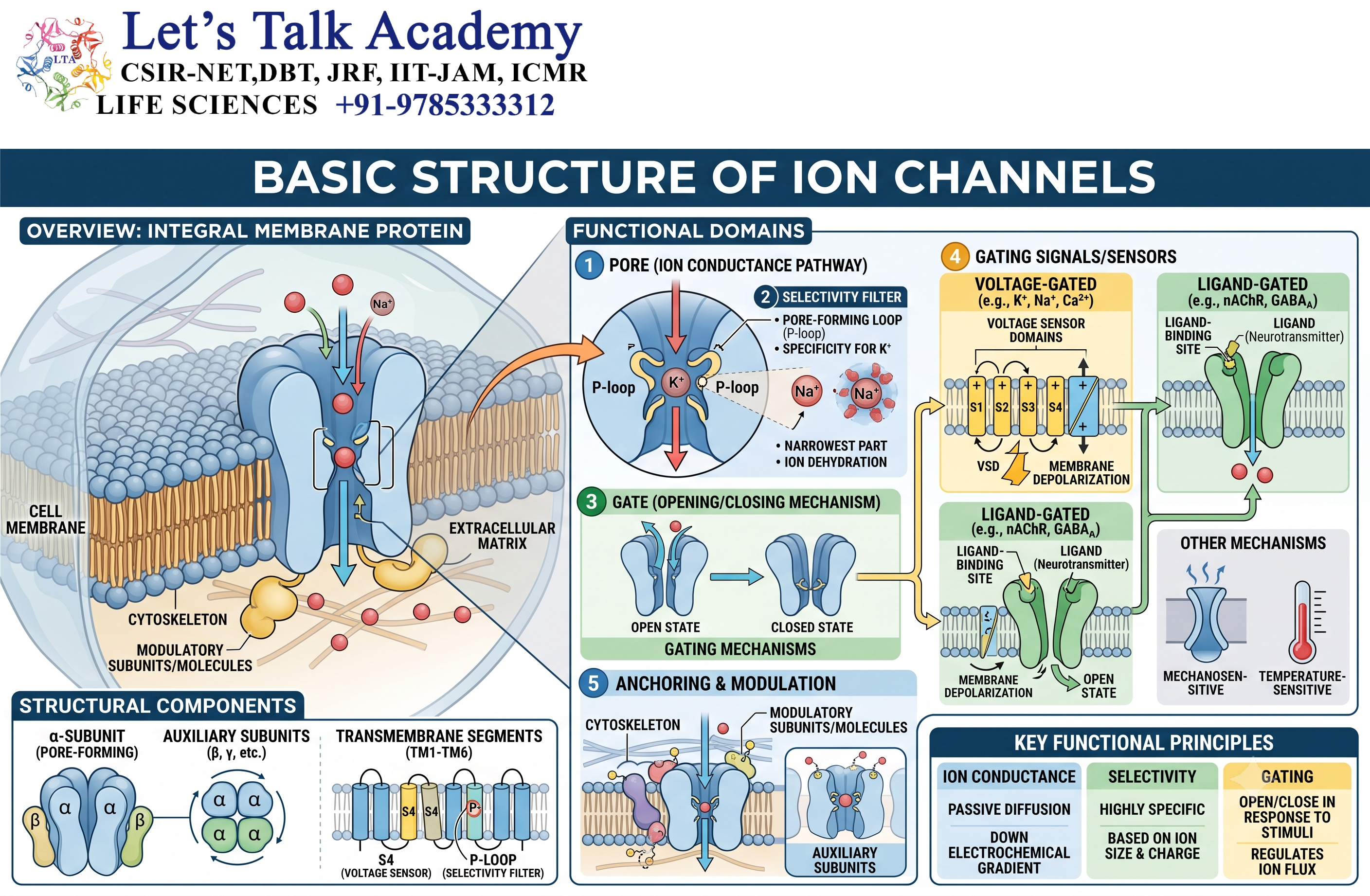
2.1 Ion Channels as Membrane Proteins
Ion channels are integral membrane proteins embedded within the lipid bilayer.
The hydrophobic interior of the membrane creates a strong barrier against charged particles. An ion cannot easily pass through this hydrophobic region because it is surrounded by water molecules and carries an electrical charge.
Ion channels overcome this barrier by forming a hydrophilic pathway through the membrane.
A simplified arrangement can be represented as:
Extracellular space → Ion channel pore → Intracellular space
When the channel is open, ions can pass through the pore.
However, the pore is not completely non-selective. Specific regions of the channel determine which ions can enter and pass through.
2.2 Channel Pore
The pore is the pathway through which ions move across the membrane.
The pore generally contains amino acid residues that interact with ions and water molecules.
Different regions of the pore may perform different functions, including:
- Ion recognition
- Ion selectivity
- Regulation of ion movement
- Channel gating
- Blocking or restricting ion passage
The physical dimensions and chemical properties of the pore strongly influence ion permeability.
2.3 Ion Selectivity
One of the most remarkable characteristics of ion channels is their ability to distinguish between different ions.
For example, a potassium-selective channel can strongly favor K⁺ over Na⁺ even though these ions have similar chemical properties.
Ion selectivity depends on several factors:
- Ionic radius
- Charge
- Hydration shell
- Chemical interactions with amino acid residues
- Geometry of the selectivity filter
The channel’s selectivity filter provides an appropriate chemical and structural environment for the preferred ion.
2.4 Selectivity Filter
The selectivity filter is a specialized region of many ion channels that determines which ions can pass through the pore.
The filter contains amino acid residues positioned in a precise three-dimensional arrangement.
For an ion to pass, it must interact favorably with the chemical groups present in the selectivity filter.
In potassium channels, for example, the selectivity filter is arranged so that K⁺ can interact efficiently with carbonyl oxygen atoms within the pore. This interaction helps compensate for the energetic cost associated with removing water molecules surrounding the ion.
Thus, ion selectivity is not simply determined by ion size. It depends on the energetic and chemical compatibility between the ion and the channel.
3. Ion Movement Through Channels
3.1 Passive Movement of Ions
Ion movement through an open channel is generally passive.
This means that the channel itself does not normally use ATP to transport ions.
Instead, ions move according to their electrochemical gradient.
The energy driving ion movement comes from existing differences in ion concentration and electrical potential across the membrane.
3.2 Chemical Gradient
A concentration difference across the membrane produces a chemical gradient.
Ions tend to move from an area of higher concentration toward an area of lower concentration.
For example, if Na⁺ concentration is greater outside a cell than inside, Na⁺ has a chemical tendency to move into the cell when a suitable channel is open.
Similarly, if K⁺ concentration is greater inside the cell, K⁺ tends to move outward through an open potassium channel.
3.3 Electrical Gradient
Ions are also affected by the electrical charge across the membrane.
If the inside of the cell is electrically negative relative to the outside, positively charged ions are electrically attracted toward the cell interior.
Therefore:
- Na⁺ is attracted toward the negative cell interior.
- Ca²⁺ is strongly attracted toward the negative cell interior.
- K⁺ experiences an inward electrical force but often has an outward chemical gradient.
- Cl⁻ experiences electrical forces opposite to those acting on cations.
3.4 Electrochemical Gradient
The electrochemical gradient is the combined influence of:
- Chemical gradient
- Electrical gradient
The actual direction of ion movement depends on the combined effect of these two forces.
This concept is extremely important for understanding membrane physiology.
For example, K⁺ is usually present at a much higher concentration inside the cell. Therefore, its chemical gradient favors outward movement. However, the negative membrane potential attracts K⁺ inward.
At a particular membrane voltage, these forces can balance each other.
4. Equilibrium Potential
4.1 Definition
The equilibrium potential of an ion is the membrane potential at which the electrical force exactly balances the chemical force acting on that ion.
At this potential, there is no net movement of that particular ion through an open channel, although individual ions may still move in both directions.
The equilibrium potential can be estimated using the Nernst equation:
Eion=RTzFln([ion]out[ion]in)E_{ion}=\frac{RT}{zF}\ln\left(\frac{[ion]_{out}}{[ion]_{in}}\right)
where:
- EionE_{ion} = equilibrium potential
- RR = gas constant
- TT = absolute temperature
- zz = ionic charge
- FF = Faraday constant
- [ion]out[ion]_{out} = extracellular ion concentration
- [ion]in[ion]_{in} = intracellular ion concentration
The Nernst equation is particularly useful for understanding the driving force acting on individual ionic species.
4.2 Driving Force
The difference between the membrane potential and the equilibrium potential of an ion determines its electrical driving force.
In simplified terms:
Driving Force=Vm−EionDriving\ Force = V_m-E_{ion}
where:
- VmV_m = membrane potential
- EionE_{ion} = equilibrium potential of the ion
The greater the difference between these values, the stronger the tendency for the ion to move through an open channel, provided the channel is permeable to that ion.
5. Ion Channels and Membrane Potential
5.1 Membrane Potential
Cells maintain a voltage difference across their plasma membranes called the membrane potential.
The membrane potential results primarily from:
- Unequal distribution of ions
- Selective membrane permeability
- Activity of ion pumps
- Activity of ion channels
In many animal cells, the inside of the cell is electrically negative relative to the extracellular environment.
5.2 Resting Membrane Potential
A resting cell has a characteristic membrane potential called the resting membrane potential.
In neurons, the resting membrane potential is commonly around −60 to −70 mV, although the exact value varies among cell types.
Potassium permeability is often particularly important in establishing the resting membrane potential because many cells possess potassium leak channels.
The Na⁺/K⁺-ATPase also plays an essential supporting role by maintaining the concentration gradients of Na⁺ and K⁺.
6. Classification of Ion Channels
Ion channels can be classified according to different criteria.
The most useful classification is based on their gating mechanism.
Major categories include:
- Voltage-gated channels
- Ligand-gated channels
- Mechanically gated channels
- Intracellularly regulated channels
- Leak channels
- Temperature-sensitive and other sensory channels
Different classification systems may overlap because a single channel can respond to more than one regulatory signal.
7. Voltage-Gated Ion Channels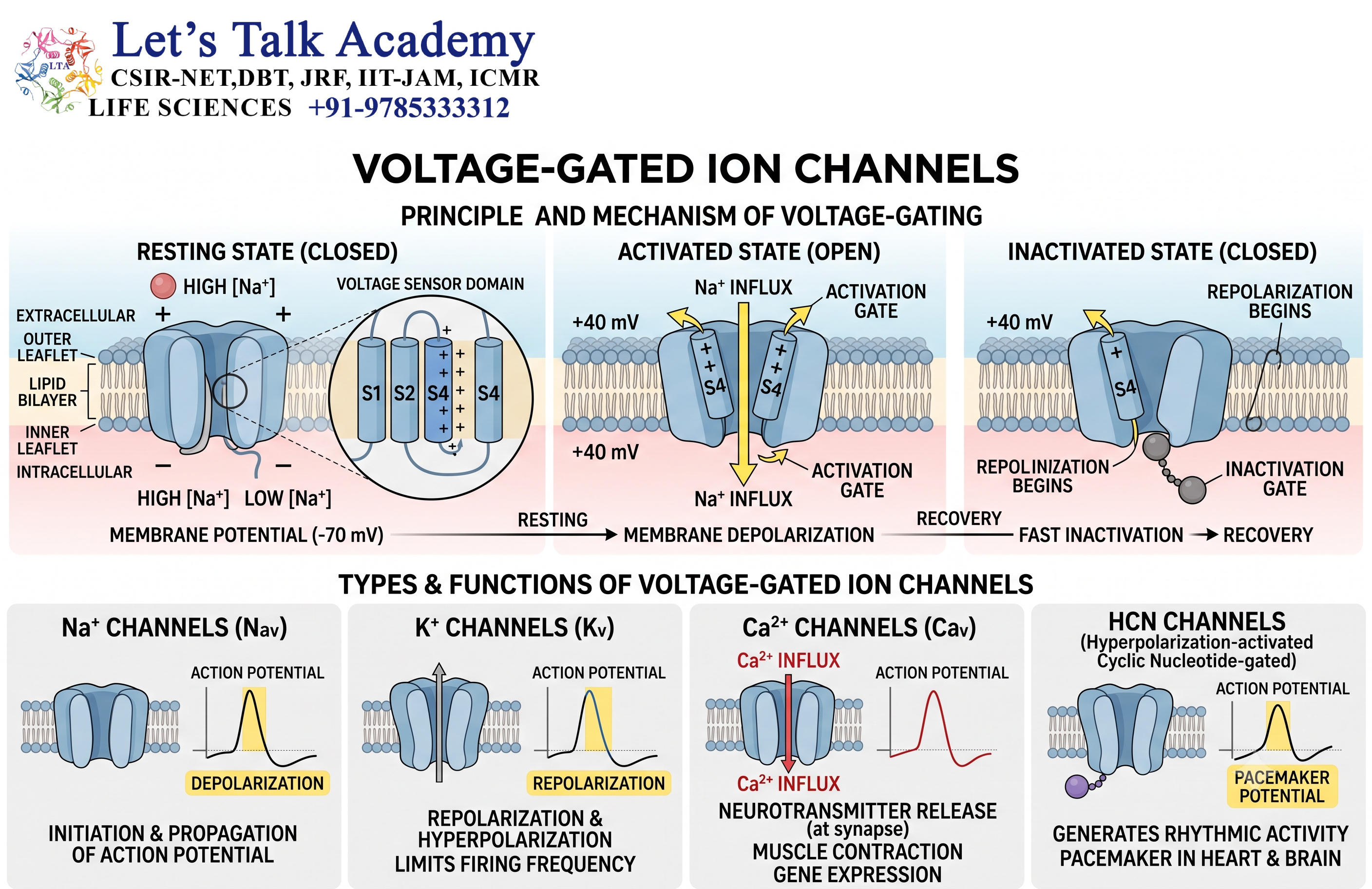
7.1 Definition
Voltage-gated ion channels are channels whose opening or closing is influenced by changes in membrane potential.
They are particularly important in electrically excitable cells.
Major examples include:
- Voltage-gated Na⁺ channels
- Voltage-gated K⁺ channels
- Voltage-gated Ca²⁺ channels
These channels are essential for electrical signaling.
7.2 Voltage Sensing
Voltage-gated channels contain specialized structural elements that detect changes in membrane voltage.
Changes in the electric field across the membrane cause movement of charged regions within the channel protein.
This structural rearrangement can result in:
Closed state → Open state
or
Open state → Inactivated/closed state
The voltage-sensing mechanism allows the channel to respond rapidly to electrical changes.
8. Voltage-Gated Sodium Channels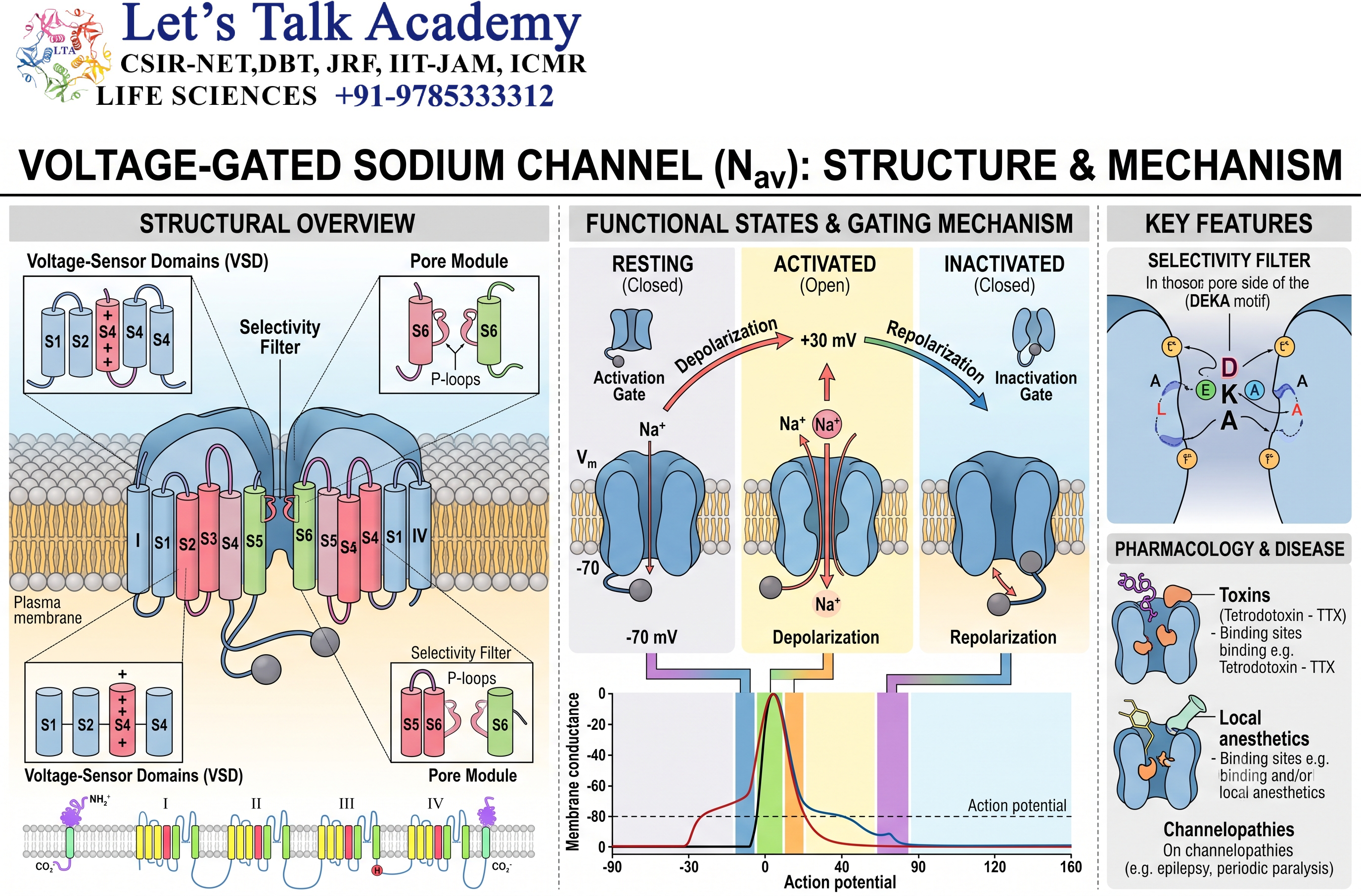
8.1 Structure
Voltage-gated sodium channels are large membrane proteins responsible for rapid Na⁺ entry during action potentials.
A typical voltage-gated sodium channel contains a single large α-subunit organized into four homologous domains.
Each domain contains several transmembrane segments.
One important segment functions as the voltage sensor, while other regions contribute to pore formation and ion selectivity.
8.2 Opening of Sodium Channels
When the membrane is sufficiently depolarized, voltage-gated Na⁺ channels rapidly transition from the closed state to the open state.
Na⁺ then enters the cell.
This produces further depolarization, which can activate additional sodium channels.
The process becomes regenerative:
Depolarization → Na⁺ channel opening → Na⁺ influx → Further depolarization
This positive-feedback mechanism is responsible for the rapid rising phase of many neuronal action potentials.
8.3 Sodium Channel Inactivation
Sodium channels do not remain open indefinitely.
After opening, they rapidly enter an inactivated state.
During inactivation, the channel becomes non-conductive even though the membrane may still be depolarized.
Recovery from inactivation generally requires membrane repolarization.
This property is essential for:
- Terminating Na⁺ influx
- Preventing continuous firing
- Establishing the refractory period
- Ensuring directional propagation of action potentials
9. Voltage-Gated Potassium Channels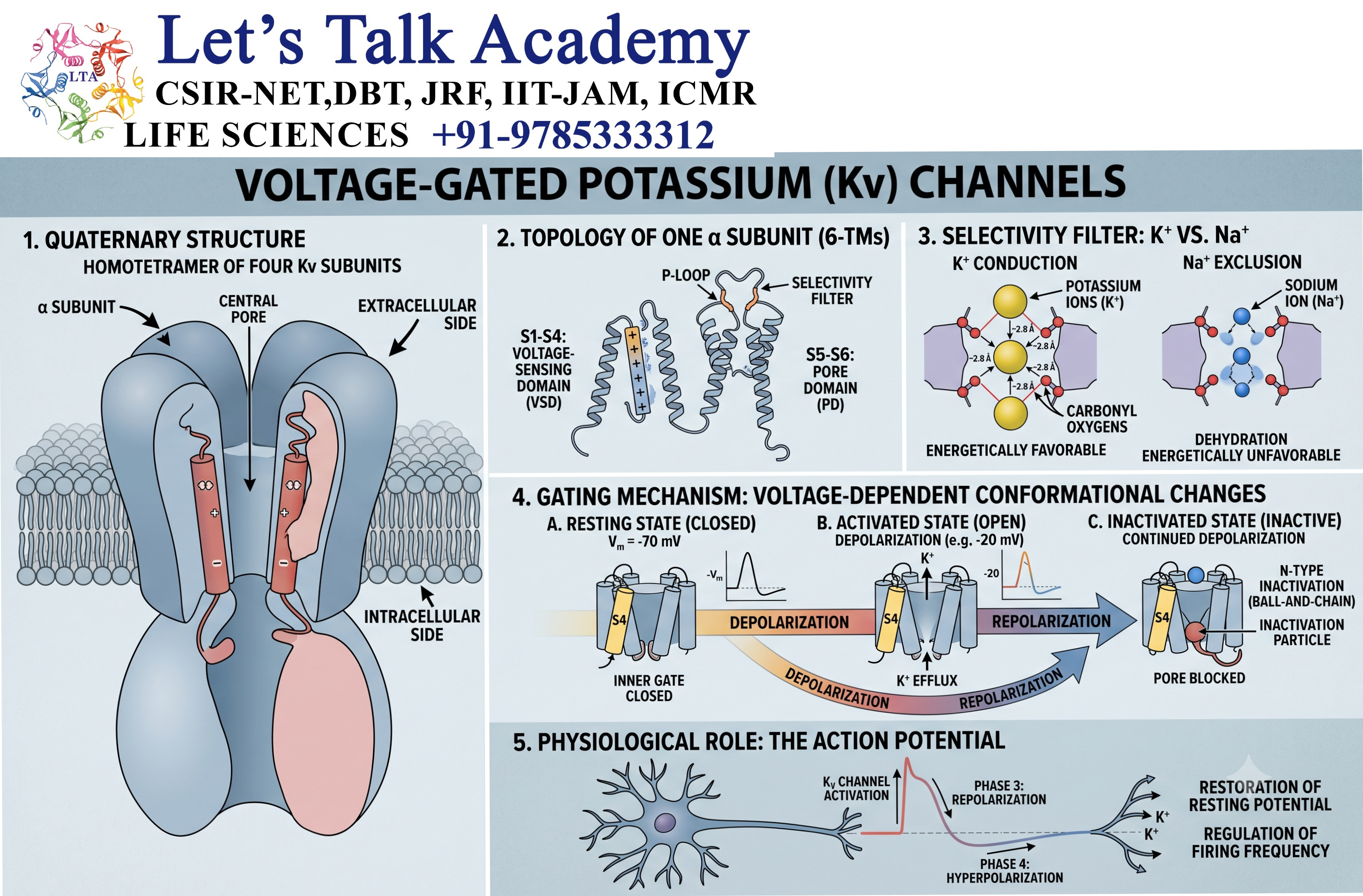
9.1 General Function
Voltage-gated potassium channels allow K⁺ to move across the membrane in response to changes in membrane potential.
They play a major role in:
- Membrane repolarization
- Restoration of resting membrane potential
- Regulation of neuronal excitability
- Control of action potential duration
9.2 Potassium Efflux
During an action potential, voltage-gated K⁺ channels open following depolarization.
Because intracellular K⁺ concentration is generally high, K⁺ tends to leave the cell.
This outward movement of positive charge contributes to membrane repolarization.
The sequence can be summarized as:
Depolarization → K⁺ channel opening → K⁺ efflux → Repolarization
9.3 Delayed Rectification
Many voltage-gated potassium channels activate more slowly than voltage-gated sodium channels.
This delayed activation allows Na⁺ influx to initiate depolarization before substantial K⁺ efflux occurs.
The subsequent K⁺ efflux helps bring the membrane potential back toward its resting value.
10. Voltage-Gated Calcium Channels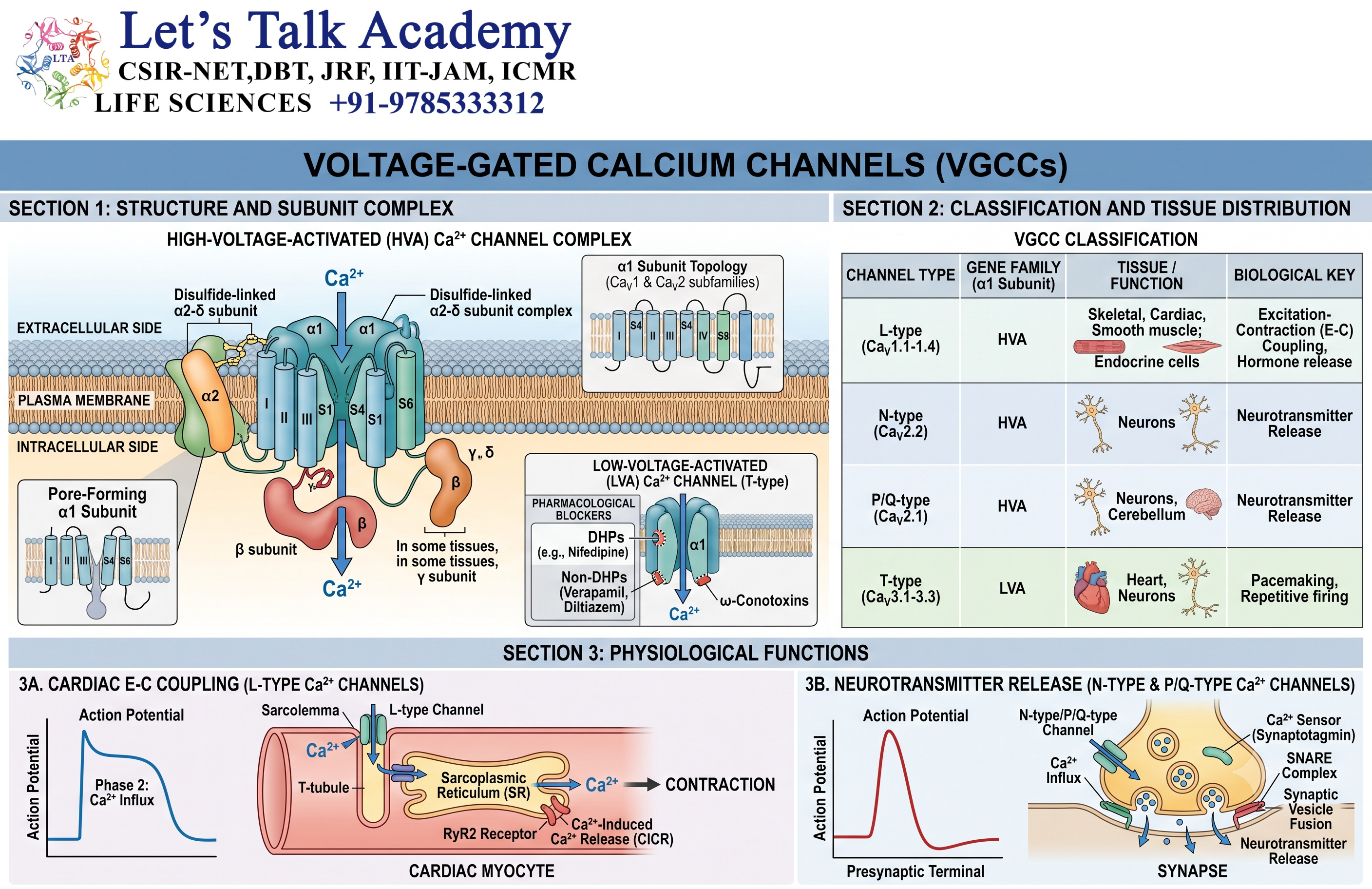
10.1 Importance of Calcium Channels
Voltage-gated calcium channels allow Ca²⁺ to enter cells in response to membrane depolarization.
Ca²⁺ has an especially important role because it functions both as an ion carrying electrical charge and as an intracellular signaling molecule.
Calcium channels participate in:
- Neurotransmitter release
- Muscle contraction
- Hormone secretion
- Gene regulation
- Enzyme activation
- Intracellular signaling
- Cardiac electrical activity
10.2 Calcium and Neurotransmitter Release
At a chemical synapse, an action potential arriving at the presynaptic terminal causes voltage-gated Ca²⁺ channels to open.
Ca²⁺ enters the presynaptic terminal.
The increase in local Ca²⁺ concentration triggers fusion of synaptic vesicles with the plasma membrane.
This results in:
Action potential → Ca²⁺ influx → Vesicle fusion → Neurotransmitter release
This is one of the most important functions of calcium channels in the nervous system.
11. Ligand-Gated Ion Channels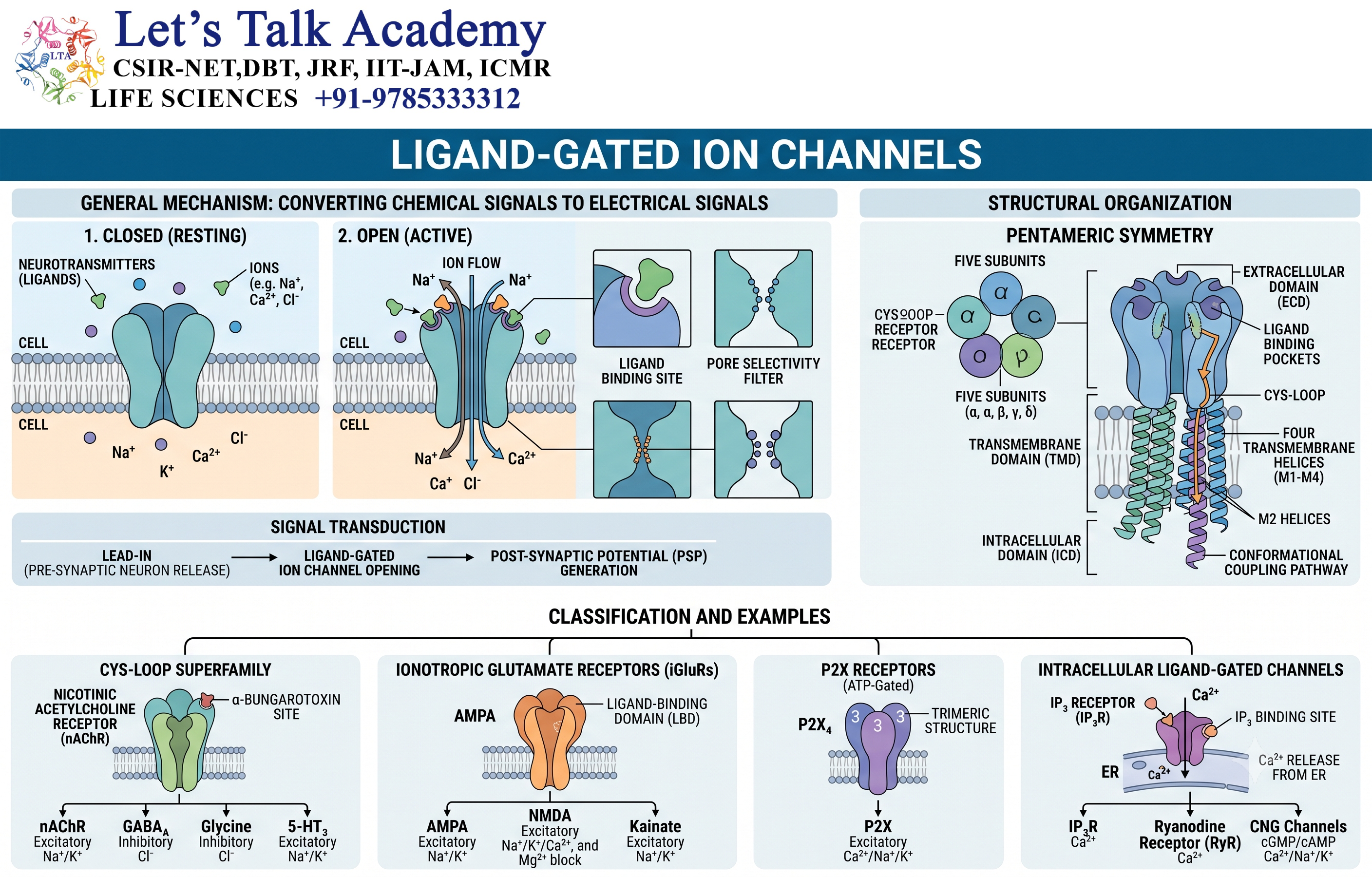
11.1 Definition
Ligand-gated ion channels open or close when a chemical messenger binds to the channel or an associated receptor.
The ligand may be:
- Neurotransmitter
- Hormone
- Intracellular signaling molecule
- Extracellular signaling molecule
These channels are particularly important in synaptic transmission.
11.2 Examples
Important ligand-gated ion channels include:
- Nicotinic acetylcholine receptor
- GABAA_A receptor
- AMPA receptor
- NMDA receptor
- Glycine receptor
- P2X receptors
11.3 Mechanism
The basic mechanism can be represented as:
Ligand binding → Conformational change → Channel opening → Ion movement → Cellular response
The response depends on which ions can pass through the channel.
12. Nicotinic Acetylcholine Receptor
The nicotinic acetylcholine receptor is a ligand-gated ion channel activated by acetylcholine.
It is found in several locations, including the neuromuscular junction and nervous system.
When acetylcholine binds to the receptor, the channel opens and permits cation movement.
At the neuromuscular junction, this produces depolarization of the muscle membrane and contributes to initiation of muscle contraction.
13. GABA-Gated Ion Channels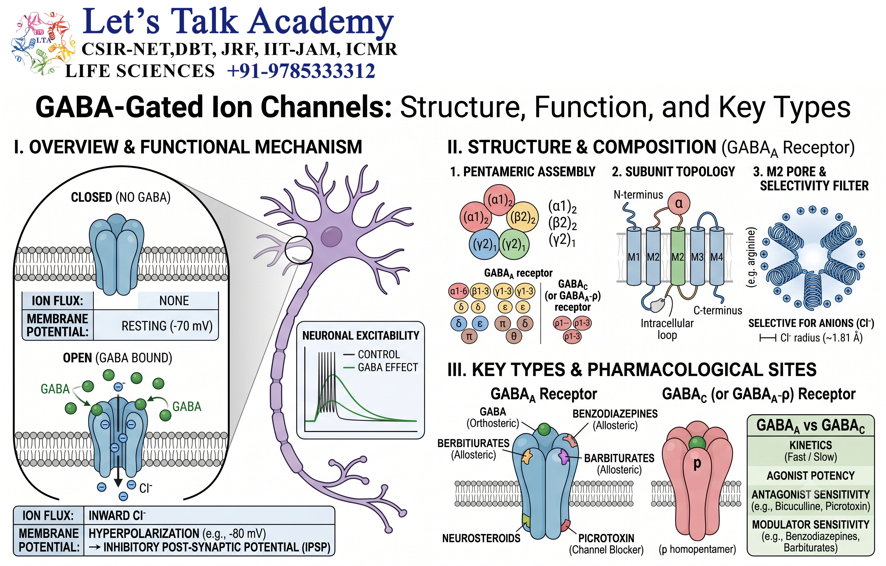
13.1 GABAA_A Receptor
The GABAA_A receptor is a ligand-gated chloride channel.
When GABA binds to the receptor, the channel opens and permits Cl⁻ movement.
GABAA_A receptor activation generally produces an inhibitory effect in mature neurons by increasing chloride conductance and reducing neuronal excitability.
14. Glutamate-Gated Ion Channels
Glutamate is one of the major excitatory neurotransmitters in the nervous system.
Important ionotropic glutamate receptors include:
- AMPA receptors
- NMDA receptors
- Kainate receptors
These receptors form ligand-gated ion channels.
14.1 AMPA Receptors
AMPA receptors primarily mediate rapid excitatory synaptic transmission.
Activation allows cations, particularly Na⁺ and K⁺, to move through the receptor-associated channel.
Na⁺ influx contributes to depolarization of the postsynaptic membrane.
14.2 NMDA Receptors
NMDA receptors are particularly interesting because their activity depends on several conditions.
They are activated by glutamate and are strongly influenced by membrane voltage and the presence of other ligands.
At resting membrane potential, Mg²⁺ can block the channel pore.
Depolarization helps relieve this Mg²⁺ block, allowing substantial ion flow, including Ca²⁺.
NMDA receptor activity is therefore important in:
- Synaptic plasticity
- Learning-related cellular mechanisms
- Memory-associated processes
- Calcium-dependent signaling
15. Mechanically Gated Ion Channels
15.1 Definition
Mechanically gated channels respond to physical deformation of the membrane or associated cellular structures.
They are activated by mechanical forces such as:
- Stretch
- Pressure
- Vibration
- Membrane tension
- Shear forces
15.2 Biological Functions
Mechanically sensitive ion channels contribute to:
- Touch
- Hearing
- Balance
- Blood-pressure sensing
- Cell-volume regulation
- Mechanical sensation
- Tissue responses to physical forces
They are especially important in sensory physiology.
16. Leak Ion Channels
16.1 Definition
Leak channels are channels that tend to remain open under resting conditions and allow ions to move across the membrane according to their electrochemical gradients.
Potassium leak channels are particularly important in many cells.
They contribute significantly to the resting membrane potential.
16.2 Importance of Potassium Leak Channels
Because intracellular K⁺ concentration is generally high, K⁺ tends to move outward through potassium leak channels.
The outward movement of positive charge makes the inside of the cell more negative.
As the membrane becomes increasingly negative, the electrical force opposing further K⁺ efflux increases.
Eventually, the system approaches a balance between chemical and electrical forces.
17. Intracellularly Regulated Ion Channels
Some ion channels are regulated by molecules inside the cell.
Important intracellular regulators include:
- Ca²⁺
- ATP
- ADP
- cAMP
- cGMP
- G-protein subunits
- pH
These channels allow the metabolic and signaling state of the cell to influence membrane permeability.
18. Calcium-Activated Potassium Channels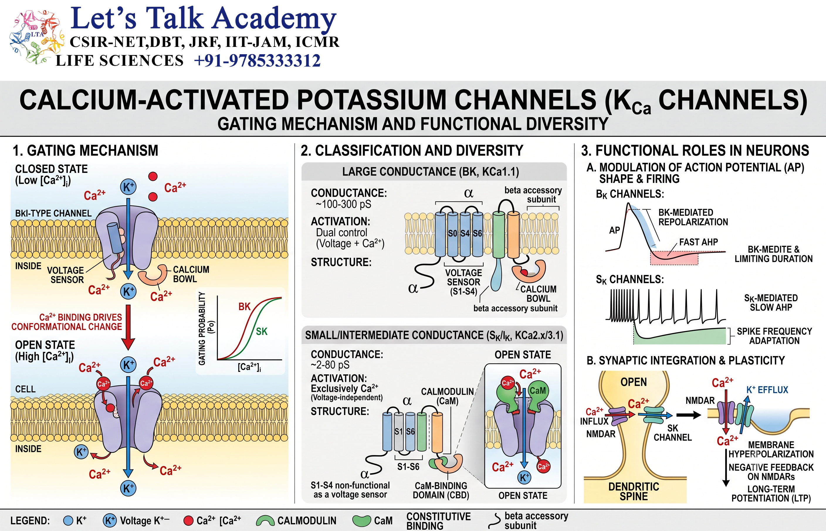
Calcium-activated potassium channels respond to intracellular Ca²⁺.
When intracellular Ca²⁺ concentration increases, these channels can open and allow K⁺ to leave the cell.
This can contribute to membrane hyperpolarization.
These channels are involved in:
- Regulation of neuronal firing
- Muscle physiology
- Hormone secretion
- Feedback regulation of intracellular calcium signaling
This provides an important example of how one ion can regulate the movement of another ion.
19. ATP-Sensitive Potassium Channels
ATP-sensitive potassium channels, commonly called KATP channels, link cellular metabolism with membrane electrical activity.
They respond to the metabolic state of the cell, particularly changes in ATP and related nucleotides.
These channels have important roles in:
- Pancreatic β-cell physiology
- Cardiac function
- Metabolic sensing
- Protection against cellular stress
In pancreatic β-cells, changes in glucose metabolism alter ATP production, which influences KATP channel activity and contributes to insulin secretion.
20. Channel Gating
20.1 Meaning of Gating
Gating refers to the process by which an ion channel changes between conductive and non-conductive states.
A channel can exist in several functional states:
- Closed
- Open
- Inactivated
The exact number and nature of states differ among channel types.
20.2 Closed State
In the closed state, the channel pore does not permit significant ion conduction.
A channel can remain closed until it receives an appropriate stimulus.
Examples include:
- Change in membrane voltage
- Ligand binding
- Mechanical force
- Intracellular signaling molecule
20.3 Open State
When the channel enters the open state, ions can pass through the pore.
Ion movement is generally rapid.
A single channel can conduct a large number of ions during a short period.
20.4 Inactivated State
Inactivation is different from simply closing the channel.
An inactivated channel is temporarily unavailable for opening even though the activating stimulus may still be present.
Inactivation is especially important for voltage-gated sodium channels.
21. Channel Conductance
Ion channels conduct ions according to their electrical and chemical properties.
The relationship between current and voltage is often described using:
I=g(Vm−Eion)I=g(V_m-E_{ion})
where:
- II = ionic current
- gg = channel conductance
- VmV_m = membrane potential
- EionE_{ion} = equilibrium potential
Conductance reflects how easily ions can pass through the channel population.
Channel opening increases membrane conductance for the ions that can pass through that channel.
22. Ion Channels and Action Potentials
22.1 Generation of an Action Potential
Action potentials are rapid changes in membrane potential that allow electrical signals to travel along excitable cells.
A simplified sequence is:
Resting state → Threshold → Depolarization → Repolarization → Hyperpolarization → Resting state
Different ion channels dominate different phases.
22.2 Depolarization
When a neuron reaches threshold, voltage-gated sodium channels open rapidly.
Na⁺ enters the cell.
The membrane potential becomes less negative and may become positive.
This phase is called depolarization.
22.3 Repolarization
Sodium channels become inactivated, while voltage-gated potassium channels are increasingly open.
K⁺ leaves the cell.
The membrane potential moves back toward negative values.
This is called repolarization.
22.4 Hyperpolarization
Potassium channels may remain open for a short period after the membrane has returned toward its resting potential.
Continued K⁺ efflux can make the membrane potential temporarily more negative than the resting potential.
This phase is called hyperpolarization or the after-hyperpolarization phase.
23. Refractory Period
23.1 Absolute Refractory Period
During the absolute refractory period, another action potential cannot normally be initiated regardless of stimulus strength.
This occurs primarily because voltage-gated sodium channels are inactivated.
23.2 Relative Refractory Period
During the relative refractory period, another action potential can occur, but a stronger-than-normal stimulus may be required.
Persistent potassium conductance contributes to this phase.
The refractory period is important because it:
- Limits the maximum firing frequency
- Helps maintain the direction of action potential propagation
- Prevents immediate repetitive firing
24. Ion Channels in Synaptic Transmission
Ion channels are central to communication between neurons.
At a chemical synapse:
- An action potential reaches the presynaptic terminal.
- Voltage-gated Ca²⁺ channels open.
- Ca²⁺ enters the presynaptic terminal.
- Synaptic vesicles fuse with the membrane.
- Neurotransmitter is released.
- Neurotransmitter binds to postsynaptic receptors.
- Ligand-gated ion channels may open.
- Ion flow changes the postsynaptic membrane potential.
This mechanism allows electrical information to be converted into a chemical signal and then back into an electrical response.
25. Ion Channels in Muscle Contraction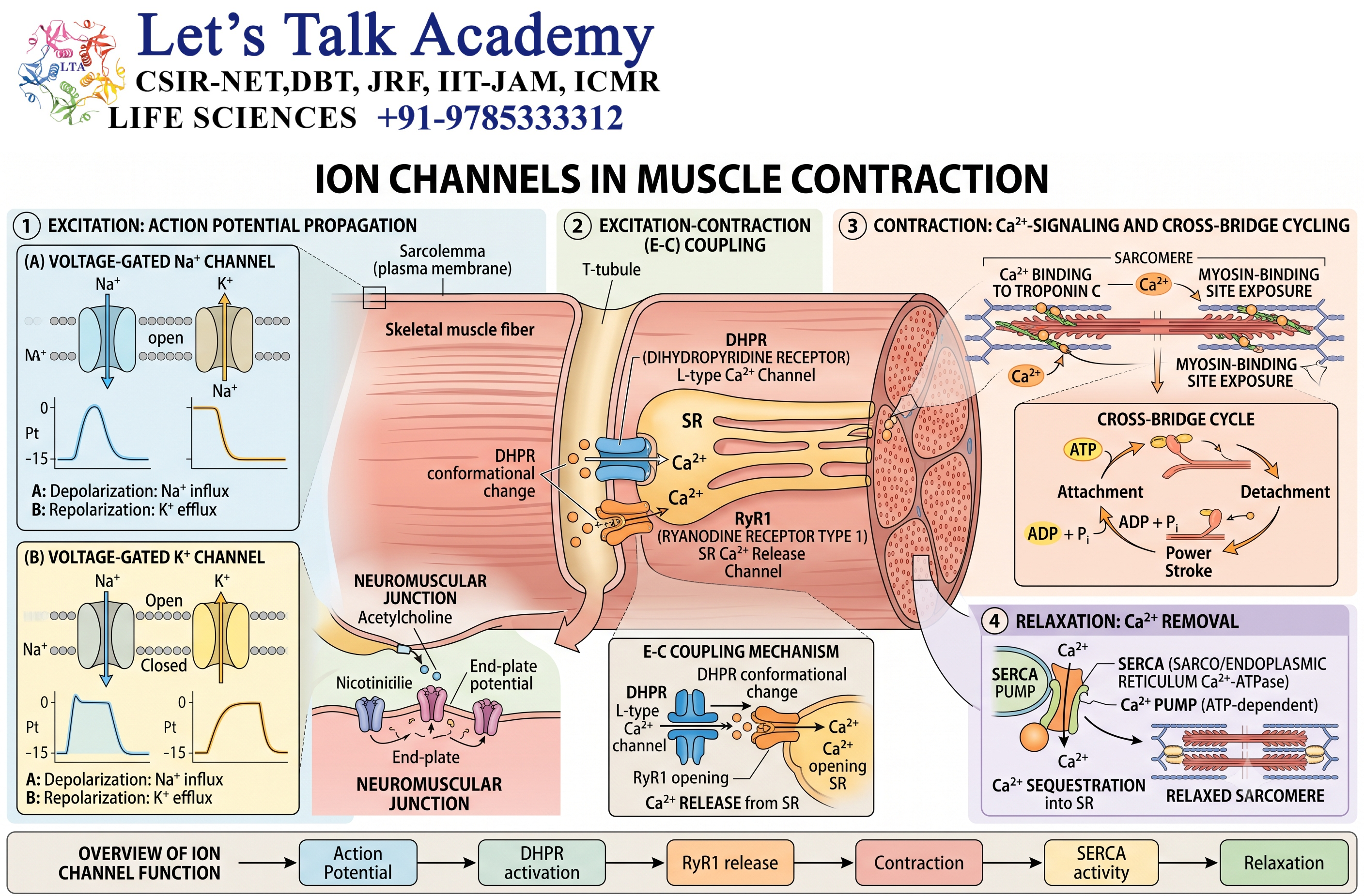
Ion channels are essential for both skeletal and cardiac muscle contraction.
In skeletal muscle, electrical excitation leads to changes in membrane potential and activation of intracellular calcium-release mechanisms.
Ca²⁺ then interacts with the contractile machinery.
The general sequence is:
Membrane excitation → Ca²⁺ signaling → Interaction with contractile proteins → Muscle contraction
Ion channels are also essential for the electrical activity that coordinates cardiac contraction.
Alterations in ion-channel function can therefore disturb normal cardiac rhythm.
26. Ion Channels in Calcium Signaling
Calcium is one of the most important intracellular signaling ions.
Under resting conditions, cytosolic free Ca²⁺ concentration is maintained at a relatively low level compared with extracellular Ca²⁺ and intracellular calcium stores.
Opening of calcium-permeable channels can cause a rapid increase in intracellular Ca²⁺.
This calcium signal can regulate:
- Protein kinases
- Protein phosphatases
- Transcription factors
- Vesicle fusion
- Muscle contraction
- Enzyme activity
- Gene expression
- Cell proliferation
- Cell death pathways
Thus, calcium channels are not merely transport proteins; they are gateways for major intracellular signaling events.
27. Ion Channels and Cell Signaling
Ion channels can directly influence intracellular signaling by changing ion concentrations.
For example:
Channel opening → Ca²⁺ influx → Activation of calcium-dependent protein → Cellular response
Similarly:
K⁺ channel opening → K⁺ efflux → Membrane hyperpolarization → Change in cellular excitability
Therefore, ion channels connect changes in the extracellular environment with intracellular biochemical responses.
28. Ion Channels and Sensory Physiology
Ion channels participate in the detection of external stimuli.
Different sensory systems use specialized channels to detect:
- Mechanical stimuli
- Temperature
- Light-associated signals
- Chemical compounds
- Pain-producing stimuli
- Changes in osmotic pressure
For example, sensory neurons contain ion channels that respond to temperature and chemical stimuli.
Activation of these channels changes membrane potential and can initiate electrical signaling toward the central nervous system.
29. Ion Channels in the Heart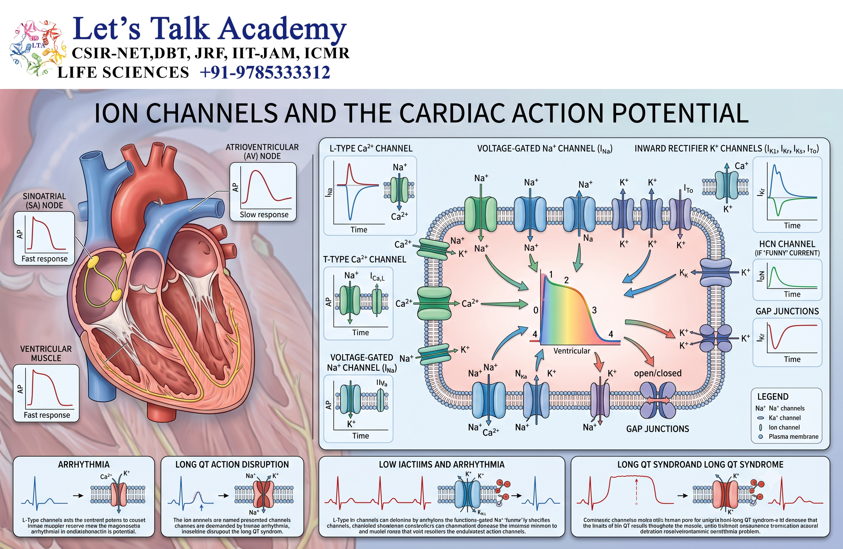
Cardiac cells depend on coordinated activity of multiple ion channels.
Major ions involved include:
- Na⁺
- K⁺
- Ca²⁺
Different channel types contribute to different phases of the cardiac action potential.
The precise timing of channel opening and closing ensures proper:
- Depolarization
- Repolarization
- Conduction
- Contraction
- Rhythm
Alterations in ion-channel function can contribute to abnormal electrical activity known as arrhythmias.
30. Ion Channelopathies
30.1 Definition
Diseases or physiological disorders caused by abnormalities in ion channels are broadly referred to as channelopathies.
A channelopathy may result from:
- Genetic mutation
- Abnormal channel expression
- Altered channel trafficking
- Defective gating
- Abnormal ion selectivity
- Autoimmune interference
- Toxin-mediated channel modification
30.2 Consequences of Channel Dysfunction
Because ion channels regulate electrical and biochemical processes, channel dysfunction can affect:
- Nervous system
- Muscles
- Heart
- Endocrine tissues
- Sensory systems
The consequences depend on which channel is affected and where it is expressed.
31. Ion Channel Blockers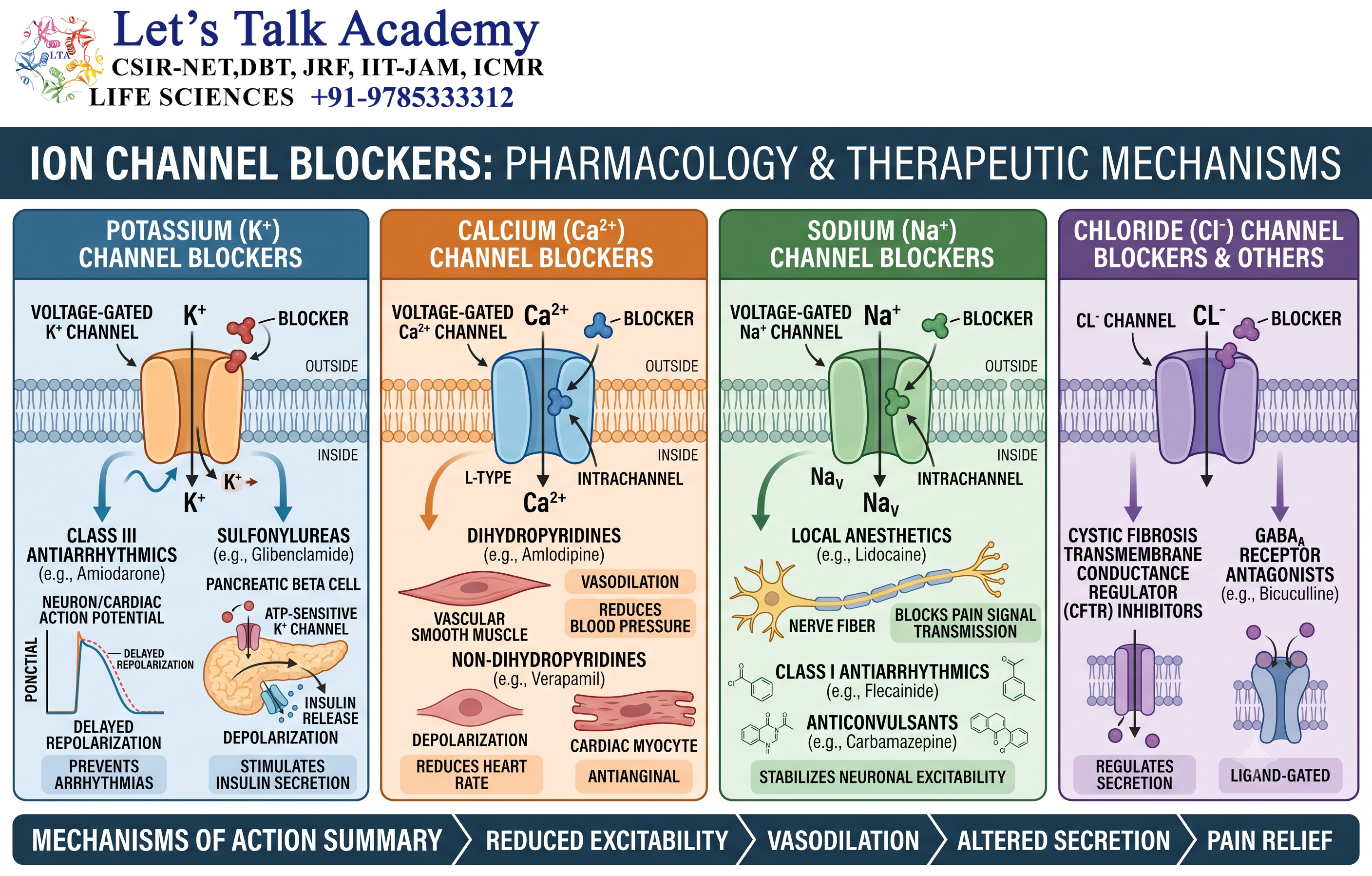
Ion channels can be inhibited by specific chemical compounds.
Channel blockers may bind:
- Within the pore
- At regulatory sites
- On extracellular regions
- On intracellular regions
Examples include compounds that block sodium, potassium, calcium, or chloride channels.
Channel blockers are important tools for studying channel function and are also used therapeutically in several areas of medicine.
32. Ion Channels as Drug Targets
Ion channels are major targets for pharmacological intervention.
Drugs can influence channel activity by:
- Blocking the channel
- Activating the channel
- Altering channel opening probability
- Delaying inactivation
- Changing channel expression
- Modifying channel trafficking
Because ion channels regulate electrical activity and signaling, modifying them can produce strong physiological effects.
33. Patch-Clamp Technique
33.1 Principle
The patch-clamp technique is one of the most important experimental methods used to study ion channels.
It allows researchers to measure tiny electrical currents passing through individual ion channels or populations of channels.
A fine glass micropipette is placed against the cell membrane to form a high-resistance seal.
Electrical current passing through the membrane patch can then be measured.
33.2 Single-Channel Recording
One of the remarkable features of the patch-clamp technique is that it can detect currents produced by individual ion channels.
A recording may show transitions such as:
Closed → Open → Closed → Open
These transitions provide direct information about channel behavior.
Researchers can determine:
- Open probability
- Conductance
- Gating kinetics
- Channel selectivity
- Response to drugs
- Voltage dependence
34. Single-Channel Conductance
The conductance of an individual ion channel is typically expressed in siemens, often in the picosiemens range for single channels.
A channel’s conductance depends on:
- Pore structure
- Ion species
- Ion concentration
- Voltage
- Channel state
Different channels have characteristic conductance properties.
35. Open Probability
The probability that an ion channel is open at a particular time is called its open probability.
It is commonly represented as:
PoP_o
A channel population may contain many individual channels, each independently transitioning between different states.
The total current depends on factors including:
- Number of channels
- Single-channel conductance
- Open probability
- Driving force
A simplified relationship is:
I=NPoiI=N P_o i
where:
- II = total current
- NN = number of channels
- PoP_o = probability of being open
- ii = current through an individual open channel
36. Ion Channels and Membrane Permeability
Membrane permeability is not constant.
It changes according to:
- Channel number
- Channel opening probability
- Channel type
- Ion concentration
- Membrane voltage
- Regulatory molecules
For example, opening many sodium channels can greatly increase sodium permeability.
Similarly, activation of potassium channels increases potassium permeability.
Changes in permeability alter the membrane potential.
37. Goldman-Hodgkin-Katz Concept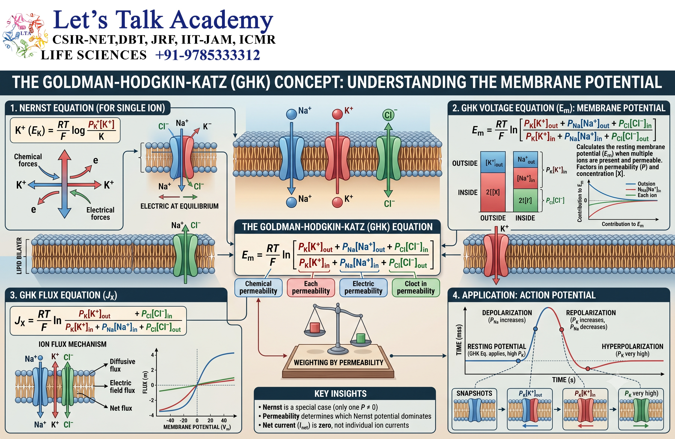
When several ion species contribute significantly to membrane potential, the membrane potential cannot be adequately described by considering only one ion.
The Goldman-Hodgkin-Katz equation considers the relative permeabilities of multiple ions, particularly:
- K⁺
- Na⁺
- Cl⁻
This provides a more realistic description of membrane potential in many physiological situations.
The equation emphasizes an important principle:
Membrane potential depends strongly on the relative permeability of the membrane to different ions.
Therefore, changing ion-channel activity can rapidly change membrane voltage.
38. Ion Channels and Active Transport Systems
Ion channels and ion pumps work together to maintain cellular ionic gradients.
The Na⁺/K⁺-ATPase, for example, uses ATP to maintain Na⁺ and K⁺ gradients.
Ion channels then provide pathways through which these ions can move down their electrochemical gradients.
Thus:
Ion pumps establish gradients → Ion channels use those gradients → Electrical and chemical signals are produced
This relationship is fundamental to cellular physiology.
39. Structural Diversity of Ion Channels
Ion channels belong to several structurally distinct protein families.
Important channel families include:
- Voltage-gated ion channels
- Ligand-gated ion channels
- Two-pore-domain potassium channels
- Transient receptor potential channels
- Mechanosensitive channels
- Cyclic nucleotide-gated channels
- Hyperpolarization-activated channels
Their structures differ significantly, but all perform the fundamental task of regulating ion movement across membranes.
40. Channel Regulation
Ion-channel activity can be regulated at multiple levels.
40.1 Regulation by Voltage
Voltage-gated channels respond to membrane potential.
40.2 Regulation by Ligands
Ligand-gated channels respond to chemical messengers.
40.3 Regulation by Phosphorylation
Protein kinases can phosphorylate channel proteins and alter their activity.
40.4 Regulation by Intracellular Ions
Ca²⁺ and other ions can regulate specific channels.
40.5 Regulation by Membrane Lipids
Membrane lipids can influence channel structure, localization, and activity.
40.6 Regulation by Accessory Proteins
Some ion channels associate with regulatory proteins that alter:
- Localization
- Gating
- Stability
- Conductance
- Cellular signaling
41. Ion Channel Distribution in Cells
Ion channels are not uniformly distributed across a cell.
Their localization is often highly organized.
For example:
- Sodium channels are concentrated at specific regions of neurons.
- Calcium channels are enriched at presynaptic terminals.
- Potassium channels occur in specific membrane domains.
- Specialized channels occur in sensory structures.
This spatial organization allows cells to generate precise electrical and biochemical responses.
42. Ion Channels and Cellular Homeostasis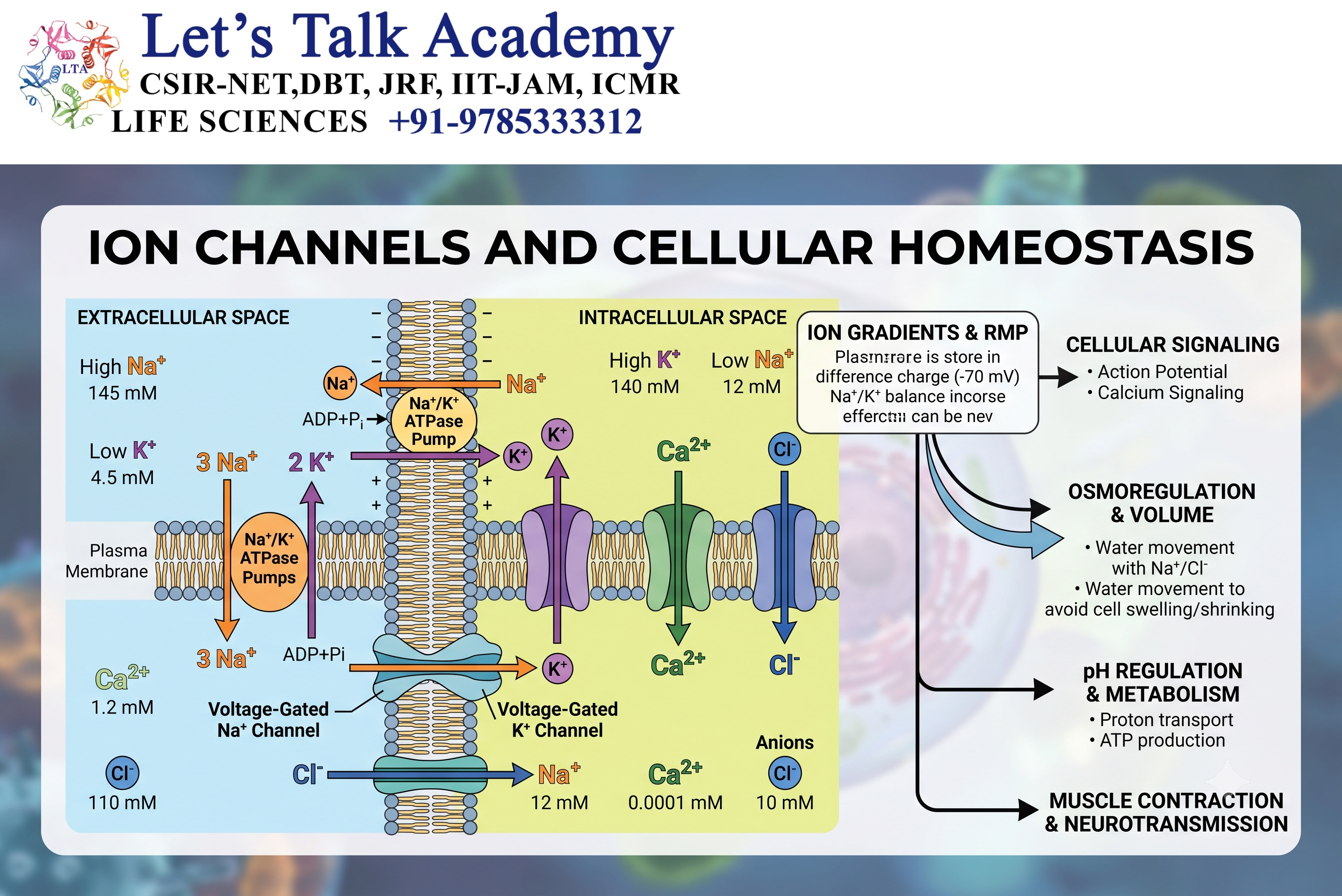
Ion channels contribute to cellular homeostasis by controlling:
- Ionic composition
- Osmotic balance
- Cell volume
- Membrane voltage
- Intracellular signaling
- pH regulation
If ion-channel activity becomes severely disrupted, the cell may lose its ability to maintain appropriate internal conditions.
43. Comparison of Major Ion Channel Types
| Channel Type | Primary Stimulus | Major Function |
|---|---|---|
| Voltage-gated | Change in membrane potential | Electrical signaling |
| Ligand-gated | Chemical ligand | Synaptic transmission |
| Mechanically gated | Mechanical force | Sensory signaling |
| Calcium-activated | Intracellular Ca²⁺ | Feedback regulation |
| ATP-sensitive | Cellular metabolic state | Metabolic sensing |
| Leak channels | Constitutive/resting activity | Resting membrane potential |
| Temperature-sensitive | Temperature changes | Thermosensation |


