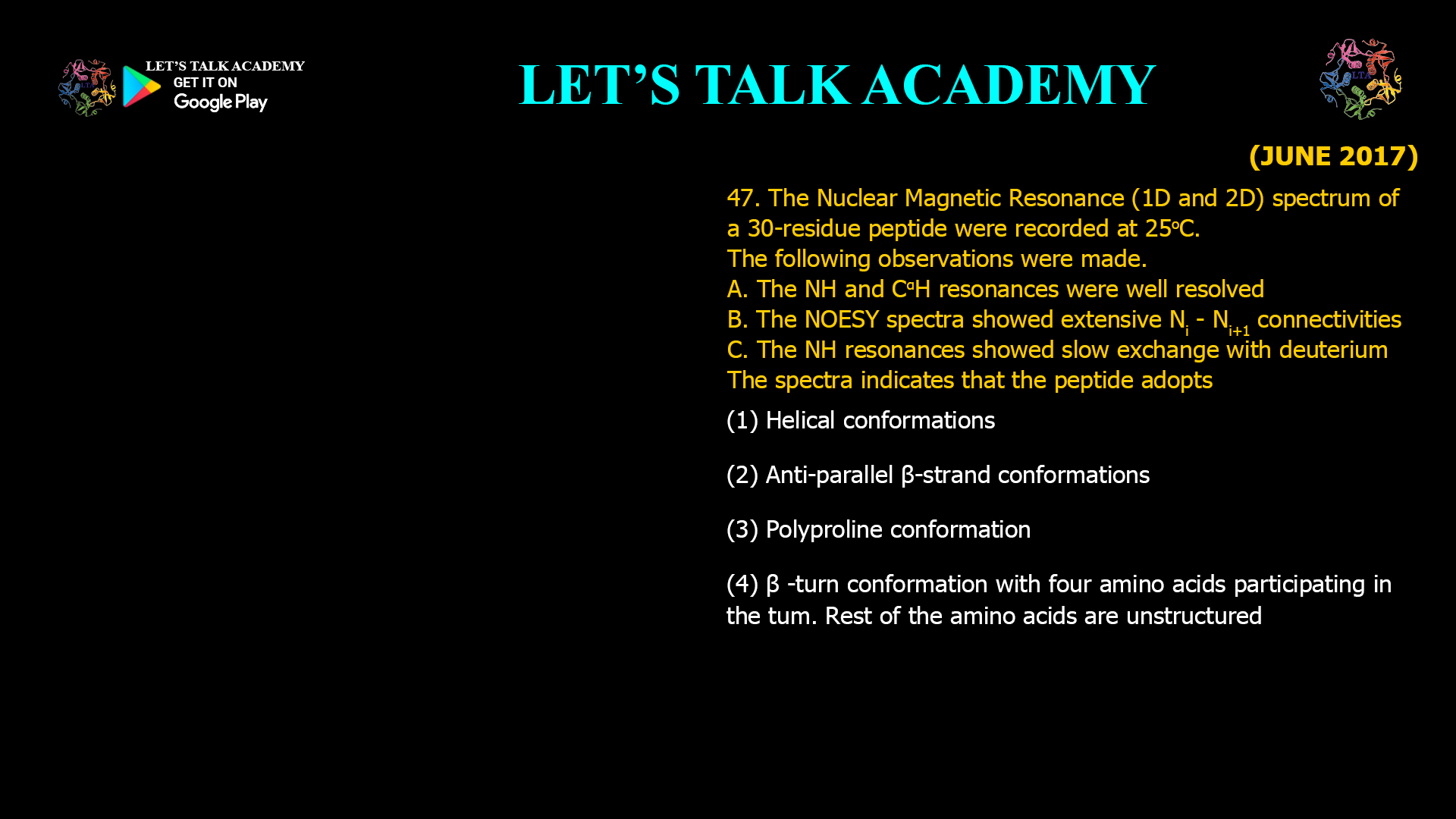
47. The Nuclear Magnetic Resonance (1D and 2D) spectrum of a 30-residue peptide were recorded at 25°C. The following observations were made.
A. The NH and CαH resonances were well resolved.
B. The NOESY spectra showed extensive Nᵢ − Nᵢ₊₁ connectivities.
C. The NH resonances showed slow exchange with deuterium.
The spectra indicates that the peptide adopts
(A) Helical conformations
(B) Anti-parallel β-strand conformations
(C) Polyproline conformation
(D) β-turn conformation with four amino acids participating in the turn. Rest of the amino acids are unstructured.
Interpreting 1D and 2D NMR Spectra to Identify Helical Peptide Conformations
Correct Answer
Option (1): Helical conformations
Explanation
Nuclear Magnetic Resonance (NMR) spectroscopy is one of the most informative techniques for determining the three-dimensional structure of peptides and proteins in solution. One-dimensional spectra provide information about chemical environments, whereas two-dimensional experiments such as NOESY reveal through-space interactions between nearby nuclei. When these observations are combined with hydrogen-deuterium exchange experiments, they provide strong evidence regarding the secondary structure adopted by a peptide.
The first observation states that the NH and CαH resonances are well resolved. Good spectral dispersion indicates that the peptide adopts a well-defined three-dimensional structure rather than a rapidly fluctuating random coil. In an unstructured peptide, many residues experience similar chemical environments, resulting in overlapping resonances. Well-separated resonances therefore suggest the presence of an ordered conformation.
The second observation is the presence of extensive Ni–Ni+1 Nuclear Overhauser Effect (NOE) connectivities in the NOESY spectrum. These sequential NOEs arise because adjacent backbone amide protons are positioned close together in space. Continuous stretches of strong sequential NH-NH connectivities are characteristic of α-helical peptides. In addition to these sequential NOEs, helical peptides frequently exhibit medium-range NOEs such as Ni–Ni+3, Ni–Ni+4, and αHi–NHi+3, reflecting the regular geometry of the helix.
The third observation states that the backbone amide protons undergo slow hydrogen-deuterium exchange. Amide hydrogens exchange slowly only when they are protected from the solvent, usually because they participate in stable intramolecular hydrogen bonds. In an α-helix, every backbone carbonyl oxygen forms a hydrogen bond with the amide proton of the residue located four positions ahead in the sequence. This extensive hydrogen-bonding network protects many amide protons from solvent exchange, resulting in the observed slow exchange rate.
Taken together, these three observations strongly indicate that the peptide adopts a stable α-helical conformation. The ordered chemical shift dispersion, characteristic NOESY connectivities, and protection of amide protons through hydrogen bonding are all hallmarks of helical secondary structure.
Why Option (1) is Correct
This option explains all three experimental observations. Well-resolved resonances indicate a folded structure, extensive sequential NOEs are characteristic of helices, and slow hydrogen-deuterium exchange demonstrates that backbone amide protons are protected by stable hydrogen bonds. The combined NMR evidence is entirely consistent with a helical conformation.
Why Option (2) is Incorrect
Anti-parallel β-strands produce a different pattern of NOEs. Their spectra are dominated by long-range inter-strand contacts between residues located on adjacent β-strands rather than continuous sequential NH-NH connectivities. Although β-sheets also contain hydrogen bonds that can slow deuterium exchange, the NOESY pattern described in the question does not correspond to an anti-parallel β-sheet.
Why Option (3) is Incorrect
Polyproline helices possess structural features that differ significantly from α-helices. In addition, proline lacks a backbone amide hydrogen, making extensive NH-NH sequential NOEs impossible. Since the observations specifically involve numerous backbone amide proton resonances and their connectivities, the peptide cannot adopt a polyproline conformation.
Why Option (4) is Incorrect
A β-turn involves only four consecutive amino acid residues and generates local NOE contacts confined to that short region. It cannot account for extensive sequential connectivities throughout a 30-residue peptide or widespread protection of amide protons from hydrogen-deuterium exchange. Therefore, the observations indicate a more extensive and continuous secondary structure rather than a single localized turn.
Role of NOESY in Secondary Structure Determination
NOESY spectroscopy detects interactions between nuclei that are separated by less than approximately 5 Å in space. Because different secondary structures position atoms differently, each structural motif produces a characteristic pattern of NOE cross-peaks. Helices generate strong sequential and medium-range NOEs, whereas β-sheets produce characteristic long-range contacts between residues located on different strands. Analysis of these interaction patterns is one of the primary methods used for determining protein structures by solution NMR.
Hydrogen-Deuterium Exchange and Structural Stability
When peptides are dissolved in D₂O, backbone amide hydrogens gradually exchange with deuterium. Amide protons exposed to the solvent exchange rapidly, whereas those involved in stable hydrogen bonds exchange much more slowly. Slow exchange therefore indicates that the peptide contains a stable hydrogen-bonded secondary structure capable of protecting backbone amides from the solvent.
Chemical Shift Dispersion in Folded Proteins
The distribution of chemical shifts in an NMR spectrum reflects the diversity of local chemical environments experienced by individual nuclei. Folded proteins and well-structured peptides display broad chemical shift dispersion because each residue occupies a unique structural environment. In contrast, unfolded polypeptides exhibit poor dispersion owing to the similarity of their rapidly fluctuating conformations.
Conclusion
The combination of well-resolved NH and CαH resonances, extensive sequential NH-NH NOESY connectivities, and slow hydrogen-deuterium exchange provides strong evidence for a stable hydrogen-bonded α-helical structure. These observations collectively identify the peptide as adopting a helical conformation. Therefore, the correct answer is Option (1).


