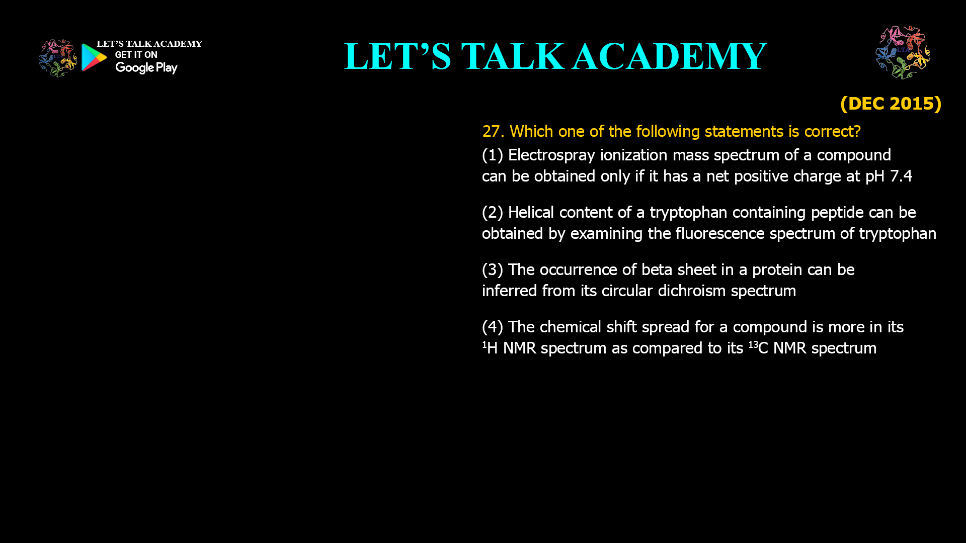
27. Which one of the following statements is correct?
(1) Electrospray ionization mass spectrum of a compound can be obtained only if it has a net positive charge at pH 7.4
(2) Helical content of a tryptophan containing peptide can be obtained by examining the fluorescence spectrum of tryptophan
(3) The occurrence of beta sheet in a protein can be inferred from its circular dichroism spectrum
(4) The chemical shift spread for a compound is more in its 1H NMR spectrum as compared to its 13C NMR spectrum
How Circular Dichroism Identifies Beta-Sheet Structure in Proteins
Modern structural biology relies on multiple analytical techniques to investigate proteins, peptides, nucleic acids, and other biological macromolecules. Each technique provides different information about molecular properties. Electrospray Ionization Mass Spectrometry (ESI-MS) measures molecular mass, fluorescence spectroscopy detects changes in the environment of fluorescent amino acids, Circular Dichroism (CD) spectroscopy reveals protein secondary structure, and Nuclear Magnetic Resonance (NMR) spectroscopy provides detailed structural information at the atomic level.
Correct Answer
Option (3): The Occurrence of β-Sheet in a Protein Can Be Inferred from Its Circular Dichroism Spectrum
The correct answer is Option (3). Circular Dichroism spectroscopy is specifically designed to analyze the secondary structure of proteins by measuring the differential absorption of left-handed and right-handed circularly polarized light. In the far-ultraviolet region, peptide bonds produce characteristic CD spectra that differ for α-helices, β-sheets, β-turns, and random coils. Because β-sheet structures generate a distinct spectral signature, their presence and approximate abundance can be inferred from the CD spectrum of a protein.
This principle forms the basis of routine secondary structure estimation in structural biology laboratories and is one of the most important applications of Circular Dichroism spectroscopy.
Understanding Circular Dichroism and Protein Secondary Structure
Circular Dichroism spectroscopy measures the difference in absorption between left-handed and right-handed circularly polarized light by chiral molecules. In proteins, the peptide backbone is responsible for characteristic electronic transitions that produce distinctive CD spectra depending on the arrangement of peptide bonds.
An α-helix typically exhibits strong negative bands near 208 nm and 222 nm together with a positive band near 190 nm. In contrast, β-sheets produce a different combination of positive and negative ellipticity, while random coils display another characteristic spectral pattern. By comparing an experimental CD spectrum with reference datasets, researchers can estimate the proportions of different secondary structural elements present in a protein.
Why Option (1) is Incorrect – Electrospray Ionization Mass Spectrometry
Electrospray Ionization (ESI) does not require a molecule to possess a permanent positive charge at physiological pH. During the ionization process, neutral molecules can acquire positive ions by protonation or negative ions by deprotonation, depending on the experimental conditions and the selected ionization mode.
Proteins, peptides, nucleic acids, carbohydrates, and many neutral organic compounds are routinely analyzed by ESI-MS even though they may not carry a net positive charge at pH 7.4. Therefore, the statement is scientifically incorrect.
Why Option (2) is Incorrect – Tryptophan Fluorescence
Tryptophan fluorescence is extremely sensitive to its local environment, including solvent polarity, protein folding, and ligand binding. However, fluorescence spectroscopy does not directly measure the percentage of α-helical secondary structure. A tryptophan residue may be buried inside either an α-helix or a β-sheet, or it may reside within a loop region, producing similar fluorescence depending on its surrounding environment.
Although fluorescence spectroscopy is valuable for monitoring conformational changes, it cannot accurately determine helical content. Circular Dichroism spectroscopy remains the preferred technique for estimating protein secondary structure.
Why Option (3) is Correct – Circular Dichroism Detects β-Sheet Structure
The far-ultraviolet Circular Dichroism spectrum originates primarily from electronic transitions within peptide bonds. Because peptide bonds adopt characteristic orientations in β-sheet structures, the resulting CD spectrum exhibits a distinct pattern that differs from α-helices and random coils.
Researchers routinely analyze these spectral features using computational deconvolution methods to estimate the proportion of β-sheet present in proteins. Consequently, Circular Dichroism spectroscopy is widely recognized as a reliable technique for identifying and quantifying β-sheet secondary structure.
Why Option (4) is Incorrect – Chemical Shift Spread in NMR
The chemical shift range of 13C NMR is substantially larger than that of 1H NMR. Proton chemical shifts usually extend from approximately 0 to 12 ppm, whereas carbon-13 chemical shifts commonly span about 0 to more than 220 ppm.
The much wider chemical shift dispersion of 13C NMR greatly reduces signal overlap and makes carbon spectra easier to interpret in many structural studies. Therefore, the statement incorrectly reverses the relationship between proton and carbon chemical shift ranges.
Why Circular Dichroism is Widely Used in Protein Research
Circular Dichroism spectroscopy has become one of the most important tools for studying protein folding, thermal denaturation, ligand binding, membrane proteins, recombinant protein quality, conformational changes, and secondary structure estimation. Since measurements can be performed rapidly in solution without crystallization, CD provides valuable structural information under conditions that closely resemble the natural biological environment.
In combination with techniques such as X-ray crystallography, Nuclear Magnetic Resonance spectroscopy, fluorescence spectroscopy, and cryo-electron microscopy, Circular Dichroism contributes significantly to understanding protein structure and function.
Comparison of the Four Techniques
Electrospray Ionization Mass Spectrometry determines molecular mass by generating gas-phase ions. Fluorescence spectroscopy measures emission from fluorescent residues such as tryptophan and is primarily used to study environmental changes rather than secondary structure. Circular Dichroism directly measures protein secondary structure through peptide bond optical activity, while NMR spectroscopy provides detailed atomic-level structural information. Among these techniques, only Circular Dichroism directly allows the inference of β-sheet content from its characteristic spectral signature.
Final Answer
Correct Option: (3) The occurrence of β-sheet in a protein can be inferred from its Circular Dichroism spectrum.
Circular Dichroism spectroscopy is specifically designed to analyze protein secondary structure by measuring the differential absorption of circularly polarized light. Because β-sheet structures generate a characteristic far-ultraviolet CD spectrum, their presence and approximate abundance can be determined from experimental CD measurements. In contrast, Electrospray Ionization Mass Spectrometry does not require molecules to possess a permanent positive charge, tryptophan fluorescence does not directly quantify α-helical content, and 13C NMR exhibits a much larger chemical shift range than 1H NMR. Therefore, Option (3) is the correct answer.


