
1. Introduction
B and T lymphocytes are the major cellular components of adaptive immunity. Although both originate from hematopoietic stem cells in the bone marrow, they undergo different developmental processes and perform distinct functions.
B cells are primarily responsible for humoral immunity, particularly antibody production, while T cells coordinate cellular immune responses, provide help to other immune cells, and directly eliminate infected or abnormal cells.
Naive B and T cells are generally not fully functional effector cells. They must first encounter appropriate antigenic signals and undergo activation, clonal expansion, and differentiation.
The overall process can be summarized as:
Antigen recognition → Activation → Signaling → Clonal expansion → Differentiation → Effector response + Memory formation
2. Definition of B-Cell Activation
B-cell activation is the process through which a mature naive B lymphocyte recognizes antigen and receives the signals necessary to proliferate and differentiate into effector and memory cells.
Activated B cells can differentiate into:
- Plasma cells
- Memory B cells
Plasma cells produce antibodies, whereas memory B cells provide long-term immunological memory.
3. Definition of T-Cell Activation
T-cell activation is the process through which a naive T lymphocyte recognizes a specific peptide–MHC complex and receives additional signals required for proliferation and differentiation.
Depending on the type of T cell and cytokine environment, activated T cells can differentiate into different effector populations.
Major populations include:
- CD4⁺ helper T cells
- CD8⁺ cytotoxic T cells
- Regulatory T cells
- Memory T cells
4. Major Requirements for Lymphocyte Activation
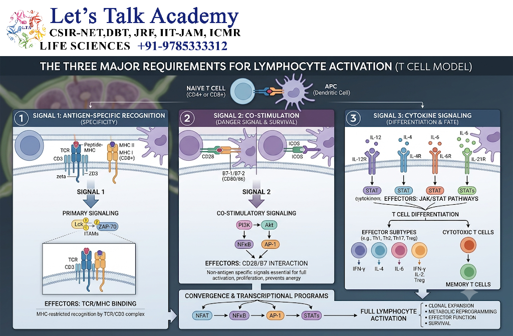
Effective activation generally requires several coordinated signals.
4.1 Antigen Recognition
The lymphocyte recognizes its specific antigen.
4.2 Co-stimulatory Signals
Additional receptor interactions provide information that supports activation.
4.3 Cytokine Signals
Cytokines influence proliferation, survival, differentiation, and functional specialization.
Thus:
Antigen recognition + Co-stimulation + Cytokine environment → Effective lymphocyte activation
5. B-Cell Receptor
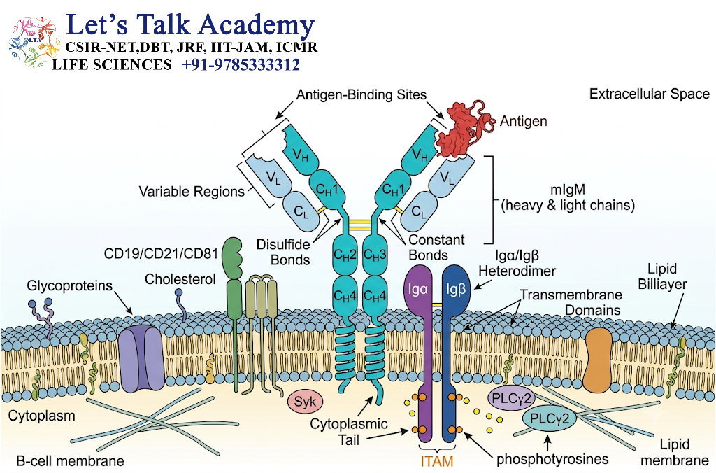
The B-cell receptor (BCR) is the antigen-recognition complex expressed on B cells.
The membrane-bound immunoglobulin component recognizes antigen.
However, membrane immunoglobulin does not efficiently transmit intracellular signals by itself. Signaling is mediated mainly through:
- CD79a
- CD79b
These molecules contain ITAMs (immunoreceptor tyrosine-based activation motifs).
6. Recognition of Antigen by B Cells
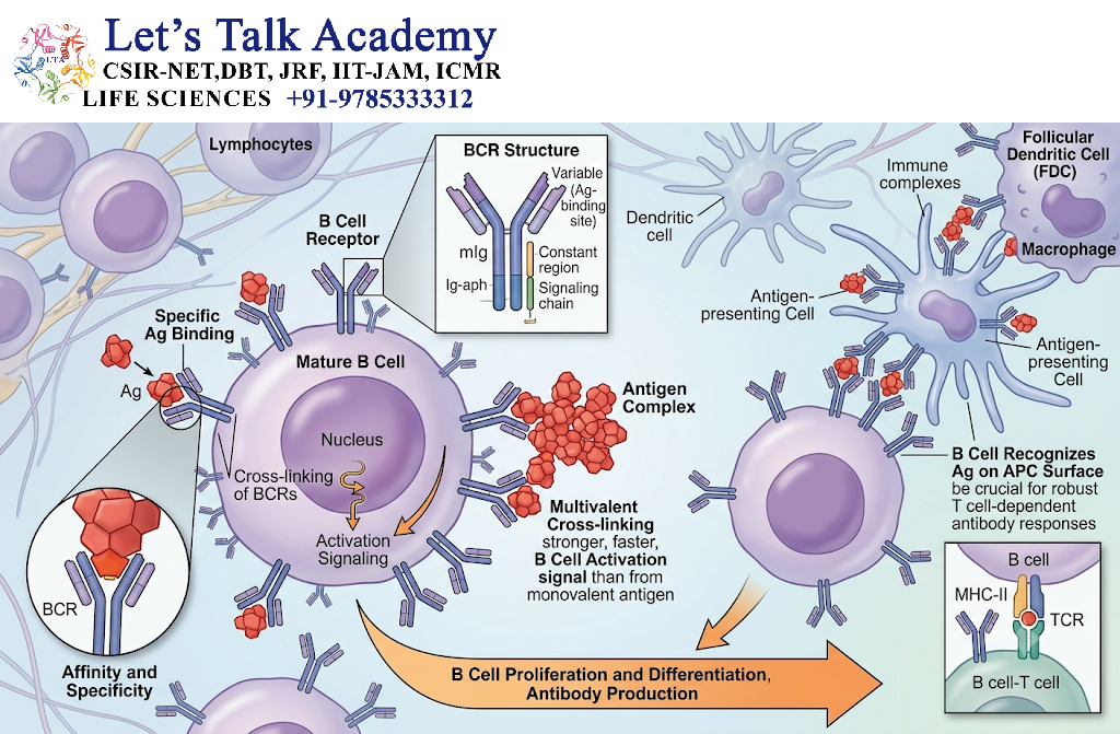
B cells can recognize antigen in its relatively intact molecular form.
This differs from T cells, which generally recognize processed peptides presented by MHC molecules.
The basic process is:
Antigen → BCR binding → BCR clustering → Intracellular signaling → B-cell activation
BCR binding can occur with:
- Proteins
- Polysaccharides
- Lipids
- Glycoproteins
- Other molecular structures
7. Initial BCR Signaling
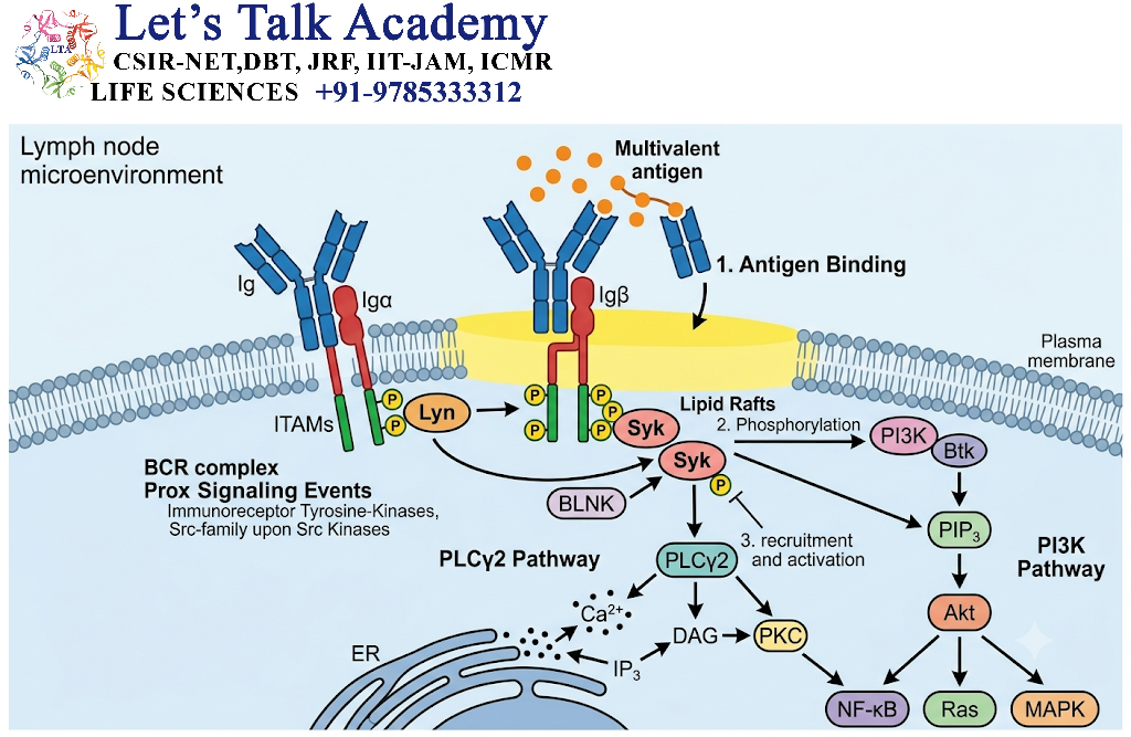
Antigen binding causes clustering and structural changes in BCR complexes.
This promotes activation of Src-family kinases such as:
- Lyn
- Fyn
- Blk
These kinases phosphorylate ITAMs within CD79a and CD79b.
Phosphorylated ITAMs recruit and activate Syk kinase.
The pathway can be simplified as:
Antigen → BCR → Src-family kinases → ITAM phosphorylation → Syk activation
8. Major BCR Signaling Pathways
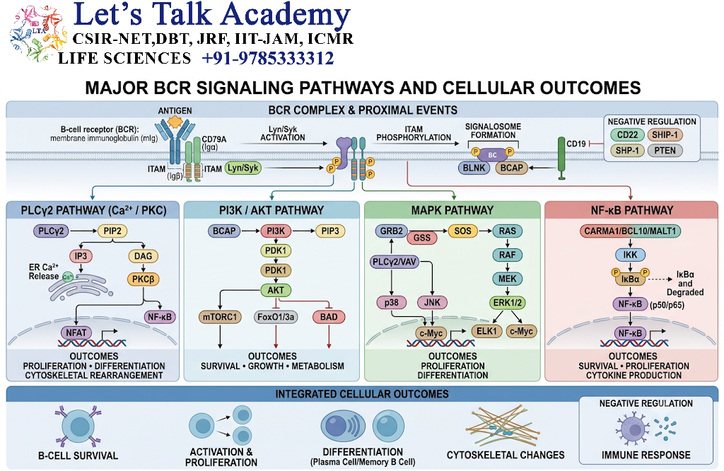
BCR signaling activates several downstream pathways.
Important pathways include:
- PLCγ2
- Calcium signaling
- NF-κB
- NFAT
- MAPK
- PI3K–AKT
These pathways ultimately alter gene expression and cellular behavior.
9. BCR Signaling Flowchart
Antigen
   ↓
BCR engagement
   ↓
Lyn / Fyn / Blk
   ↓
CD79a/CD79b ITAM phosphorylation
   ↓
Syk activation
   ↓
Signaling complexes
   ↓
 ┌──────────┬───────────┬──────────┬──────────┐
 ↓          ↓           ↓          ↓
PLCγ2      PI3K       MAPK       NF-κB
 ↓          ↓           ↓
Ca²⁺       AKT        ERK/JNK
 ↓
NFAT
   ↓
Gene expression
   ↓
B-cell activation
10. Role of Co-Receptors in B-Cell Activation
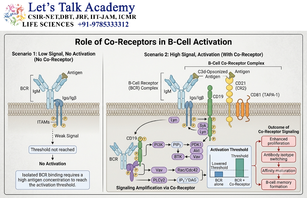
B-cell activation is influenced by several co-receptors.
An important positive co-receptor complex contains:
- CD19
- CD21
- CD81
CD21 can recognize complement fragments associated with antigen.
Co-engagement of the BCR and CD19-containing co-receptor complex can amplify BCR signaling.
11. Antigen Internalization by B Cells
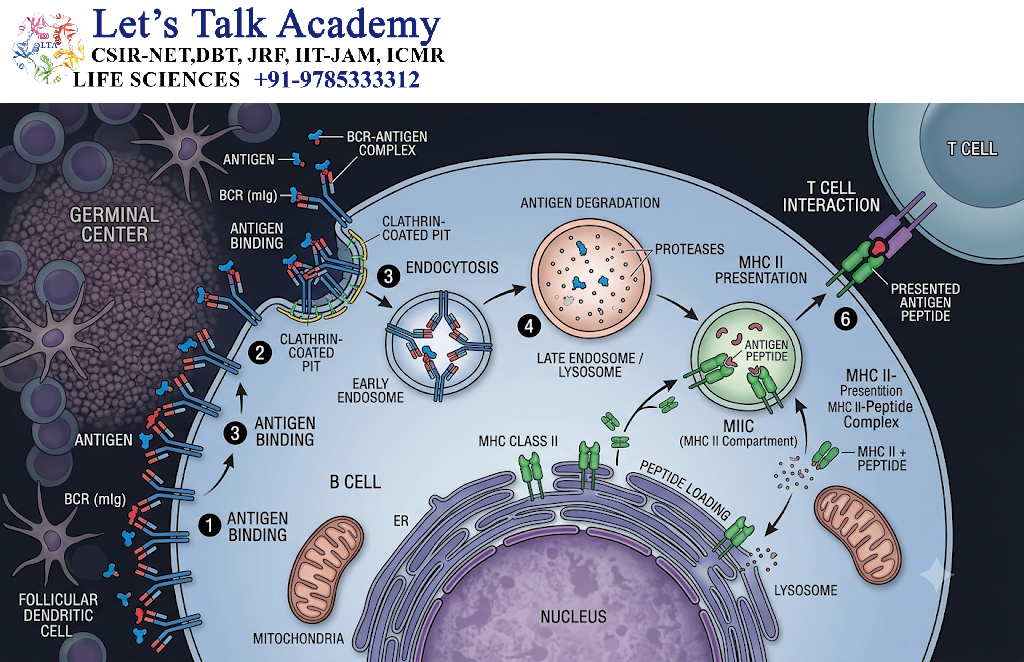
After BCR binding, B cells can internalize the antigen.
The antigen is then processed into peptide fragments.
These peptides can be loaded onto MHC Class II molecules and displayed on the B-cell surface.
Therefore, B cells can function as antigen-presenting cells for CD4⁺ helper T cells.
12. T-Cell Help for B-Cell Activation
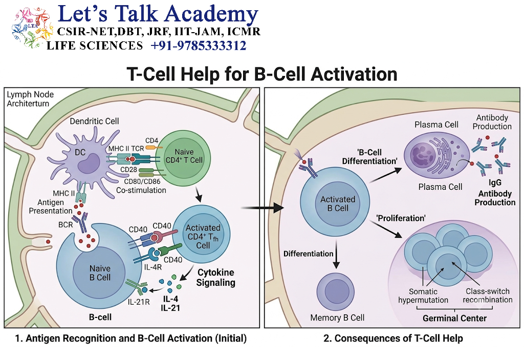
Many protein antigens generate strong antibody responses through T-cell-dependent B-cell activation.
The process involves:
BCR binds antigen → Antigen internalization → Peptide processing → MHC II presentation → CD4⁺ T-cell recognition → T-cell help → B-cell proliferation and differentiation
An important interaction is:
CD40 on B cell ↔ CD40L on activated T cell
This interaction is essential for many T-cell-dependent B-cell responses.
13. Cytokines in B-Cell Differentiation
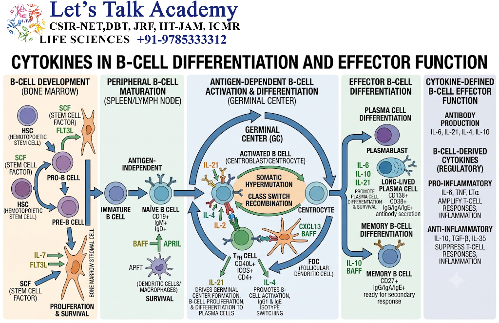
Cytokines produced by helper T cells and other immune cells influence B-cell differentiation.
They can affect:
- Plasma-cell formation
- Memory-cell formation
- Antibody class switching
- Survival
- Proliferation
Different cytokine environments can therefore produce different antibody responses.
14. T-Independent B-Cell Activation
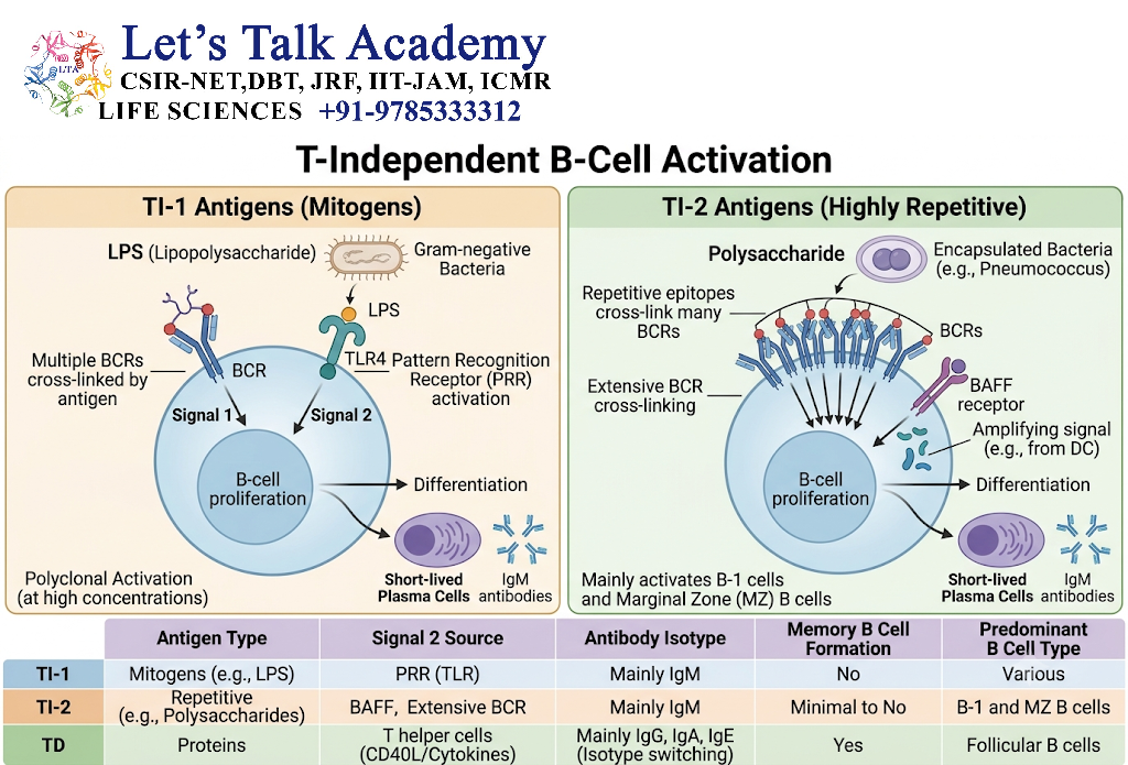
Some antigens can activate B cells without conventional T-cell help.
These are called T-independent antigens.
Common examples include certain:
- Polysaccharides
- Repetitive microbial surface structures
- Bacterial components
T-independent responses often produce:
- Rapid antibody production
- Predominantly IgM responses
- Limited affinity maturation compared with strong germinal-center responses
- Variable memory responses
15. T-Dependent vs T-Independent B-Cell Activation
| Feature | T-Dependent | T-Independent |
|---|---|---|
| Main antigen | Usually protein | Often repetitive non-protein structures |
| T-cell help | Required | Not required |
| CD40–CD40L | Important | Not essential in the conventional pathway |
| Germinal center | Common | Limited or absent |
| Class switching | Strong | More limited |
| Affinity maturation | Prominent | Usually limited |
| Memory | Strong | Generally weaker |
16. Clonal Selection of B Cells
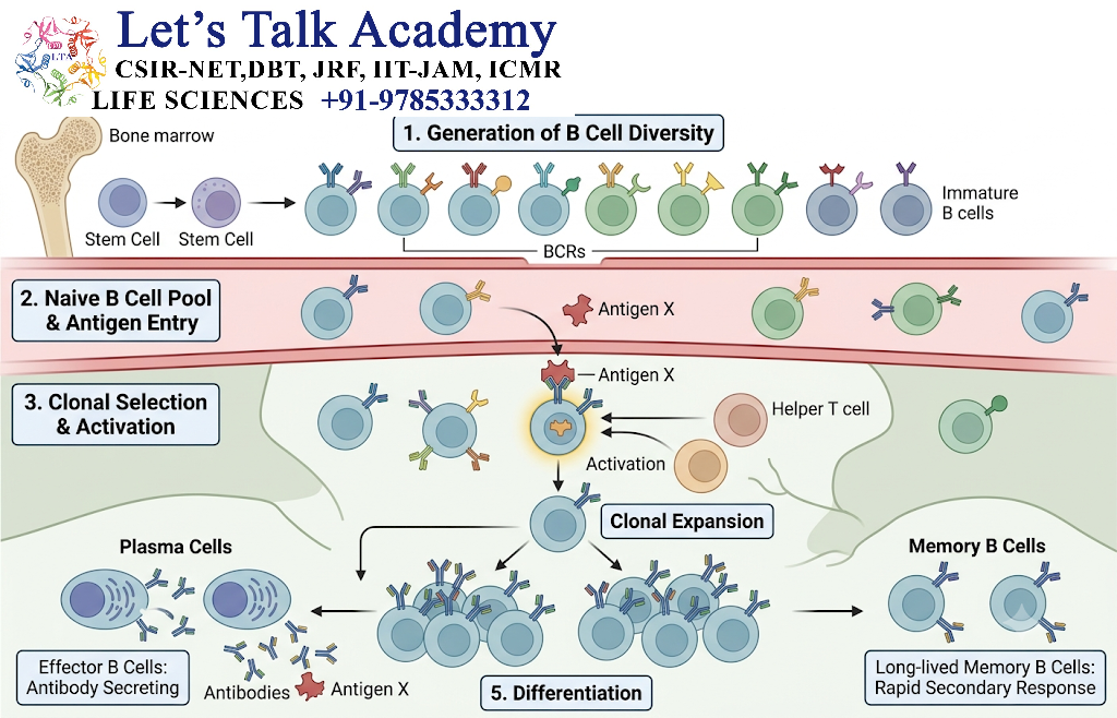
The adaptive immune system contains many B-cell clones with different antigen receptors.
When an antigen enters the body, it preferentially activates B cells whose BCRs recognize that antigen.
This principle is called clonal selection.
Many B-cell clones
       ↓
Antigen enters
       ↓
Specific BCR recognizes antigen
       ↓
Selected B-cell clone
       ↓
Clonal expansion
       ↓
Effector + Memory B cells
17. Clonal Expansion of B Cells
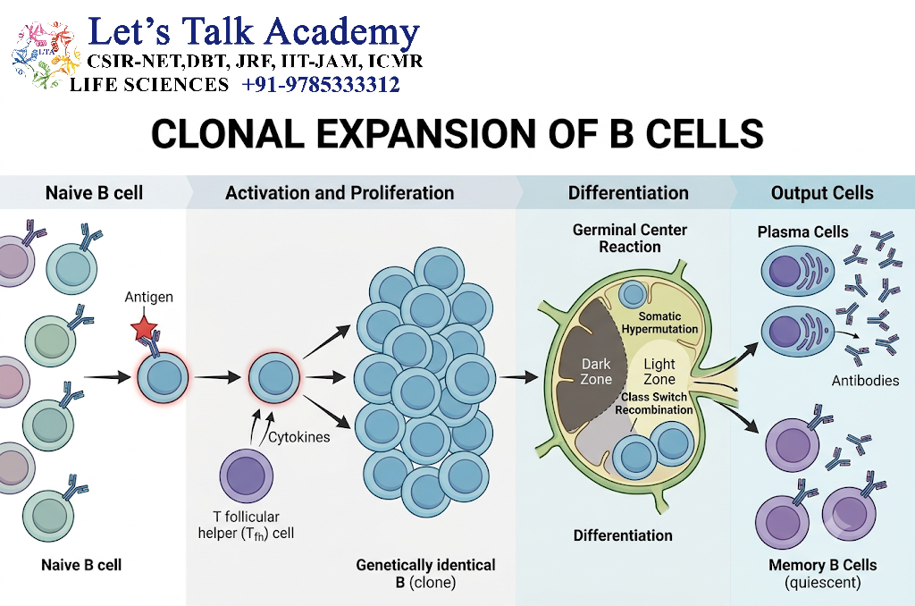
After activation, selected B cells undergo rapid proliferation.
This produces many daughter cells carrying the same antigen specificity.
This process is called clonal expansion.
The expanded population can then differentiate into different functional cell types.
18. B-Cell Differentiation
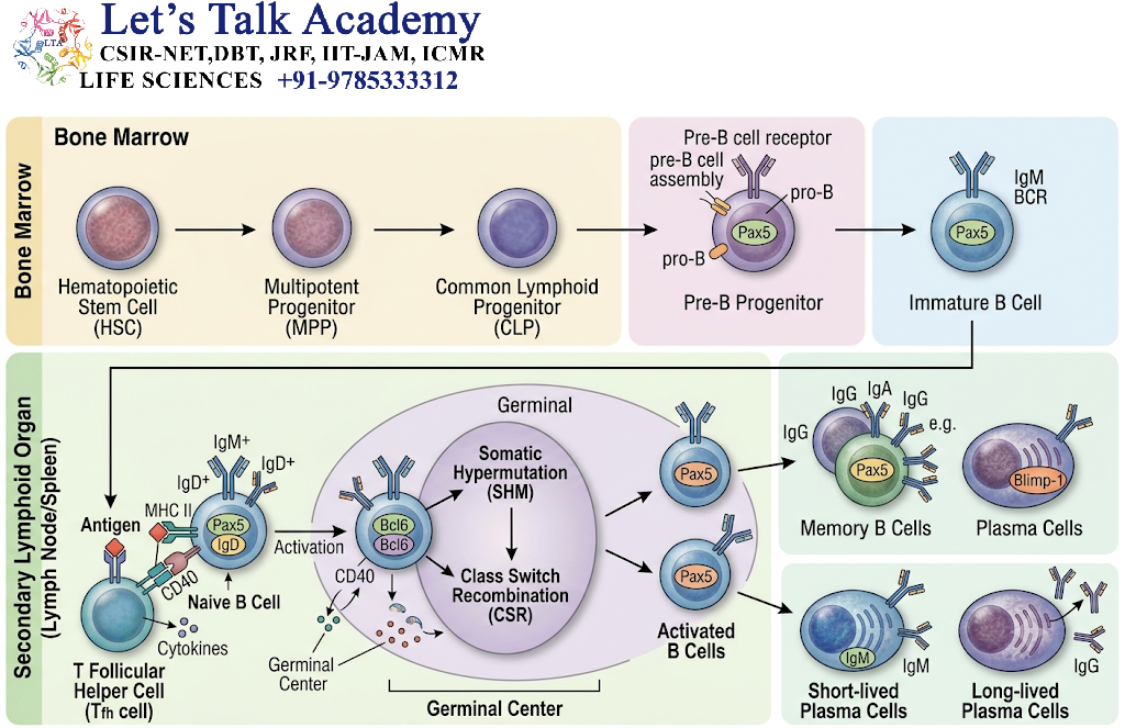
Activated B cells can differentiate primarily into:
18.1 Plasma Cells
Plasma cells are specialized antibody-secreting cells.
They contain extensive rough endoplasmic reticulum and Golgi apparatus to support high levels of antibody production.
18.2 Memory B Cells
Memory B cells survive after the initial immune response and allow a faster response during subsequent exposure to the same antigen.
19. Plasma Cell Formation
The differentiation process involves major changes in gene expression.
Important transcriptional regulators include:
- BLIMP-1
- XBP-1
- IRF4
BLIMP-1 promotes the plasma-cell transcriptional program, while XBP-1 supports the high secretory capacity required for antibody production.
20. Germinal Center Reaction
For many T-cell-dependent antibody responses, activated B cells enter specialized structures called germinal centers within secondary lymphoid organs.
Germinal centers contain two major functional regions:
- Dark zone
- Light zone
The germinal center reaction supports:
- Rapid B-cell proliferation
- Somatic hypermutation
- Affinity maturation
- Class-switch recombination
- Selection of B cells
- Memory-cell formation
- Plasma-cell formation
21. Somatic Hypermutation
Activated B cells can introduce mutations into immunoglobulin variable-region genes.
This process is called somatic hypermutation.
The enzyme activation-induced cytidine deaminase (AID) is essential for this process.
The resulting B-cell population contains antibodies with different binding properties.
22. Affinity Maturation
Following somatic hypermutation, B cells with improved antigen-binding properties can be preferentially selected.
This results in affinity maturation.
Simplified sequence:
Activation → Somatic hypermutation → Variation in affinity → Selection → Higher-affinity B-cell clones
23. Class-Switch Recombination
Activated B cells can change the antibody isotype they produce while maintaining the same basic antigen specificity.
This is called class-switch recombination (CSR).
For example:
IgM → IgG
or:
IgM → IgA
or:
IgM → IgE
AID is also essential for class-switch recombination.
The variable region remains responsible for antigen recognition, while the constant region of the heavy chain changes.
24. B-Cell Activation and Differentiation Flowchart
Antigen
   ↓
BCR recognition
   ↓
BCR signaling
   ↓
Antigen internalization
   ↓
MHC II presentation
   ↓
CD4⁺ T-cell help
   ↓
CD40–CD40L + cytokines
   ↓
Clonal expansion
   ↓
Germinal center reaction
   ↓
 ┌──────────────────────────┐
 ↓                          ↓
Somatic hypermutation     Class switching
 ↓                          ↓
Affinity maturation        Different isotypes
 └────────────┬─────────────┘
              ↓
       Differentiation
        ┌─────────────┐
        ↓             ↓
   Plasma cells   Memory B cells
        ↓             ↓
   Antibodies     Rapid secondary
                  response
25. Definition of T-Cell Activation
T-cell activation occurs when a T cell recognizes a specific peptide–MHC complex and receives appropriate co-stimulatory and cytokine signals.
The three major conceptual signals are:
- TCR recognition of peptide–MHC
- Co-stimulation
- Cytokine signaling
These signals determine whether the T cell becomes activated and what functional state it adopts.
26. T-Cell Receptor
The T-cell receptor (TCR) recognizes antigen-derived peptides presented by MHC molecules.
The TCR itself is associated with the CD3 signaling complex.
CD3 contains signaling chains with ITAMs that transmit signals into the cell.
27. Signal 1: Antigen Recognition
The first major signal is produced by interaction between:
TCR ↔ Peptide–MHC
For CD4⁺ T cells:
TCR ↔ Peptide–MHC II
For CD8⁺ T cells:
TCR ↔ Peptide–MHC I
The appropriate co-receptor also participates:
CD4 → MHC II
CD8 → MHC I
28. TCR Signaling Pathway
TCR engagement activates Src-family kinases, particularly:
Lck
Lck phosphorylates ITAMs associated with the CD3 complex.
This promotes activation of:
ZAP-70
ZAP-70 then activates adaptor proteins and signaling pathways that ultimately regulate transcription factors.
Important pathways include:
- Calcium–NFAT
- Ras–MAPK–AP-1
- PKCθ–NF-κB
29. TCR Signaling Flowchart
Peptide–MHC
     ↓
TCR + CD4/CD8
     ↓
Lck activation
     ↓
CD3 ITAM phosphorylation
     ↓
ZAP-70 activation
     ↓
Adaptor proteins
     ↓
 ┌────────────┬─────────────┬─────────────┐
 ↓            ↓             ↓
Ca²⁺         Ras/MAPK      PKCθ
 ↓            ↓             ↓
NFAT         AP-1          NF-κB
 └────────────┴─────────────┘
              ↓
       Gene transcription
              ↓
       T-cell activation
30. Signal 2: Co-Stimulation
TCR recognition alone is generally insufficient for full activation of a naive T cell.
An important co-stimulatory interaction is:
CD28 on T cell ↔ B7 molecules (CD80/CD86) on APC
This provides an important positive signal supporting T-cell activation.
Without appropriate co-stimulation, antigen recognition can result in a state of functional unresponsiveness called anergy in appropriate contexts.
31. Signal 3: Cytokines
Cytokines provide additional information that influences T-cell proliferation and differentiation.
The cytokine environment helps determine which functional T-cell lineage develops.
Thus:
Signal 1 → What antigen is recognized
Signal 2 → Is appropriate activation occurring
Signal 3 → What type of T-cell response develops
32. IL-2 and T-Cell Proliferation
One of the most important cytokines for activated T-cell proliferation is interleukin-2 (IL-2).
T-cell activation increases expression of:
- IL-2
- IL-2 receptor α chain (CD25)
IL-2 signaling promotes proliferation and survival of activated T cells.
The process is:
T-cell activation → IL-2 production → IL-2 receptor signaling → Clonal expansion
33. Clonal Expansion of T Cells
Once activated, antigen-specific T cells proliferate extensively.
The resulting daughter cells carry the same antigen receptor specificity.
This creates a large population capable of responding to the antigen.
Naive T cell
     ↓
Antigen recognition
     ↓
Activation
     ↓
IL-2 production
     ↓
Clonal expansion
     ↓
Large antigen-specific T-cell population
34. CD4⁺ T-Cell Differentiation
Activated CD4⁺ T cells can differentiate into several functional subsets.
Major populations include:
- Th1
- Th2
- Th17
- Tfh
- Treg
The differentiation pathway depends strongly on the cytokine environment and transcriptional programs.
35. Th1 Cells
Th1 cells are particularly associated with cellular immune responses against intracellular pathogens.
Important cytokine:
IFN-γ
Major transcription factor:
T-bet
Functions include:
- Macrophage activation
- Support of cell-mediated immunity
- Promotion of inflammatory responses appropriate to intracellular pathogens
36. Th2 Cells
Th2 cells are associated with immune responses involving:
- Helminths
- Type 2 inflammation
- IgE-associated responses
Important cytokines include:
- IL-4
- IL-5
- IL-13
Important transcription factor:
GATA-3
Functions include:
- Support of B-cell responses
- Eosinophil-associated immunity
- Promotion of IgE responses in appropriate contexts
37. Th17 Cells
Th17 cells produce cytokines that contribute to protection against certain extracellular bacteria and fungi, particularly at barrier tissues.
Important cytokines include:
- IL-17
- IL-22
Important transcription factor:
RORγt
Functions include:
- Recruitment and activation of neutrophil-associated responses
- Barrier defense
- Mucosal immunity
Excessive or dysregulated Th17 responses can contribute to inflammatory and autoimmune conditions.
38. T Follicular Helper Cells
T follicular helper (Tfh) cells specialize in providing help to B cells within lymphoid follicles and germinal centers.
Important features include:
- CXCR5 expression
- BCL-6 transcription factor
- IL-21 production
- CD40L expression
Tfh cells support:
- B-cell proliferation
- Class switching
- Affinity maturation
- Plasma-cell formation
- Memory B-cell formation
39. Regulatory T Cells
Regulatory T cells (Treg) suppress excessive immune responses and help maintain immune tolerance.
Important characteristics include:
- CD4 expression
- CD25 expression
- FOXP3 transcription factor
Tregs can suppress immune responses through:
- Inhibitory cytokines
- Cell-contact mechanisms
- Modulation of APC function
- Consumption of growth-promoting cytokines
40. CD4⁺ T-Cell Differentiation Table
| T-cell subset | Major cytokines | Important transcription factor | Major function |
|---|---|---|---|
| Th1 | IFN-γ | T-bet | Intracellular pathogen defense |
| Th2 | IL-4, IL-5, IL-13 | GATA-3 | Type 2 immunity |
| Th17 | IL-17, IL-22 | RORγt | Barrier and extracellular pathogen defense |
| Tfh | IL-21 | BCL-6 | B-cell help |
| Treg | IL-10, TGF-β and others | FOXP3 | Immune regulation |
These are simplified functional associations; T-cell phenotypes can be influenced by context and may overlap.
41. CD8⁺ T-Cell Activation
CD8⁺ T cells recognize peptide–MHC Class I complexes.
After activation, they can differentiate into cytotoxic T lymphocytes (CTLs).
CTLs can kill target cells displaying appropriate antigen.
Important mechanisms include:
Perforin–Granzyme Pathway
CTL → Perforin release → Granzyme entry → Caspase activation → Apoptosis
Fas–FasL Pathway
FasL on CTL → Fas on target cell → Death signaling → Apoptosis
42. CD8⁺ T-Cell Differentiation
Activated CD8⁺ T cells can produce:
- Effector cytotoxic T cells
- Memory CD8⁺ T cells
Effector CTLs eliminate infected or abnormal cells.
Memory CD8⁺ T cells persist and can respond rapidly after subsequent antigen exposure.
43. T-Cell Memory Formation
After an immune response, most activated effector T cells eventually decline.
However, a subset survives as memory T cells.
Major memory populations include:
- Central memory T cells
- Effector memory T cells
- Tissue-resident memory T cells
These populations differ in their location, migration properties, and functional behavior.
44. B-Cell Memory Formation
Memory B cells can persist for long periods after antigen exposure.
During a later exposure:
Antigen → Memory B-cell activation → Rapid expansion/differentiation → Faster antibody response
Secondary responses are often faster and can involve antibodies with improved affinity and different isotypes compared with the initial response.
45. Primary and Secondary Immune Responses
Primary Response
Occurs during the first encounter with an antigen.
Characteristics include:
- Activation of naive lymphocytes
- Clonal expansion
- Effector-cell formation
- Memory-cell generation
Secondary Response
Occurs after re-exposure to the same or related antigen.
Memory cells respond more rapidly and efficiently.
Primary exposure
      ↓
Naive lymphocytes
      ↓
Activation
      ↓
Effector response
      ↓
Memory cells
      ↓
Second exposure
      ↓
Rapid and enhanced response
46. B-Cell and T-Cell Activation: Integrated View
                    ANTIGEN
                       ↓
          ┌────────────┴────────────┐
          ↓                         ↓
        B Cell                    APC
          ↓                         ↓
       BCR binds               Peptide–MHC
          ↓                         ↓
      BCR signaling               TCR
          ↓                         ↓
   Antigen internalization     T-cell signaling
          ↓                         ↓
        MHC II                Co-stimulation
          ↓                         ↓
     CD4⁺ T-cell help             IL-2
          ↓                         ↓
     CD40–CD40L             Clonal expansion
          ↓                         ↓
   B-cell expansion        Differentiation
          ↓                  ┌──────┴──────┐
   ┌──────┴──────┐           ↓             ↓
   ↓             ↓        Effector       Memory
Plasma        Memory        T cells       T cells
cells         B cells
   ↓
Antibodies
47. Comparison of B-Cell and T-Cell Activation
| Feature | B Cells | T Cells |
|---|---|---|
| Main receptor | BCR | TCR |
| Antigen recognition | Can recognize intact antigen | Usually recognizes peptide–MHC |
| Major co-receptor/help | CD19/CD21/CD81; T-cell help for many protein antigens | CD28 and other co-stimulatory pathways |
| Major signaling proteins | Syk, BTK, PLCγ2 | Lck, ZAP-70, PLCγ1 |
| Major effector cells | Plasma cells | Helper and cytotoxic effector cells |
| Main soluble effector | Antibodies | Cytokines |
| Memory | Memory B cells | Memory T cells |
| Major antigen presentation role | B cells can present antigen on MHC II | T cells do not generally function as professional APCs |
| Major immune function | Humoral immunity | Cellular immunity and immune coordination |
48. Regulation of Lymphocyte Activation
Activation must be tightly controlled to prevent excessive immune responses.
Important regulatory mechanisms include:
- Inhibitory receptors
- Regulatory T cells
- Cytokine signaling
- Receptor downregulation
- Apoptosis of excess effector cells
- Immune checkpoints
- Inhibitory signaling pathways
Important inhibitory receptors include:
- CTLA-4
- PD-1
These mechanisms help limit unnecessary or prolonged immune activation.
49. Immune Checkpoints
Immune checkpoint molecules regulate T-cell activity.
CTLA-4
CTLA-4 can compete with CD28 for B7 molecules and generally provides inhibitory signaling.
PD-1
PD-1 signaling can reduce T-cell activity following interaction with its ligands.
These pathways are important for maintaining immune balance and preventing excessive tissue damage.
50. Differentiation Depends on the Cellular Environment
Lymphocyte differentiation is not determined by antigen recognition alone.
It depends on the combination of:
- Antigen receptor signaling
- Co-stimulation
- Cytokines
- Transcription factors
- Metabolic state
- Tissue environment
- Duration and strength of stimulation
Therefore:
Same antigen → Different environment → Different immune outcome
51. Metabolic Changes During Activation
Resting lymphocytes have relatively low metabolic requirements.
Following activation, they undergo major metabolic changes to support:
- DNA synthesis
- Protein synthesis
- Cell division
- Cytokine production
- Effector functions
Activated lymphocytes increase nutrient uptake and alter pathways involving:
- Glucose
- Amino acids
- Lipids
- Mitochondrial metabolism
Metabolic programming therefore contributes to immune-cell differentiation and function.
52. Termination of Immune Responses
After elimination or control of the antigen, the immune response must contract.
Many effector lymphocytes undergo apoptosis.
This process:
Antigen clearance → Reduced stimulation → Effector-cell contraction → Survival of selected memory cells
The remaining memory population can provide long-term protection.


