16.
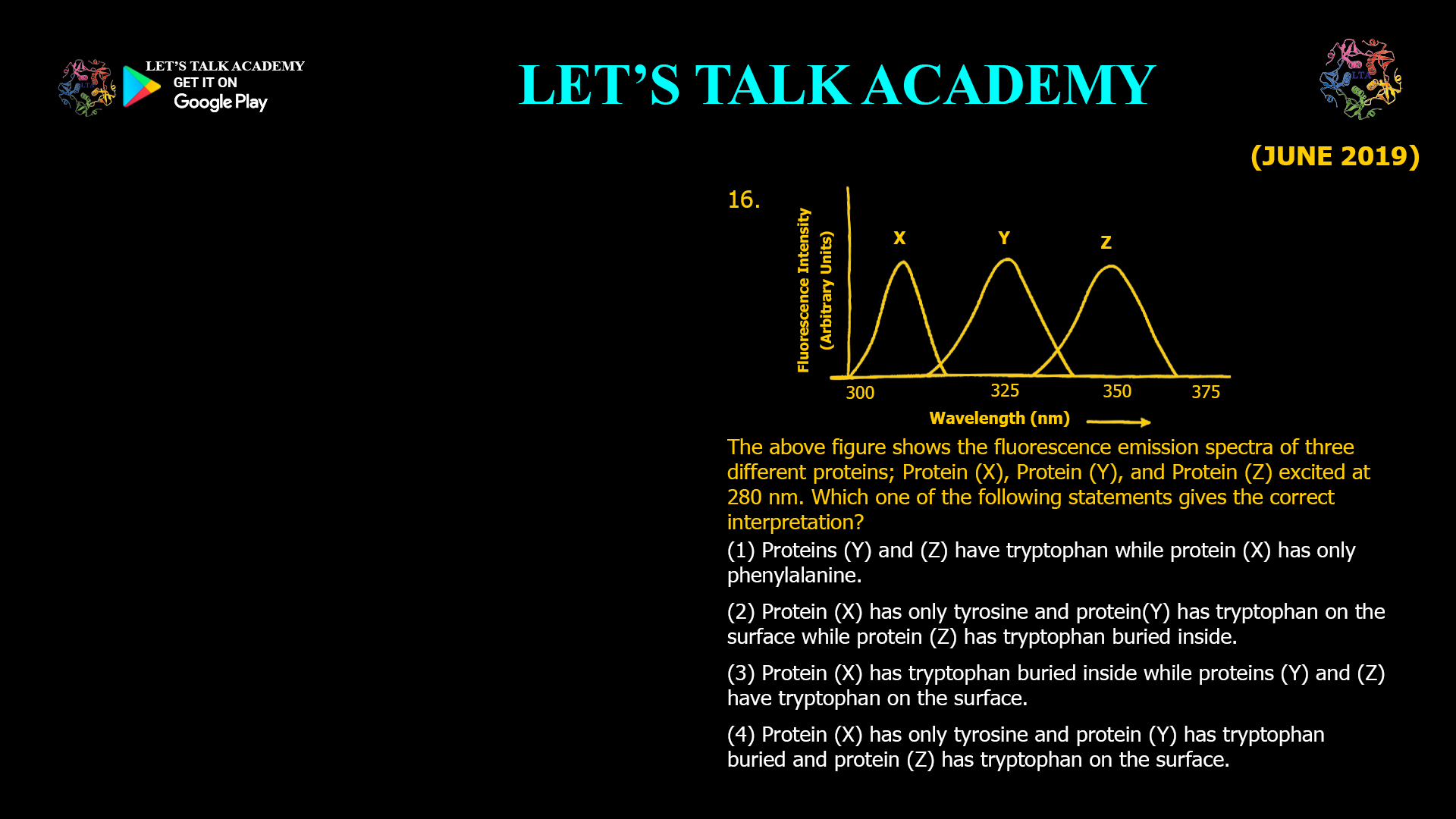
The above figure shows the fluorescence emission spectra of three different proteins; Protein (X), Protein (Y), and Protein (Z) excited at 280 nm. Which one of the following statements gives the correct interpretation?
(1) Proteins (Y) and (Z) have tryptophan while protein (X) has only phenylalanine.
(2) Protein (X) has only tyrosine and protein (Y) has tryptophan on the surface while protein (Z) has tryptophan buried inside.
(3) Protein (X) has tryptophan buried inside while proteins (Y) and (Z) have tryptophan on the surface.
(4) Protein (X) has only tyrosine and protein (Y) has tryptophan buried and protein (Z) has tryptophan on the surface.
How to Interpret Protein Fluorescence Emission Spectra of Tyrosine and Tryptophan
Intrinsic protein fluorescence is an extremely useful technique for understanding protein structure, folding, stability, solvent accessibility, and conformational changes. Proteins contain aromatic amino acids capable of absorbing ultraviolet radiation, particularly tryptophan, tyrosine, and phenylalanine. However, these aromatic amino acids do not contribute equally to the fluorescence spectrum of a protein. Tryptophan generally provides the strongest and most environmentally sensitive fluorescence signal, while tyrosine has a characteristic emission at a considerably shorter wavelength.
The position of the fluorescence emission maximum provides valuable information about the identity and molecular environment of an aromatic amino acid. In particular, the emission maximum of tryptophan changes significantly depending on whether its indole side chain is buried inside the hydrophobic core of a protein or exposed to the polar aqueous environment at its surface. This makes tryptophan fluorescence an important experimental probe for studying protein folding and structural changes.
Correct Answer
Option (4): Protein X Has Only Tyrosine, Protein Y Has Buried Tryptophan, and Protein Z Has Surface-Exposed Tryptophan
The correct answer is Option (4). The three fluorescence peaks can be interpreted according to the characteristic emission properties of aromatic amino acids and the sensitivity of tryptophan fluorescence to its surrounding environment.
Protein X produces an emission maximum close to approximately 303 nm, which corresponds well to the characteristic fluorescence emission of tyrosine. Protein Y produces an emission maximum at an intermediate wavelength around 325–330 nm, indicating that its tryptophan residue is located within a relatively non-polar or hydrophobic environment and is therefore buried within the protein structure. Protein Z emits near approximately 350 nm, indicating that its tryptophan residue is exposed to the polar aqueous environment on the protein surface.
Understanding Intrinsic Protein Fluorescence
Proteins can exhibit natural fluorescence without requiring an externally attached fluorescent probe. This phenomenon is known as intrinsic protein fluorescence and arises primarily from aromatic amino acids. Among the naturally occurring amino acids, tryptophan is generally the dominant contributor because it has strong fluorescence and its emission properties are extremely sensitive to environmental polarity.
Tyrosine also contributes to intrinsic protein fluorescence, although its emission occurs at a shorter wavelength and its fluorescence can be influenced by interactions with other residues. Phenylalanine has a much weaker fluorescence contribution under typical protein spectroscopy conditions. Therefore, fluorescence spectra recorded after excitation near 280 nm are commonly interpreted primarily in terms of tyrosine and tryptophan.
Interpretation of Protein X Fluorescence
Why Does Protein X Represent Tyrosine Fluorescence?
Protein X shows an emission maximum near approximately 300–305 nm. This wavelength range closely corresponds to the characteristic fluorescence emission of tyrosine, whose emission maximum is typically around 303 nm under many conditions.
Therefore, among the alternatives provided in the question, the short-wavelength fluorescence peak of Protein X is most consistent with a protein whose observed intrinsic fluorescence is dominated by tyrosine rather than tryptophan. This observation is essential for distinguishing Protein X from Proteins Y and Z.
Interpretation of Protein Y Fluorescence
Why Does Protein Y Contain Buried Tryptophan?
Protein Y displays a fluorescence maximum around approximately 325–330 nm. Tryptophan fluorescence is strongly dependent upon the polarity of the environment surrounding its indole ring. When tryptophan becomes buried within the hydrophobic interior of a folded protein, it has limited interaction with surrounding water molecules.
This non-polar environment results in fluorescence emission at a shorter wavelength than would occur if the same residue were completely exposed to water. Consequently, tryptophan buried inside the hydrophobic core of a protein commonly produces a blue-shifted fluorescence emission maximum in the approximate 320–335 nm region, although the exact wavelength depends on the particular microenvironment.
The position of the Protein Y peak therefore strongly supports the interpretation that it contains tryptophan located in a relatively hydrophobic and buried environment.
Interpretation of Protein Z Fluorescence
Why Does Protein Z Contain Surface-Exposed Tryptophan?
Protein Z produces its emission maximum close to approximately 350 nm. This longer-wavelength fluorescence is characteristic of tryptophan exposed to a polar environment such as water. When the indole group of tryptophan is accessible to surrounding solvent molecules, interactions between the excited fluorophore and polar solvent stabilize the excited state.
This stabilization decreases the energy difference associated with fluorescence emission. Because photon energy is inversely related to wavelength, the reduction in emission energy results in fluorescence at a longer wavelength. The resulting movement toward longer wavelengths is described as a red shift.
Therefore, an emission maximum close to 350 nm provides strong evidence that the tryptophan residue of Protein Z is relatively solvent exposed and situated on or near the protein surface.
Why Hydrophobic Tryptophan Shows a Blue Shift
The relationship between tryptophan fluorescence and its surrounding environment can be understood through interactions between the excited indole group and neighboring molecules. When tryptophan is located within the hydrophobic core of a protein, relatively few polar molecules are available to stabilize its excited electronic state.
Consequently, the energy difference associated with fluorescence remains comparatively large. The emitted photon therefore possesses relatively higher energy and a shorter wavelength, producing the characteristic blue-shifted fluorescence associated with buried tryptophan.
Why Surface-Exposed Tryptophan Shows a Red Shift
The opposite phenomenon occurs when tryptophan is exposed to water. Water is highly polar and can interact with the excited indole group. Solvent reorientation and other interactions stabilize the excited state, decreasing the energy of fluorescence emission.
The emitted photons consequently possess lower energy and longer wavelengths. This produces a red shift in the fluorescence spectrum, often moving the emission maximum toward approximately 350 nm or beyond depending upon the specific molecular environment.
Why Option (1) is Incorrect
Option (1) states that Proteins Y and Z contain tryptophan while Protein X contains only phenylalanine. The identification of Y and Z with tryptophan is reasonable, but assigning the approximately 303 nm emission peak of Protein X to phenylalanine is incorrect in the context of the choices provided. Phenylalanine has comparatively weak fluorescence and characteristically emits at substantially shorter wavelengths, whereas tyrosine shows a prominent emission maximum near approximately 303 nm. Therefore, Protein X is better interpreted as showing tyrosine-dominated fluorescence.
Why Option (2) is Incorrect
Option (2) correctly associates Protein X with tyrosine but reverses the environmental interpretation of Proteins Y and Z. It states that Protein Y contains surface-exposed tryptophan while Protein Z contains buried tryptophan. This contradicts the expected effect of solvent polarity on tryptophan fluorescence.
Protein Y emits at the shorter wavelength and is therefore more consistent with buried tryptophan in a hydrophobic environment. Protein Z emits at the longer wavelength near 350 nm and consequently represents tryptophan exposed to the polar solvent.
Why Option (3) is Incorrect
Option (3) incorrectly identifies Protein X as containing buried tryptophan. The emission maximum of Protein X is located close to approximately 303 nm, which is much more characteristic of tyrosine fluorescence than conventional tryptophan fluorescence.
Furthermore, Option (3) classifies both Protein Y and Protein Z as having surface-exposed tryptophan despite the substantial difference between their fluorescence emission maxima. The blue-shifted peak of Protein Y indicates a less polar environment, whereas the red-shifted peak of Protein Z indicates greater solvent exposure.
Why Option (4) is Correct
Option (4) correctly interprets all three fluorescence spectra. Protein X exhibits an emission maximum around 303 nm and is therefore consistent with tyrosine-dominated fluorescence. Protein Y has an emission maximum around 325–330 nm, indicating tryptophan situated within a hydrophobic or relatively buried environment. Protein Z emits close to 350 nm, demonstrating that its tryptophan is exposed to the polar aqueous environment on the protein surface.
Therefore, Option (4) is the only choice that simultaneously explains the identity of the fluorophore contributing to Protein X and the environmental sensitivity of tryptophan fluorescence observed in Proteins Y and Z.
How Fluorescence Can Reveal Protein Folding and Unfolding
The same principle demonstrated in this question can be applied experimentally to monitor protein folding. A properly folded globular protein often contains tryptophan residues buried within its hydrophobic core. These residues can display relatively blue-shifted fluorescence because they are protected from water.
If the protein is denatured using heat, extreme pH, or chemical denaturants, its hydrophobic core can become disrupted and previously buried tryptophan residues become exposed to water. The resulting increase in environmental polarity can shift the fluorescence emission maximum toward longer wavelengths. Researchers can therefore monitor changes in tryptophan emission spectra to follow protein unfolding and conformational transitions.
Final Answer
Correct Option: (4) Protein X has only tyrosine, Protein Y has tryptophan buried inside, and Protein Z has tryptophan on the surface.
Protein X shows fluorescence near approximately 303 nm, which is characteristic of tyrosine. Protein Y exhibits a blue-shifted tryptophan fluorescence maximum around 325–330 nm, indicating that the tryptophan is located in a relatively hydrophobic environment and buried within the protein. Protein Z displays fluorescence near approximately 350 nm, characteristic of tryptophan exposed to a polar aqueous environment. Thus, the progressive shift from X to Y to Z reflects the characteristic emission of tyrosine followed by the environmental sensitivity of tryptophan fluorescence, confirming Option (4) as the correct answer.


