
1. Strategies of Cell Division
Cell division is one of the most fundamental biological processes through which living organisms grow, develop, reproduce, repair damaged tissues, and maintain cellular organization. A cell cannot continue increasing in size indefinitely because larger cells face difficulties in transporting nutrients, removing wastes, communicating with other cells, and maintaining an efficient surface-area-to-volume ratio. Cell division provides a solution by producing new cells while preserving the continuity of genetic information.
The strategies of cell division are not identical in all organisms or even in all cells of the same organism. Depending on the biological requirement, cells may divide to produce genetically similar daughter cells, genetically different cells, or specialized reproductive cells. The two major strategies in eukaryotic organisms are mitosis and meiosis, while prokaryotic organisms generally reproduce through binary fission.
Cell division is therefore not simply a mechanical separation of one cell into two. It is a highly coordinated process involving DNA replication, chromosome condensation, chromosome movement, cytoplasmic division, cell-cycle regulation, and communication with the cellular environment.
1.1 Meaning of Cell Division
Cell division refers to the process by which a parent cell produces one or more daughter cells. Before a typical eukaryotic cell divides, its genetic material must be accurately duplicated so that the daughter cells receive the appropriate chromosome complement.
A successful division requires coordination between several events:
- Replication of DNA
- Duplication of centrosomes or other microtubule-organizing structures where applicable
- Chromosome condensation
- Assembly of the spindle apparatus
- Alignment of chromosomes
- Accurate segregation of chromosomes
- Division of the cytoplasm
- Re-establishment of daughter-cell organization
The ultimate outcome depends on the type of division.
In mitosis, one parent cell generally produces two genetically similar daughter cells.
In meiosis, one parent cell undergoes two successive divisions following a single round of DNA replication and produces haploid cells with genetic variation.
In binary fission, commonly observed in bacteria, the replicated chromosome is segregated as the cell elongates and the cell divides into two daughter cells.
1.2 Why Cells Divide
Cells divide for several important biological reasons.
1.2.1 Growth
Multicellular organisms begin as a single cell. Repeated cell divisions increase the number of cells and allow the developing organism to become larger and more complex.
Growth is not simply an increase in cell size. In many tissues, growth primarily results from an increase in cell number through controlled cell proliferation.
1.2.2 Development
Cell division is essential during embryonic development. Repeated divisions transform a single fertilized cell into an organism containing billions of cells organized into tissues and organs.
During development, cell division is coordinated with:
- Cell differentiation
- Cell migration
- Cell death
- Tissue patterning
- Cell-cell communication
Thus, cell division contributes not only to the number of cells but also to the organization of the developing organism.
1.2.3 Tissue Repair and Regeneration
Many tissues continuously replace old or damaged cells. For example, cells of the skin and intestinal epithelium undergo regular proliferation.
Following tissue injury, cell division can increase to replace damaged cells and restore tissue integrity.
1.2.4 Asexual Reproduction
Cell division can serve as a reproductive mechanism in unicellular organisms. A single cell can divide and generate new individuals.
Examples include binary fission in bacteria and several forms of cell division associated with asexual reproduction in unicellular eukaryotes.
1.2.5 Sexual Reproduction
Sexual reproduction requires specialized cells called gametes. Meiosis produces haploid gametes or reproductive cells in many sexually reproducing organisms.
The reduction in chromosome number during meiosis is essential because fertilization subsequently restores the diploid chromosome number.
2. Major Strategies of Cell Division
The major strategies can be grouped into three broad categories:
- Binary fission
- Mitosis
- Meiosis
Although these processes differ considerably, they share a fundamental requirement: genetic material must be distributed accurately to newly formed cells.
3. Binary Fission
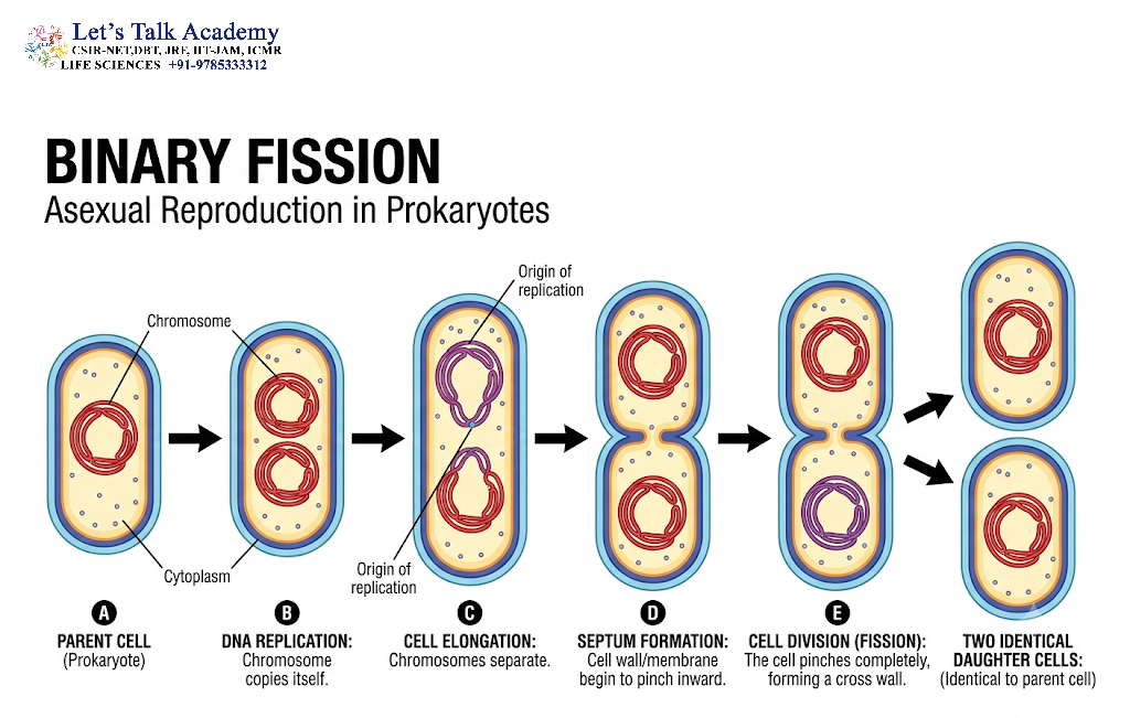
Binary fission is a common mode of reproduction in prokaryotic organisms such as bacteria and archaea.
Unlike eukaryotic cells, prokaryotes generally do not possess a membrane-bound nucleus or mitotic spindle. Their genetic material is usually organized as a circular chromosome located in the nucleoid region.
3.1 Basic Mechanism of Binary Fission
The process begins with replication of the bacterial chromosome. DNA replication generally starts at a specific origin and proceeds so that two chromosome copies are produced.
As replication continues, the chromosomes become associated with different regions of the growing cell. The cell elongates, and the chromosome copies become separated.
A division structure then develops near the middle of the cell. In many bacteria, the protein FtsZ plays a central role in forming a ring-like structure that contributes to cytokinesis.
The cell membrane and cell wall eventually constrict inward, producing two daughter cells.
3.2 Important Features of Binary Fission
Binary fission is characterized by:
- Absence of a typical mitotic spindle
- Direct replication of the bacterial chromosome
- Cell elongation during chromosome segregation
- Formation of a division site
- Cytoplasmic separation
- Production of daughter cells that are usually genetically similar to the parent, except when mutations or genetic exchange have occurred
Binary fission is therefore a highly efficient strategy for rapid population growth under favorable conditions.
4. Mitosis

Mitosis is a form of nuclear division in eukaryotic cells in which duplicated chromosomes are separated so that daughter nuclei receive equivalent sets of chromosomes.
It is particularly important for:
- Growth
- Development
- Tissue maintenance
- Cell replacement
- Wound healing
- Asexual reproduction in some organisms
The key feature of mitosis is the equal distribution of replicated chromosomes.
4.1 Relationship Between Mitosis and the Cell Cycle
Mitosis is only one part of the complete cell cycle.
The cell cycle is broadly divided into:
- G₁ phase
- S phase
- G₂ phase
- M phase
During the G₁ phase, the cell grows and performs its normal metabolic activities.
During the S phase, DNA replication occurs.
During the G₂ phase, the cell continues growing and prepares the molecular machinery required for division.
The M phase includes mitosis and cytokinesis.
4.2 Stages of Mitosis
Mitosis is conventionally divided into several stages:
- Prophase
- Prometaphase
- Metaphase
- Anaphase
- Telophase
Cytokinesis generally overlaps with the later stages of mitosis.
4.3 Prophase
During prophase, chromatin becomes highly condensed to form visible chromosomes.
Each replicated chromosome consists of two sister chromatids joined at the centromere region.
Other important events include:
- Chromosome condensation
- Formation of the mitotic spindle
- Changes in the centrosome position
- Progressive reorganization of the nucleus
Chromosome condensation is important because highly compact chromosomes can be transported more efficiently during segregation.
4.4 Prometaphase
During prometaphase, the nuclear envelope breaks down in typical open mitosis.
Microtubules of the spindle gain access to the chromosomes. Specialized protein complexes called kinetochores assemble at centromeric regions.
Spindle microtubules attach to kinetochores and begin positioning chromosomes.
4.5 Metaphase
During metaphase, chromosomes become aligned approximately at the equatorial region of the cell, forming the metaphase plate.
Proper chromosome alignment is critical because each sister chromatid must ultimately move toward the opposite pole.
The cell contains mechanisms that monitor whether chromosomes are correctly attached to spindle microtubules.
4.6 Anaphase
Anaphase begins when sister chromatids separate.
The separated chromatids are then considered individual daughter chromosomes.
They move toward opposite poles of the cell through coordinated spindle activity.
This step is one of the most important events in maintaining chromosome-number stability.
4.7 Telophase
During telophase, chromosomes arrive at opposite poles and begin to decondense.
New nuclear envelopes form around the separated chromosome sets.
The cell therefore develops two daughter nuclei.
4.8 Cytokinesis
Cytokinesis is the physical division of the cytoplasm.
In animal cells, a contractile ring containing actin and myosin generates a cleavage furrow that constricts the cell.
In plant cells, the rigid cell wall prevents a typical cleavage furrow. Instead, vesicles accumulate at the center of the cell and form a cell plate, which develops into the new cell wall separating the daughter cells.
4.9 Outcome of Mitosis
A typical mitotic division produces:
- Two daughter cells
- Approximately the same chromosome number as the parent cell
- Genetically similar cells, barring mutations and other sources of genetic variation
Mitosis therefore maintains chromosome number across successive generations of somatic cell division.
5. Meiosis
Meiosis is a specialized form of cell division associated with sexual reproduction.
Unlike mitosis, meiosis involves two consecutive nuclear divisions after a single round of DNA replication.
These divisions are called:
- Meiosis I
- Meiosis II
The major biological purposes of meiosis are:
- Reduction of chromosome number
- Generation of genetic variation
- Production of haploid reproductive cells or their precursors
5.1 Meiosis I
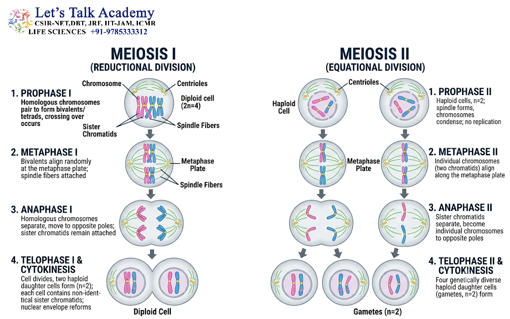
Meiosis I is known as the reductional division because homologous chromosomes are separated.
DNA replication occurs before meiosis I, during the preceding S phase.
However, DNA replication does not occur between meiosis I and meiosis II.
5.2 Prophase I
Prophase I is the longest and most complex stage of meiosis.
Homologous chromosomes pair with one another through a process called synapsis.
The paired homologues form structures called bivalents or tetrads.
During this stage, homologous chromosomes can exchange corresponding DNA segments through crossing over.
Crossing over generates new combinations of genetic material.
Prophase I is traditionally divided into:
- Leptotene
- Zygotene
- Pachytene
- Diplotene
- Diakinesis
5.2.1 Leptotene
Chromosomes begin to condense and become visible as thin structures.
5.2.2 Zygotene
Homologous chromosomes begin pairing through synapsis.
5.2.3 Pachytene
Synapsis is well established, and crossing over occurs between homologous chromosomes.
5.2.4 Diplotene
Homologous chromosomes begin separating while remaining connected at visible crossover regions called chiasmata.
5.2.5 Diakinesis
Chromosomes become maximally condensed, and the cell prepares for metaphase I.
5.3 Metaphase I
Homologous chromosome pairs align at the equatorial region.
The orientation of each homologous pair is not predetermined. This random orientation contributes to independent assortment, another major source of genetic variation.
5.4 Anaphase I
Homologous chromosomes separate and move toward opposite poles.
Importantly, sister chromatids remain together during anaphase I.
This distinguishes meiosis I from mitotic anaphase.
5.5 Telophase I and Cytokinesis
Chromosomes reach the poles and the cell may undergo cytokinesis.
The resulting cells contain one chromosome from each homologous pair.
They are therefore haploid with respect to chromosome sets, although each chromosome still consists of two sister chromatids.
6. Meiosis II
Meiosis II resembles mitosis in several aspects because sister chromatids are separated.
However, there is no additional round of DNA replication before meiosis II.
6.1 Prophase II
Chromosomes condense again if they had partially decondensed, and a new spindle apparatus forms.
6.2 Metaphase II
Chromosomes align at the equatorial region.
6.3 Anaphase II
Sister chromatids finally separate and move toward opposite poles.
6.4 Telophase II
Nuclear envelopes may reform around the chromosome sets, followed by cytokinesis.
6.5 Outcome of Meiosis
A typical meiotic division produces four haploid products from one diploid starting cell, although the exact developmental outcome varies among organisms and sexes.
The products are genetically different because of:
- Crossing over
- Independent assortment
- Random chromosome segregation
7. Comparison Between Mitosis and Meiosis
| Feature | Mitosis | Meiosis |
|---|---|---|
| Number of divisions | One | Two |
| DNA replication | Once before division | Once before meiosis I |
| Daughter cells | Usually two | Usually four |
| Chromosome number | Generally maintained | Reduced by half |
| Homologous pairing | Absent | Present in prophase I |
| Crossing over | Generally absent | Characteristic of prophase I |
| Main function | Growth, repair, maintenance | Sexual reproduction |
| Genetic similarity | Usually high | Genetically diverse |
| Homologous chromosomes separate | No | Yes, in meiosis I |
| Sister chromatids separate | Yes | In meiosis II |
8. Regulation of Cell Division
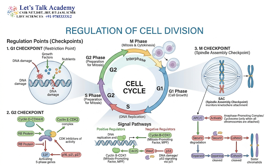
Cell division must be tightly regulated. Uncontrolled division can disrupt tissue organization and contribute to disease.
The cell cycle contains several important checkpoints.
8.1 G₁ Checkpoint
The G₁ checkpoint assesses whether the cell is sufficiently prepared to enter S phase.
Factors considered include:
- Cell size
- Nutrient availability
- Growth signals
- DNA integrity
- Cellular environment
If conditions are unfavorable, the cell may pause the cycle.
8.2 G₂ Checkpoint
The G₂ checkpoint determines whether DNA replication has been completed correctly and whether significant DNA damage remains.
Cells with unresolved DNA damage should not proceed normally into mitosis.
8.3 Spindle Assembly Checkpoint
The spindle assembly checkpoint operates during mitosis.
It ensures that chromosomes are appropriately attached to spindle microtubules before sister chromatids are separated.
Failure of this control can result in unequal chromosome distribution.
9. Molecular Control of Cell Division
Cell-cycle progression is regulated by a network of proteins.
Among the most important regulators are cyclins and cyclin-dependent kinases (CDKs).
CDKs require association with appropriate cyclins to become active.
Different cyclin-CDK combinations regulate different transitions of the cell cycle.
Important regulatory proteins include:
- Cyclins
- CDKs
- Retinoblastoma protein (Rb)
- p53
- CDK inhibitors
- Anaphase-promoting complex/cyclosome (APC/C)
These regulators ensure that events occur in the correct order.
10. Role of DNA Replication in Cell Division
Accurate DNA replication is essential before chromosome segregation.
During S phase, each chromosome is duplicated to produce two sister chromatids.
The duplicated DNA must remain associated until the appropriate stage of division.
Proteins involved in chromosome organization and cohesion ensure that sister chromatids remain connected until their separation is triggered.
Errors in replication can introduce mutations, while errors in chromosome segregation can produce abnormal chromosome numbers.
11. Chromosome Segregation

Chromosome segregation is one of the most critical aspects of cell division.
The spindle apparatus consists primarily of microtubules and associated proteins.
Microtubules can be broadly classified into:
- Kinetochore microtubules
- Interpolar microtubules
- Astral microtubules
Kinetochore microtubules interact with chromosome-associated kinetochores.
Interpolar microtubules interact with microtubules from the opposite spindle pole.
Astral microtubules contribute to spindle positioning and interaction with the cell cortex.
The coordinated behavior of these structures allows chromosomes to be distributed accurately.
12. Strategies That Maintain Genetic Stability
Successful cell division requires several safeguards.
12.1 Accurate DNA Replication
DNA must be replicated once and only once during each cell cycle.
12.2 Chromosome Condensation
Condensation prevents chromosomes from becoming excessively tangled during segregation.
12.3 Sister Chromatid Cohesion
Sister chromatids remain connected until the correct stage of chromosome segregation.
12.4 Spindle Attachment
Chromosomes must establish appropriate connections with spindle microtubules.
12.5 Cell-Cycle Checkpoints
Checkpoints prevent progression when essential cellular events have not been completed correctly.
Together, these mechanisms minimize the transmission of genetic errors to daughter cells.
13. Asymmetric Cell Division

Not all cell divisions produce two identical daughter cells.
In asymmetric cell division, daughter cells differ in their developmental potential, size, cellular components, or molecular composition.
This strategy is particularly important during development and stem-cell biology.
An asymmetric division can generate:
- One self-renewing stem cell
- One differentiating daughter cell
This allows tissues to maintain a stem-cell population while simultaneously producing specialized cells.
14. Symmetric Cell Division
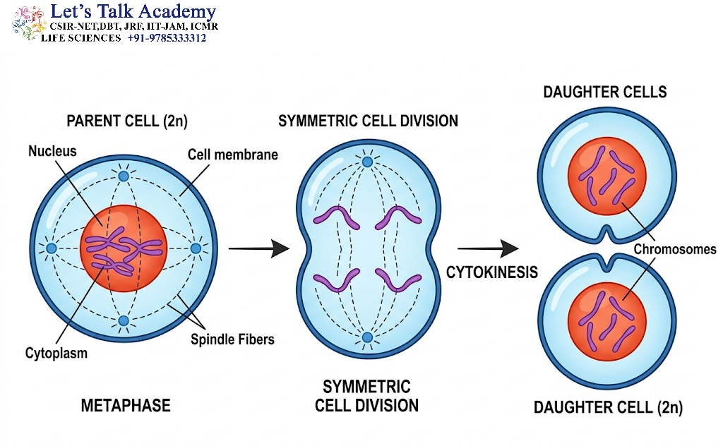
In symmetric division, daughter cells receive broadly similar cellular components and developmental potential.
Symmetric divisions are especially important when tissues need to increase their cell number rapidly.
The balance between symmetric and asymmetric division is crucial for tissue development and homeostasis.
15. Cell Division and Differentiation
Cell division and differentiation are closely connected but are not the same process.
Cell division increases cell number, whereas cell differentiation changes cellular structure and function.
During development, a proliferating population of cells may gradually become specialized into:
- Neurons
- Muscle cells
- Blood cells
- Epithelial cells
- Connective-tissue cells
The timing of cell-cycle exit and differentiation is therefore an important developmental decision.
16. Cell Division and Stem Cells
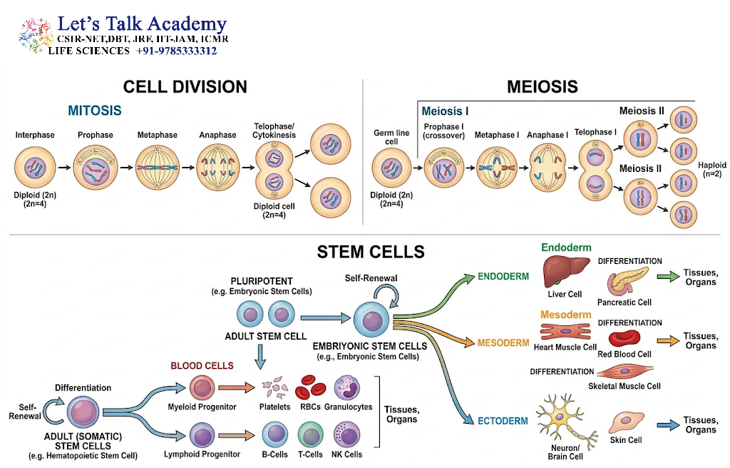
Stem cells possess the ability to self-renew and, depending on the type of stem cell, produce differentiated descendants.
Their division strategies must be carefully regulated.
Two broad outcomes are possible:
Symmetric self-renewal: both daughter cells retain stem-cell characteristics.
Asymmetric division: one daughter remains a stem cell while the other begins differentiation.
This balance prevents both excessive depletion and uncontrolled expansion of stem-cell populations.
17. Cell Division and Apoptosis

Cell division is balanced by controlled cell death.
Apoptosis is a programmed form of cell death that removes unnecessary, damaged, or potentially harmful cells.
During development and tissue maintenance, cell proliferation and apoptosis work together to determine the final number of cells.
A disruption in either process can affect tissue homeostasis.
18. Errors in Cell Division
Cell division is highly accurate, but errors can occur.
One important type of error is nondisjunction, in which chromosomes fail to separate properly.
Nondisjunction can lead to cells containing abnormal chromosome numbers, a condition called aneuploidy.
Other possible abnormalities include:
- Incorrect spindle attachment
- Chromosome breakage
- DNA replication errors
- Failure of cytokinesis
- Abnormal centrosome numbers
- Chromosome missegregation
Persistent chromosome-segregation errors can contribute to genomic instability.
19. Cell Division and Cancer
Normal cell proliferation is controlled by signals from within the cell and from the surrounding tissue.
Cancer can arise when regulatory mechanisms controlling proliferation, survival, and genome stability become disrupted.
Mutations affecting genes involved in:
- Cell-cycle regulation
- DNA repair
- Growth signaling
- Apoptosis
- Chromosome segregation
can promote abnormal cellular proliferation.
Tumor cells may therefore display altered cell-cycle control, increased proliferation, genomic instability, and reduced sensitivity to normal growth-limiting signals.
20. Cell Division in Plants and Animals
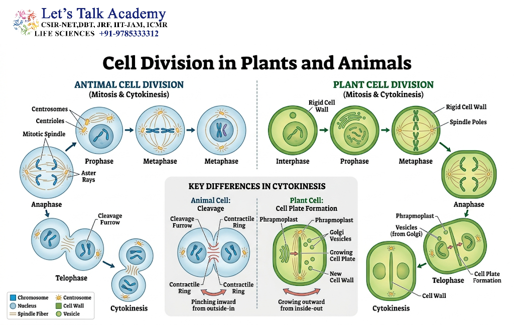
Although the fundamental principles of chromosome segregation are conserved, cytokinesis differs between plants and animals.
Animal Cells
Animal cells generally undergo cytokinesis through formation of a cleavage furrow.
An actin-myosin contractile ring constricts the plasma membrane.
Plant Cells
Plant cells have rigid cell walls and therefore cannot simply pinch inward.
Instead, vesicles accumulate at the center of the cell and form a cell plate.
The cell plate expands outward and eventually develops into a new cell wall.
21. Cell Division in Different Biological Contexts
The strategy of division depends strongly on biological requirements.
In unicellular organisms, division may directly increase population size.
In multicellular organisms, division supports growth, tissue replacement, and development.
In reproductive tissues, meiosis generates haploid cells and genetic diversity.
In developing tissues, asymmetric division can establish different cell lineages.
Therefore, cell division should be understood as a collection of coordinated strategies rather than a single universal mechanism.
22. Important Conceptual Points
Several concepts are particularly important for understanding cell division.
DNA replication occurs before mitosis and meiosis I, not before every nuclear division.
Mitosis generally separates sister chromatids.
Meiosis I separates homologous chromosomes.
Meiosis II separates sister chromatids.
Crossing over occurs during prophase I of meiosis.
Independent assortment contributes to genetic variation.
Cytokinesis is different from nuclear division.
Cell-cycle checkpoints help prevent propagation of damaged or incorrectly replicated DNA.
Aneuploidy can result from chromosome-segregation errors.
Cell division must be coordinated with growth, differentiation, and cell death.
23. Integrated View of Cell Division
The strategies of cell division can be understood as a sequence of interconnected biological decisions.
First, the cell must determine whether division is necessary. If division is initiated, the cell prepares its molecular machinery and replicates its DNA. The duplicated genetic material is then organized into chromosomes and connected to the appropriate segregation machinery.
The chromosomes are subsequently positioned and separated. Finally, cytokinesis divides the cytoplasm and establishes daughter cells.
In mitosis, the central goal is generally to preserve chromosome number and produce genetically similar daughter cells.
In meiosis, the goal is different. Homologous chromosomes are separated during the first division, chromosome number is reduced, and genetic variation is generated through recombination and independent assortment.
In prokaryotes, binary fission provides a simpler but highly effective strategy for chromosome duplication and cell separation.
These different mechanisms demonstrate how evolution has developed distinct solutions to the common biological problem of distributing genetic information into new cells.


