
1. Signal Transduction Pathways
1.1 Introduction
Cells continuously receive information from their surrounding environment. They must detect changes in nutrients, hormones, growth factors, neurotransmitters, temperature, osmotic conditions, stress, and signals from neighboring cells. To respond appropriately, cells use highly organized communication systems called signal transduction pathways.
Signal transduction is the process by which a cell converts an external or internal signal into a specific cellular response.
A signaling molecule, called a ligand, first interacts with a receptor. The activated receptor then initiates a series of molecular events involving signaling proteins, enzymes, second messengers, protein kinases, phosphatases, and transcription factors. Ultimately, these events produce a cellular response.
The general principle can be represented as:
Signal → Receptor → Signal Transduction → Amplification → Cellular Response → Signal Termination
Signal transduction is fundamental to almost every aspect of cell biology. It controls cell growth, differentiation, metabolism, movement, survival, immune responses, secretion, gene expression, and adaptation to environmental changes.
1.2 Definition of Signal Transduction
Signal transduction is the molecular process through which a cell detects a signal and converts it into intracellular biochemical events that produce a specific cellular response.
The signal may originate:
- outside the cell,
- at the cell surface,
- within the cytoplasm,
- or inside the nucleus.
1.3 Importance of Signal Transduction
Signal transduction allows cells to:
- communicate with other cells,
- detect environmental changes,
- regulate metabolism,
- control cell division,
- differentiate into specialized cell types,
- respond to hormones,
- respond to growth factors,
- regulate gene expression,
- control programmed cell death,
- maintain homeostasis.
Without signal transduction, cells would be unable to coordinate their activities with their environment.
2. Basic Components of Cell Signaling
A typical signaling system contains several major components.
2.1 Signaling Molecule
The signaling molecule is the molecule that carries information.
Examples include:
- hormones,
- growth factors,
- neurotransmitters,
- cytokines,
- lipids,
- gases,
- extracellular metabolites.
A signaling molecule may be called a ligand when it binds to a receptor.
2.2 Receptor
A receptor is a protein that recognizes a specific signaling molecule.
Receptors may be located:
- on the plasma membrane,
- in the cytoplasm,
- in the nucleus.
2.3 Intracellular Signaling Proteins
Once a receptor is activated, it interacts with intracellular signaling proteins.
These may include:
- G proteins,
- adaptor proteins,
- protein kinases,
- protein phosphatases,
- small GTPases,
- scaffold proteins.
2.4 Second Messengers
Second messengers are small intracellular signaling molecules produced or released after receptor activation.
Important second messengers include:
- cAMP,
- cGMP,
- Ca²⁺,
- IP₃,
- DAG.
2.5 Effector Proteins
Effector proteins are molecules whose activities are changed by signaling pathways.
They may include:
- enzymes,
- ion channels,
- cytoskeletal proteins,
- transcription factors.
2.6 Cellular Response
The final response may involve:
- altered enzyme activity,
- gene expression,
- secretion,
- movement,
- metabolism,
- cell growth,
- cell division,
- differentiation,
- survival or apoptosis.
3. General Steps of Signal Transduction
Signal transduction can be divided into several major stages.
3.1 Signal Reception
The signaling molecule interacts with its receptor.
Ligand + Receptor → Receptor activation
3.2 Signal Transmission
The activated receptor communicates with intracellular signaling proteins.
3.3 Signal Amplification
One receptor activation can activate many downstream molecules.
3.4 Signal Integration
Different signaling pathways can interact and combine information from several signals.
3.5 Cellular Response
The signaling pathway ultimately changes cellular activity.
3.6 Signal Termination
The signal must eventually be stopped or reduced.
A complete conceptual sequence is:
Signal → Reception → Transduction → Amplification → Integration → Response → Termination
4. Types of Cell Signaling
4.1 Endocrine Signaling
In endocrine signaling, hormones are released into the bloodstream and act on distant target cells.
Endocrine cell → bloodstream → distant target cell
Examples include many hormones produced by endocrine glands.
4.2 Paracrine Signaling
Paracrine signals act on nearby cells.
Signaling cell → nearby target cell
Growth factors and many local mediators act through paracrine mechanisms.
4.3 Autocrine Signaling
In autocrine signaling, a cell responds to a signaling molecule released by itself.
Cell → signal → same cell
This mechanism is important in several regulatory processes, including some immune and growth responses.
4.4 Synaptic Signaling
Neurons release neurotransmitters across a synaptic junction.
The neurotransmitter binds receptors on the postsynaptic cell.
4.5 Contact-Dependent Signaling
Some signals require direct physical contact between neighboring cells.
A membrane-bound signaling molecule on one cell interacts with a receptor on another cell.
5. Receptor Classes
Cell-surface receptors can be divided into several major groups.
5.1 G-Protein-Coupled Receptors
GPCRs activate heterotrimeric G proteins.
5.2 Receptor Tyrosine Kinases
RTKs possess or recruit tyrosine kinase activity and regulate pathways such as:
- Ras-MAPK,
- PI3K-AKT.
5.3 Cytokine Receptors
Many cytokine receptors signal through JAK-STAT proteins.
5.4 Receptor Serine/Threonine Kinases
These receptors commonly signal through SMAD proteins.
5.5 Receptor Guanylyl Cyclases
These receptors generate cGMP.
6. G-Protein-Coupled Receptor Signaling
6.1 Structure of GPCRs
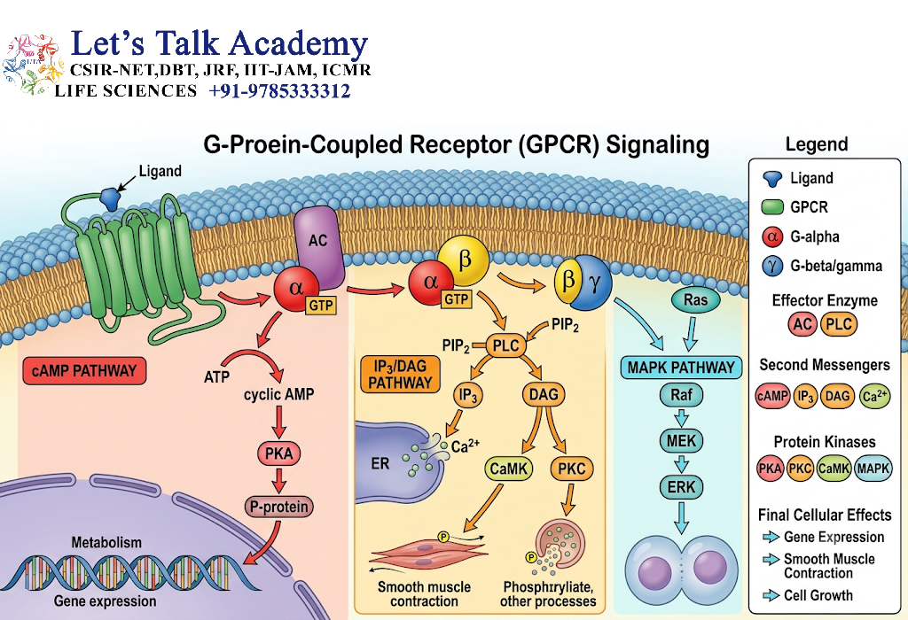
GPCRs are membrane proteins containing seven transmembrane α-helices.
They have:
- an extracellular region,
- seven transmembrane segments,
- an intracellular region.
6.2 Heterotrimeric G Proteins
Heterotrimeric G proteins contain:
- Gα,
- Gβ,
- Gγ.
The Gα subunit binds GDP in the inactive state.
6.3 Activation
When a ligand binds to a GPCR:
Ligand → GPCR → GDP-GTP exchange → G protein activation
The activated Gα-GTP and/or Gβγ complex can regulate downstream effectors.
6.4 Signal Termination
Gα possesses intrinsic GTPase activity.
GTP → GDP
This returns the G protein toward its inactive state.
7. cAMP Signal Transduction Pathway
7.1 Formation of cAMP
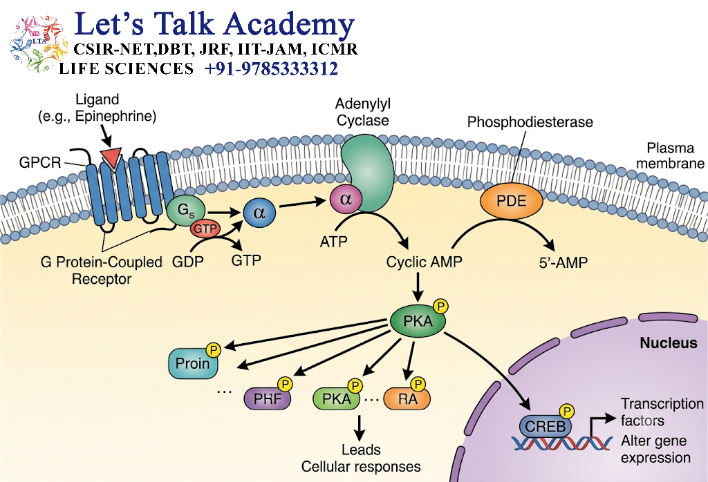
Adenylyl cyclase converts ATP into cyclic AMP.
ATP → cAMP
7.2 PKA Activation
cAMP activates protein kinase A (PKA).
PKA phosphorylates target proteins.
7.3 CREB Activation
PKA can phosphorylate the transcription factor CREB.
Activated CREB can regulate transcription of target genes.
7.4 Complete Pathway
Ligand → GPCR → Gs → adenylyl cyclase → cAMP → PKA → CREB → gene expression
7.5 Biological Functions
The cAMP pathway can regulate:
- glycogen metabolism,
- lipolysis,
- cardiac activity,
- secretion,
- gene transcription.
8. PLC-IP₃-DAG Pathway
8.1 Activation of Phospholipase C
Some GPCRs activate Gq.
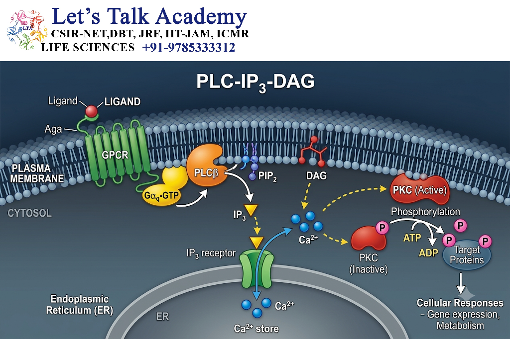
Gq activates phospholipase C-β (PLCβ).
8.2 PIP₂ Cleavage
PLC cleaves the membrane phospholipid PIP₂ into:
- IP₃,
- DAG.
8.3 IP₃ Function
IP₃ binds to receptors on the endoplasmic reticulum and stimulates Ca²⁺ release.
8.4 DAG Function
DAG remains associated with the plasma membrane and participates in activation of protein kinase C.
8.5 Complete Pathway
Ligand → GPCR → Gq → PLC → PIP₂ → IP₃ + DAG → Ca²⁺ + PKC → cellular response
9. Calcium Signaling
9.1 Calcium as a Second Messenger
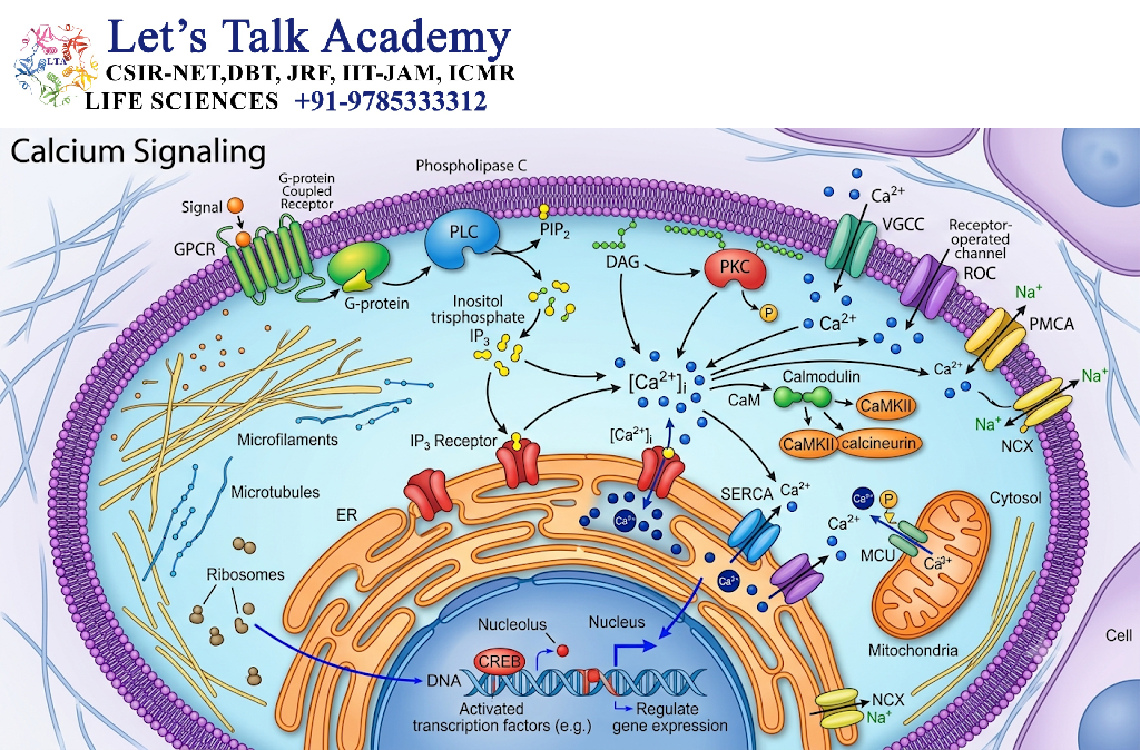
Ca²⁺ is an important intracellular messenger.
Changes in cytosolic calcium concentration can occur rapidly and can produce powerful cellular responses.
9.2 Sources of Calcium
Calcium can enter the cytoplasm from:
- extracellular fluid,
- endoplasmic reticulum,
- other intracellular stores.
9.3 Calmodulin
Ca²⁺ binds to calmodulin, producing a complex that can regulate many proteins.
9.4 Cellular Effects
Calcium signaling regulates:
- muscle contraction,
- secretion,
- metabolism,
- enzyme activity,
- gene expression.
10. Receptor Tyrosine Kinase Signaling
10.1 Definition
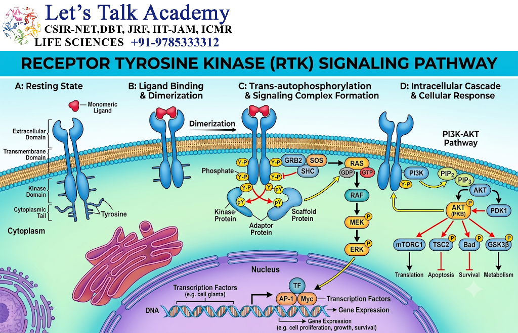
Receptor tyrosine kinases are transmembrane receptors that regulate intracellular signaling through tyrosine phosphorylation.
10.2 Activation
Ligand binding generally promotes receptor dimerization or rearrangement and activation of kinase domains.
The receptor phosphorylates tyrosine residues.
10.3 Docking Proteins
Phosphorylated tyrosine residues provide binding sites for signaling proteins containing domains such as:
- SH2,
- PTB.
10.4 Major Pathways
RTKs can activate:
- Ras-MAPK,
- PI3K-AKT,
- PLCγ pathways.
11. Ras-MAPK Pathway
11.1 Ras Protein
Ras is a small GTP-binding protein.
It acts as a molecular switch.
Ras-GDP → inactive
Ras-GTP → active
11.2 Activation
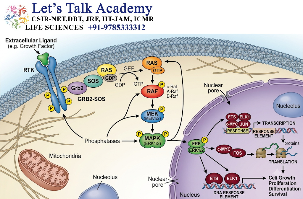
Activated RTK recruits adaptor proteins and a guanine nucleotide exchange factor.
This promotes:
Ras-GDP → Ras-GTP
11.3 Kinase Cascade
Activated Ras stimulates Raf.
The pathway continues:
Ras → Raf → MEK → ERK
11.4 Nuclear Response
Activated ERK can enter the nucleus and regulate transcription factors.
11.5 Functions
The Ras-MAPK pathway regulates:
- cell proliferation,
- differentiation,
- growth,
- development,
- gene expression.
12. PI3K-AKT Pathway
12.1 PI3K
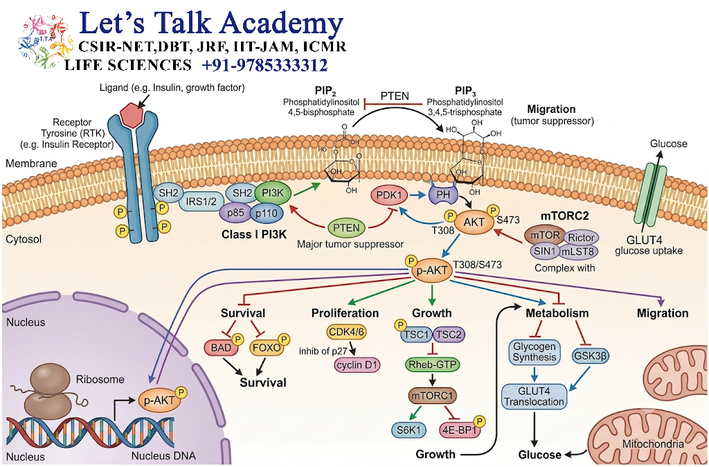
Phosphoinositide 3-kinase, or PI3K, is activated downstream of several receptors.
It converts PIP₂ into PIP₃.
12.2 AKT Activation
PIP₃ recruits signaling proteins including AKT to the plasma membrane.
AKT becomes activated through phosphorylation by upstream kinases.
12.3 Functions
AKT regulates:
- cell survival,
- glucose metabolism,
- protein synthesis,
- cell growth,
- nutrient utilization.
12.4 mTOR Connection
AKT can promote activity of mTOR signaling, which regulates cell growth and protein synthesis.
A simplified pathway is:
RTK → PI3K → PIP₃ → AKT → mTOR → growth and metabolism
13. JAK-STAT Signaling
13.1 General Mechanism
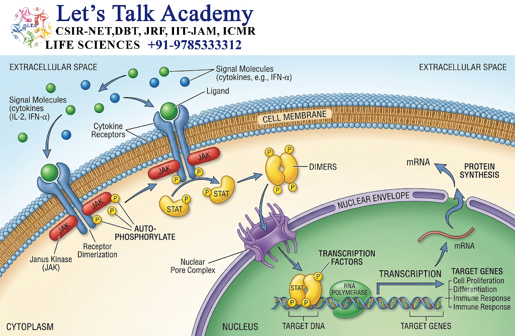
Some receptors activate Janus kinases, or JAKs.
Ligand binding promotes receptor-associated JAK activation.
13.2 STAT Proteins
JAKs phosphorylate STAT proteins.
Phosphorylated STAT proteins form dimers.
13.3 Nuclear Entry
STAT dimers enter the nucleus and regulate transcription.
13.4 Complete Pathway
Ligand → receptor → JAK → STAT phosphorylation → STAT dimer → nucleus → gene expression
13.5 Biological Functions
JAK-STAT signaling participates in:
- immune regulation,
- cell growth,
- differentiation,
- hematopoiesis,
- responses to extracellular regulatory molecules.
14. TGF-β-SMAD Pathway
14.1 Receptor Activation
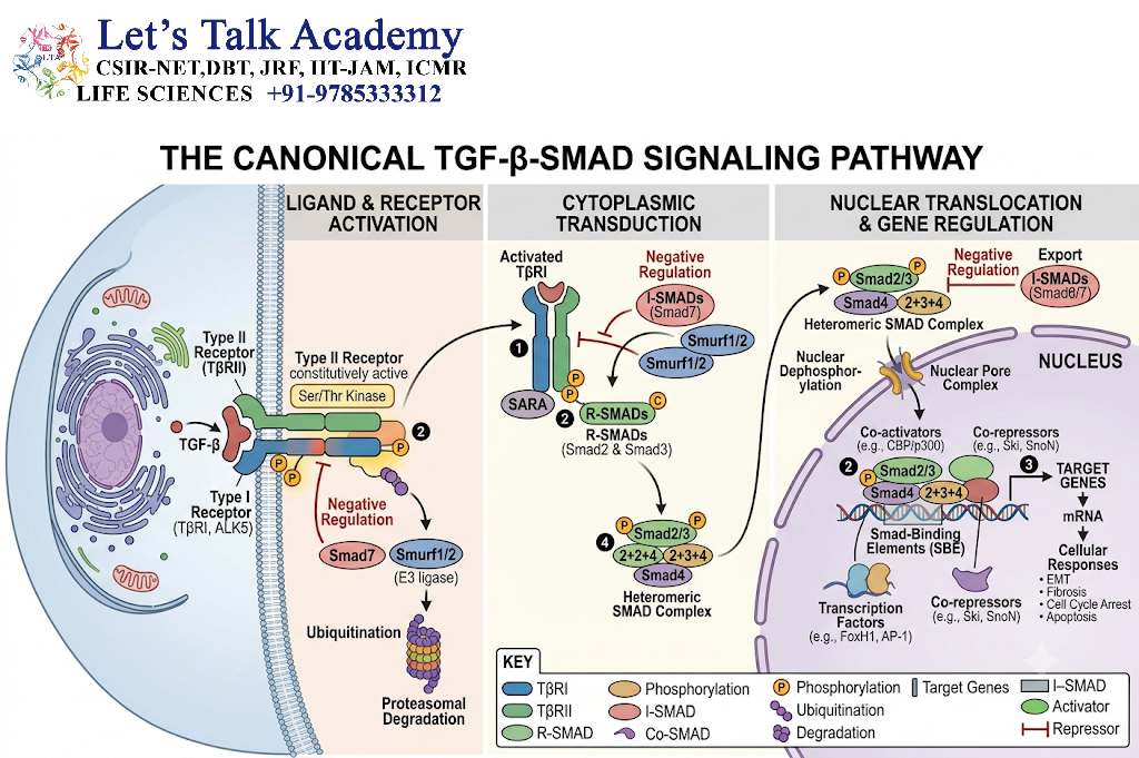
TGF-β family ligands bind receptor complexes containing serine/threonine kinase activity.
14.2 SMAD Activation
Receptor activation phosphorylates receptor-regulated SMAD proteins.
14.3 Nuclear Signaling
Activated SMAD proteins form complexes and enter the nucleus.
They regulate target gene transcription.
14.4 Functions
The pathway regulates:
- development,
- differentiation,
- cell growth,
- extracellular matrix formation,
- tissue homeostasis.
15. cGMP Signaling
15.1 Formation of cGMP
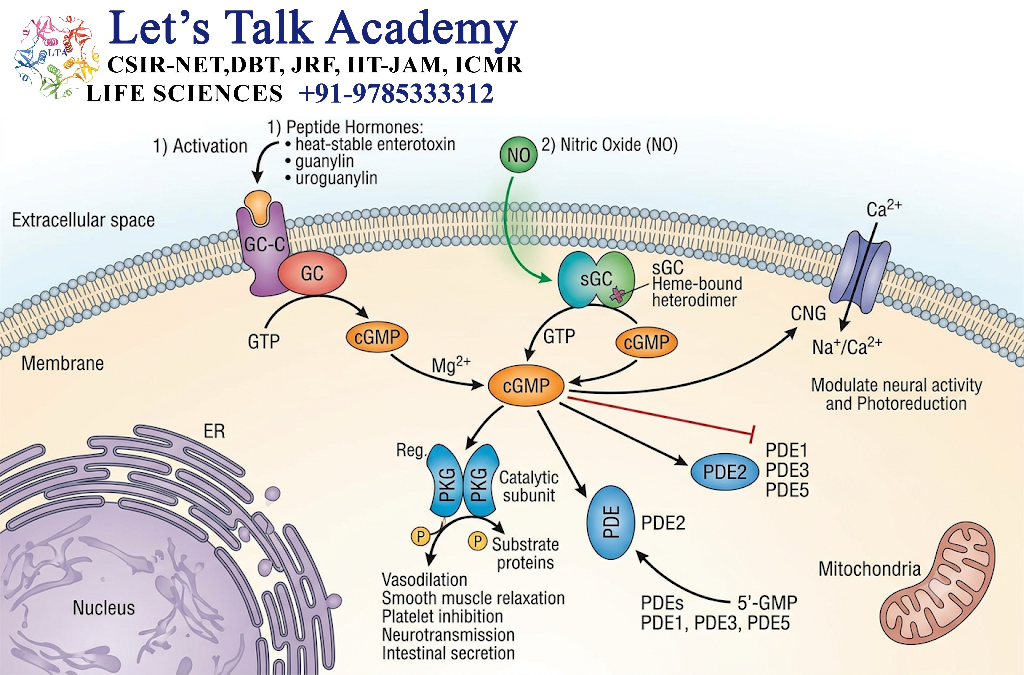
Guanylyl cyclase converts GTP into cGMP.
15.2 Protein Kinase G
cGMP can activate protein kinase G (PKG).
15.3 Biological Functions
cGMP signaling contributes to:
- smooth-muscle relaxation,
- ion transport,
- cellular signaling,
- regulation of vascular function.
16. Nitric Oxide Signaling
16.1 Nitric Oxide
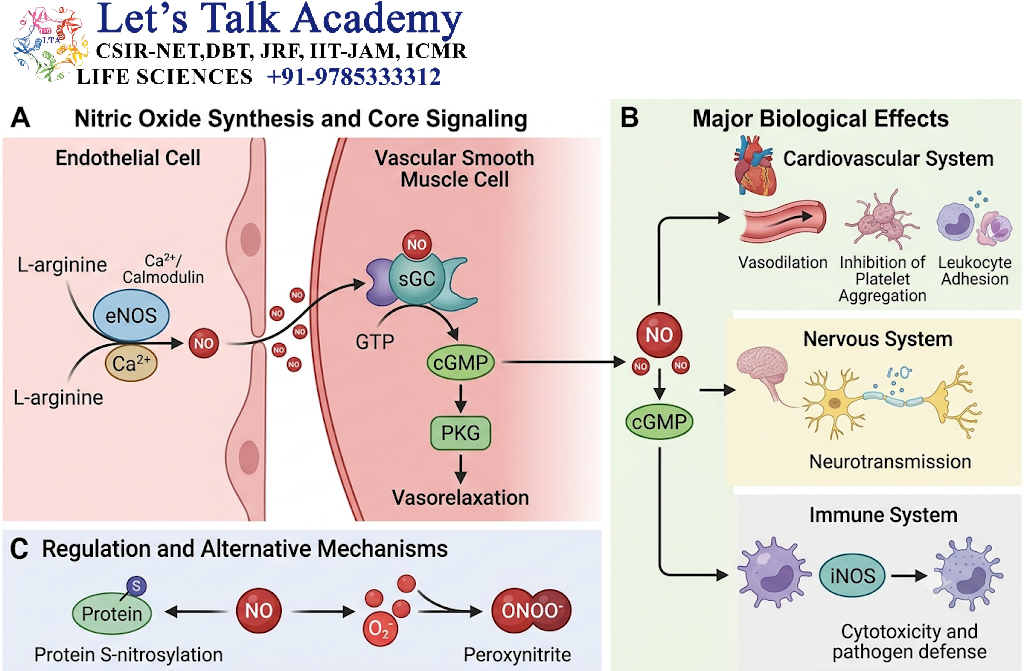
Nitric oxide, or NO, is a small gaseous signaling molecule.
Because it can diffuse across membranes, it does not require a conventional membrane receptor to enter target cells.
16.2 Soluble Guanylyl Cyclase
NO activates soluble guanylyl cyclase.
This increases cGMP production.
16.3 Pathway
NO → soluble guanylyl cyclase → cGMP → PKG → cellular response
This pathway is particularly important in vascular smooth-muscle signaling.
17. Nuclear Receptor Signaling
17.1 General Mechanism
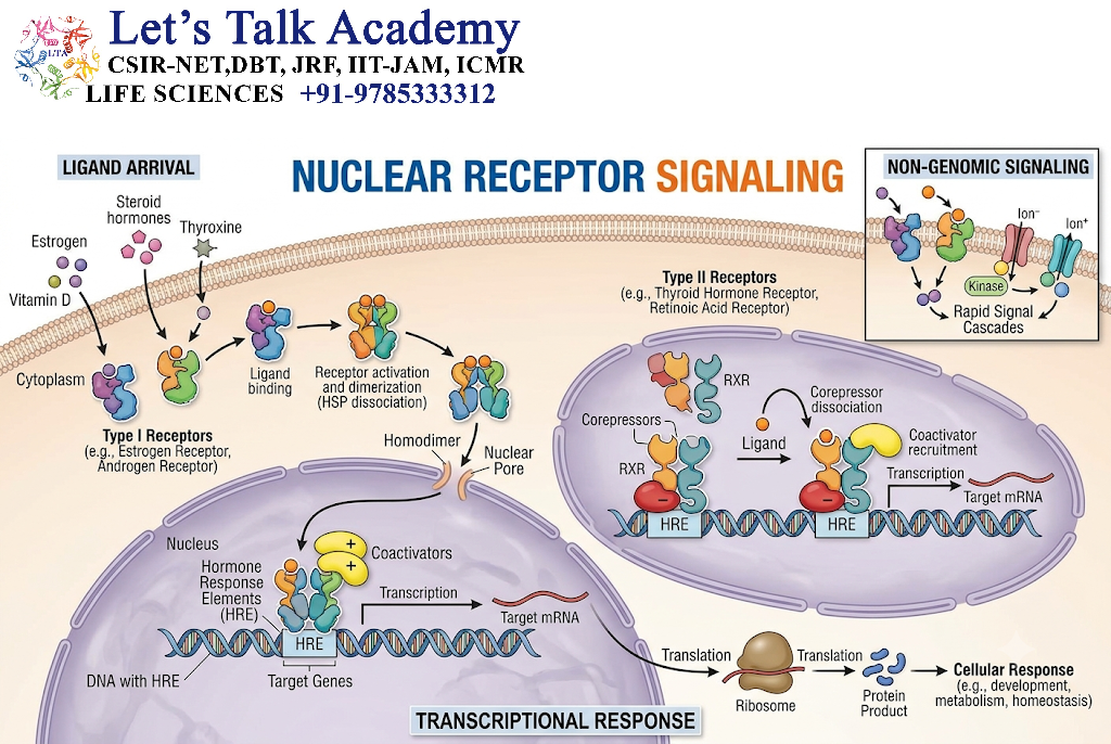
Lipid-soluble molecules can enter cells and interact with intracellular receptors.
Examples include receptors for:
- steroid hormones,
- thyroid hormones,
- vitamin D,
- retinoids.
17.2 Gene Regulation
Activated receptors interact with DNA regulatory sequences and transcriptional machinery.
17.3 Pathway
Hormone → intracellular receptor → DNA regulatory region → transcriptional regulation → protein synthesis → cellular response
This type of signaling often produces slower but longer-lasting effects compared with many second-messenger pathways.
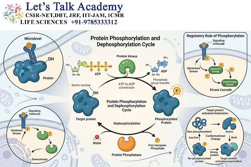
18. Protein Phosphorylation
18.1 Protein Kinases
Protein kinases transfer phosphate groups, usually from ATP, to proteins.
Phosphorylation can change:
- enzyme activity,
- protein localization,
- protein stability,
- protein interactions,
- transcription-factor activity.
18.2 Protein Phosphatases
Protein phosphatases remove phosphate groups.
They provide an important mechanism for controlling and terminating signaling pathways.
18.3 Reversible Regulation
Therefore:
Kinase → phosphorylation
Phosphatase → dephosphorylation
This reversible system allows rapid regulation of cellular proteins.
19. Second Messenger Systems
Second messengers transmit information from receptors to intracellular targets.
19.1 cAMP
Generated by adenylyl cyclase.
Major target:
PKA
19.2 cGMP
Generated by guanylyl cyclase.
Major target:
PKG
19.3 IP₃
Promotes Ca²⁺ release from intracellular stores.
19.4 DAG
Participates in PKC activation.
19.5 Ca²⁺
Regulates many enzymes and calcium-binding proteins.
20. Signal Amplification
20.1 Principle
Signal amplification occurs when one signaling event produces many downstream signaling molecules.
For example:
One ligand
→ receptor activation
→ multiple G proteins
→ multiple adenylyl cyclase molecules
→ many cAMP molecules
→ multiple PKA molecules
→ phosphorylation of many target proteins.
20.2 Biological Importance
Amplification allows cells to respond strongly even when extracellular signaling molecules are present at very low concentrations.
21. Signal Integration
Cells rarely receive only one signal.
A cell may simultaneously receive signals from:
- hormones,
- growth factors,
- nutrients,
- neurotransmitters,
- stress signals.
These pathways interact and determine the final response.
For example:
Growth signal + nutrient availability + survival signal → integrated cellular response
Thus, signal transduction is not simply a series of isolated pathways.
22. Signal Specificity
Different cells can respond differently to the same signaling molecule.
This occurs because cells differ in:
- receptor expression,
- receptor subtype,
- signaling proteins,
- transcription factors,
- metabolic state.
Therefore:
Same signal + different cellular machinery = different cellular response
23. Scaffold Proteins
23.1 Definition
Scaffold proteins organize signaling proteins into functional complexes.
23.2 Importance
They can:
- bring pathway components together,
- increase signaling efficiency,
- improve pathway specificity,
- prevent inappropriate interactions.
For example, scaffold proteins can organize components of kinase cascades.
24. Small GTPases
Small GTPases act as molecular switches.
They generally alternate between:
GTP-bound active state
and
GDP-bound inactive state
Important examples include:
- Ras,
- Rho,
- Rab,
- Ran,
- Arf.
These proteins regulate diverse processes such as:
- cell proliferation,
- cytoskeletal organization,
- vesicle trafficking,
- nuclear transport.
25. Regulation of Signal Transduction
Signal transduction pathways must be tightly regulated.
Major regulatory mechanisms include:
- receptor desensitization,
- receptor internalization,
- protein dephosphorylation,
- GTP hydrolysis,
- second-messenger degradation,
- protein degradation,
- feedback inhibition.
26. Receptor Desensitization
26.1 Definition
Desensitization occurs when a cell becomes less responsive to persistent stimulation.
26.2 GPCR Desensitization
Activated GPCRs can be phosphorylated by receptor kinases.
β-arrestin can bind to phosphorylated receptors and reduce G-protein activation.
26.3 Importance
Desensitization prevents excessive responses to continuous stimulation.
27. Receptor Internalization
Some activated receptors are removed from the plasma membrane through endocytosis.
The internalized receptor may:
- be recycled,
- remain in an endosomal compartment,
- undergo degradation.
This changes the sensitivity and duration of signaling.
28. Negative Feedback
Negative feedback reduces pathway activity.
For example:
Signal → pathway activation → inhibitory protein expression → pathway suppression
Negative feedback prevents uncontrolled activation.
29. Positive Feedback
Positive feedback increases pathway activity.
A signaling pathway can activate molecules that further enhance the original signal.
Positive feedback can produce:
- rapid amplification,
- switch-like behavior,
- sustained activation.
30. Signal Termination
Signal termination is essential for cellular homeostasis.
Mechanisms include:
Ligand removal
The signaling molecule may be degraded or removed from circulation.
Receptor inactivation
The receptor may become inactive or internalized.
GTP hydrolysis
GTP-binding proteins return to their GDP-bound state.
Second-messenger degradation
cAMP and cGMP can be degraded by phosphodiesterases.
Dephosphorylation
Protein phosphatases remove phosphate groups.
31. Signal Transduction and Gene Expression
Many signaling pathways eventually reach the nucleus.
For example:
Extracellular signal
→ receptor
→ kinase cascade
→ transcription factor
→ DNA regulatory region
→ altered gene expression
→ new protein production
This mechanism allows extracellular signals to produce long-term changes in cell behavior.
32. Signal Transduction and Cell Growth
Growth factors activate pathways such as:
- Ras-MAPK,
- PI3K-AKT,
- mTOR.
These pathways regulate:
- cell-cycle progression,
- protein synthesis,
- metabolism,
- survival,
- cell proliferation.
33. Signal Transduction and Cell Survival
The PI3K-AKT pathway is an important regulator of cell survival.
Activated AKT can inhibit or regulate several proteins involved in pro-survival and pro-death processes.
This allows extracellular signals to influence whether a cell survives under particular conditions.
34. Signal Transduction and Apoptosis
Signal transduction can either promote survival or activate apoptosis depending on the signaling context.
For example:
Survival signals → PI3K-AKT → pro-survival signaling
Whereas certain death-receptor pathways can activate:
Death receptor → adaptor proteins → caspases → apoptosis
Therefore, signaling pathways participate in determining cell fate.
35. Signal Transduction and Cell Differentiation
During development, signaling pathways determine which genes are expressed in particular cells.
Pathways such as:
- MAPK,
- JAK-STAT,
- TGF-β-SMAD,
- Wnt,
- Hedgehog,
- Notch,
contribute to cell differentiation and tissue development.
36. Signal Transduction and Metabolism
Hormonal signals regulate metabolism through pathways involving:
- cAMP,
- PKA,
- PI3K-AKT,
- AMPK,
- mTOR.
For example, insulin signaling through PI3K-AKT promotes glucose uptake and anabolic processes in responsive tissues.
37. Signal Transduction and the Cytoskeleton
Signaling pathways regulate the cytoskeleton through proteins such as Rho-family GTPases.
These pathways influence:
- cell shape,
- cell movement,
- cell adhesion,
- intracellular transport,
- cell division.
38. Signal Transduction and Vesicle Trafficking
Small GTPases such as Rab proteins regulate vesicle movement and membrane trafficking.
A simplified pathway is:
Signal → Rab activation → vesicle targeting → membrane fusion → cargo delivery
This is important for secretion and membrane-protein transport.
39. Major Signal Transduction Pathways
| Pathway | Main receptor type | Major signaling components | Major functions |
|---|---|---|---|
| cAMP-PKA | GPCR | Gs, adenylyl cyclase, cAMP, PKA | Metabolism, secretion, gene regulation |
| PLC-IP₃/DAG | GPCR/RTK | PLC, IP₃, DAG, Ca²⁺, PKC | Contraction, secretion, metabolism |
| Ras-MAPK | RTK | Ras, Raf, MEK, ERK | Growth, proliferation, differentiation |
| PI3K-AKT | RTK and others | PI3K, PIP₃, AKT, mTOR | Survival, metabolism, growth |
| JAK-STAT | Cytokine-associated receptor | JAK, STAT | Gene expression, immune regulation |
| TGF-β-SMAD | Serine/threonine kinase receptor | SMAD proteins | Differentiation, development |
| cGMP-PKG | Guanylyl cyclase receptor | cGMP, PKG | Smooth-muscle and ion regulation |
| NO-cGMP | Soluble guanylyl cyclase | NO, cGMP, PKG | Vascular signaling |
| Nuclear receptor | Intracellular receptor | Nuclear receptor, DNA response elements | Gene regulation |
40. Integrated Signal Transduction Flowchart
Extracellular signal
↓
Receptor recognition
↓
Receptor activation
↓
Intracellular signaling proteins
↓
Second messengers / protein kinases
↓
Signal amplification
↓
Signal integration
↓
Effector proteins / transcription factors
↓
Cellular response
↓
Signal termination
41. Example: Insulin Signal Transduction
Insulin provides a clear example of receptor-mediated signal transduction.
Insulin
↓
Insulin receptor
↓
Tyrosine phosphorylation
↓
IRS proteins
↓
PI3K
↓
PIP₃
↓
AKT
↓
GLUT4 translocation
↓
Increased glucose uptake
At the same time, insulin signaling influences glycogen synthesis, protein synthesis, lipid metabolism, and other cellular processes.
42. Example: Epinephrine Signal Transduction
Epinephrine can activate β-adrenergic GPCRs.
Epinephrine
↓
β-adrenergic receptor
↓
Gs protein
↓
Adenylyl cyclase
↓
cAMP
↓
PKA
↓
Phosphorylation of metabolic enzymes
↓
Metabolic response
This pathway allows rapid changes in cellular metabolism.
43. Example: Growth Factor Signaling
A growth factor can activate an RTK.
Growth factor
↓
RTK
↓
Ras
↓
Raf
↓
MEK
↓
ERK
↓
Transcription factors
↓
Gene expression
↓
Cell growth / proliferation / differentiation
44. Signal Transduction and Disease
Abnormal signal transduction can contribute to disease.
Problems can occur because of:
- receptor mutations,
- excessive receptor activation,
- defective receptor degradation,
- abnormal kinase activity,
- altered phosphatase activity,
- abnormal second-messenger production,
- constitutively active GTPases,
- defective feedback regulation.
44.1 Cancer
Abnormal activation of pathways such as:
- Ras-MAPK,
- PI3K-AKT-mTOR,
can promote uncontrolled proliferation and survival.
44.2 Metabolic Disorders
Defects in insulin receptor signaling can impair normal glucose regulation.
44.3 Immune Disorders
Abnormal JAK-STAT signaling can disturb immune and inflammatory regulation.
45. Signal Transduction Crosstalk
45.1 Definition
Crosstalk occurs when one signaling pathway influences another.
For example:
RTK signaling → PI3K-AKT
and
RTK signaling → Ras-MAPK
may occur simultaneously.
The cell integrates both pathways to produce an appropriate response.
45.2 Importance
Crosstalk provides:
- flexibility,
- coordination,
- signal integration,
- pathway specificity.
46. Signal Transduction and Cellular Decision-Making
Cells use signaling pathways to make decisions such as:
- divide,
- differentiate,
- migrate,
- survive,
- secrete molecules,
- alter metabolism,
- undergo apoptosis.
The final decision depends on the intensity, duration, location, and combination of signals.
47. Duration and Intensity of Signaling
The same signaling pathway can produce different effects depending on how long it remains active.
Short signal
May produce a rapid metabolic response.
Prolonged signal
May alter gene expression and cellular differentiation.
Therefore, signaling information is encoded not only by whether a pathway is activated but also by:
- signal strength,
- duration,
- frequency,
- location.
48. Spatial Regulation of Signaling
Signaling molecules do not necessarily act uniformly throughout the cell.
Some signaling events are restricted to specific cellular compartments.
Examples include:
- plasma membrane,
- endosomes,
- cytoplasm,
- mitochondria,
- nucleus.
Spatial organization increases signaling specificity.
49. Temporal Regulation of Signaling
Temporal regulation refers to changes in signaling activity over time.
A pathway can be:
- transient,
- sustained,
- oscillatory,
- pulsatile.
Different temporal patterns can produce different cellular outcomes.
50. Importance of Signal Transduction in Multicellular Organisms
Signal transduction enables coordination between cells and tissues.
It contributes to:
- embryonic development,
- tissue organization,
- nervous-system function,
- immune responses,
- endocrine regulation,
- reproduction,
- metabolism,
- tissue repair,
- homeostasis.
A multicellular organism depends on continuous communication between its cells.


