
1. Introduction to Second Messengers
Cells continuously receive information from their surrounding environment. Hormones, neurotransmitters, growth factors, cytokines and other signaling molecules bind to specific receptors on or inside the cell. However, many extracellular signaling molecules do not directly enter the cell and control intracellular processes themselves.
Instead, receptor activation generates or releases small intracellular signaling molecules known as second messengers. These molecules rapidly transmit and amplify signals from activated receptors to intracellular target proteins.
The concept can be understood as:
First messenger → Receptor → Second messenger → Target protein → Cellular response
Here, the extracellular signaling molecule is often called the first messenger, while the intracellular signaling molecule is called the second messenger.
Important second messengers include:
- Cyclic AMP (cAMP)
- Cyclic GMP (cGMP)
- Inositol 1,4,5-trisphosphate (IP₃)
- Diacylglycerol (DAG)
- Calcium ions (Ca²⁺)
Second-messenger systems are fundamental to processes such as metabolism, muscle contraction, secretion, gene expression, cell growth, differentiation and cell survival.
2. Definition of Second Messengers
2.1 Definition
Second messengers are small intracellular signaling molecules whose concentrations or activities change in response to receptor activation and which transmit signals from activated receptors to intracellular effector proteins.
The term “second” refers to their position in the signaling sequence:
First messenger: extracellular signaling molecule
Second messenger: intracellular signaling molecule
For example:
Adrenaline → β-adrenergic receptor → G protein → Adenylyl cyclase → cAMP → PKA → cellular response
Here, adrenaline is the first messenger and cAMP is the second messenger.
3. Characteristics of Second Messengers
Second messengers have several important properties.
3.1 Small and Rapidly Acting
Most second messengers are small molecules or ions that can be produced or released rapidly.
3.2 Intracellular
They function inside the cell rather than acting as extracellular signaling molecules.
3.3 Signal Amplification
A small number of activated receptors can generate large amounts of second messenger.
3.4 Short-Lived
Many second messengers are rapidly degraded, removed or sequestered. This allows signaling to be terminated quickly.
3.5 Regulated Concentration
Their intracellular concentration is tightly controlled by enzymes, pumps, channels and binding proteins.
3.6 Specific Cellular Effects
The same second messenger can produce different effects in different cell types because cells contain different receptors, enzymes and downstream targets.
4. First Messengers and Second Messengers
| Feature | First Messenger | Second Messenger |
|---|---|---|
| Location | Usually extracellular | Intracellular |
| Examples | Hormones, neurotransmitters, growth factors | cAMP, cGMP, IP₃, DAG, Ca²⁺ |
| Main function | Activates receptor | Transmits intracellular signal |
| Receptor interaction | Usually directly binds receptor | Usually generated/released after receptor activation |
| Signal amplification | Indirect | Often strong |
| Duration | Variable | Often short-lived |
The distinction is useful because the first messenger initiates signaling, while the second messenger helps distribute and amplify that signal within the cell.
5. Major Types of Second Messengers
The major second-messenger systems are:
- cAMP
- cGMP
- IP₃
- DAG
- Ca²⁺
Each has different sources, targets and biological effects.
| Second Messenger | Major Source | Major Target/Effect |
|---|---|---|
| cAMP | ATP | Protein kinase A |
| cGMP | GTP | Protein kinase G |
| IP₃ | PIP₂ | Ca²⁺ release from ER |
| DAG | PIP₂ | Protein kinase C |
| Ca²⁺ | Extracellular space/ER | Calmodulin, PKC and other proteins |
6. cAMP as a Second Messenger
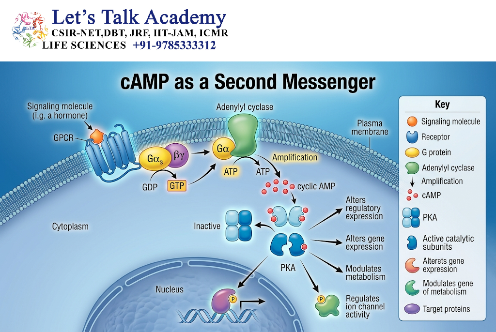
6.1 Formation of cAMP
Cyclic AMP or cAMP is produced from ATP by the enzyme adenylyl cyclase.
The general reaction is:
ATP → cAMP + PPi
Adenylyl cyclase is commonly activated by G-protein-coupled receptors.
6.2 GPCR-cAMP Pathway
A typical pathway is:
Ligand → GPCR → Gs protein → Adenylyl cyclase → cAMP → PKA → Cellular response
For example, adrenaline can activate β-adrenergic receptors.
6.3 Activation of Protein Kinase A
cAMP binds to regulatory subunits of protein kinase A (PKA).
This releases the catalytic subunits, allowing PKA to phosphorylate target proteins.
cAMP → PKA activation → Protein phosphorylation → Cellular response
6.4 Biological Functions of cAMP
cAMP participates in:
- Glycogen metabolism
- Lipolysis
- Cardiac activity
- Ion-channel regulation
- Gene expression
- Hormonal responses
- Neuronal signaling
6.5 Regulation of cAMP
The intracellular level of cAMP is controlled mainly by:
Adenylyl cyclase: produces cAMP
Phosphodiesterases (PDEs): degrade cAMP into AMP
Therefore:
Adenylyl cyclase ↑ → cAMP ↑
Phosphodiesterase ↑ → cAMP ↓
7. cGMP as a Second Messenger
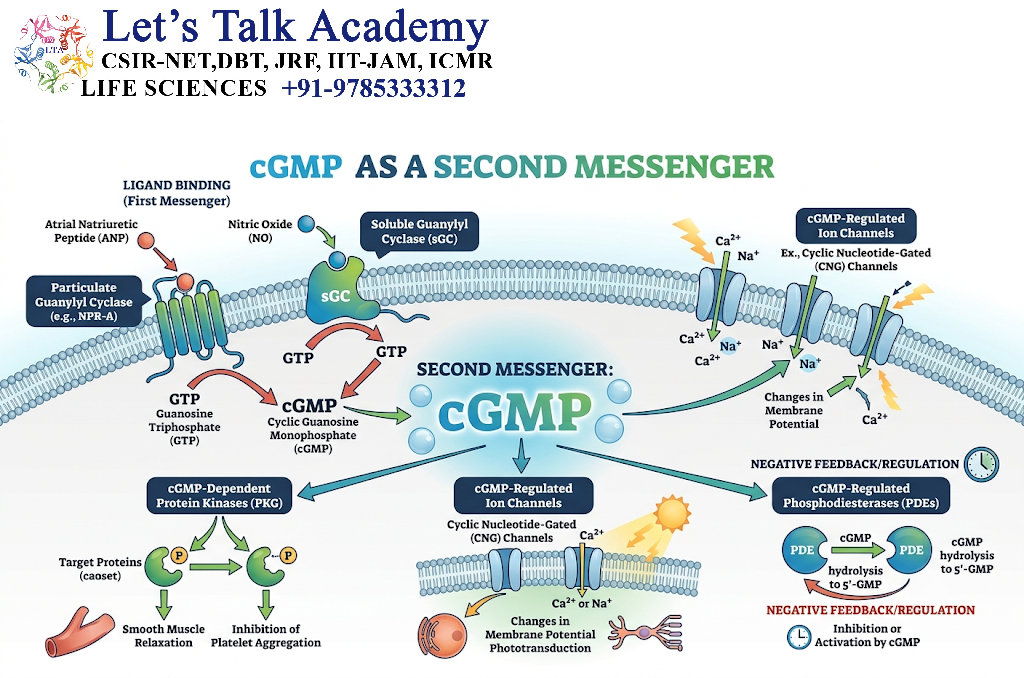
Cyclic GMP (cGMP) is another important cyclic nucleotide second messenger.
It is synthesized from GTP by guanylyl cyclase.
GTP → cGMP
7.1 Types of Guanylyl Cyclase
Two important forms are:
- Membrane-associated receptor guanylyl cyclases
- Soluble guanylyl cyclase
7.2 Nitric Oxide-cGMP Pathway
Nitric oxide (NO) can diffuse into nearby cells and activate soluble guanylyl cyclase.
The pathway is:
NO → Soluble guanylyl cyclase → cGMP → PKG → Cellular response
One important effect occurs in vascular smooth muscle.
Increased cGMP signaling promotes smooth-muscle relaxation and contributes to vasodilation.
7.3 cGMP Functions
cGMP participates in:
- Smooth-muscle relaxation
- Vascular signaling
- Phototransduction
- Ion-channel regulation
- Hormonal signaling
7.4 Regulation of cGMP
Phosphodiesterases degrade cGMP and therefore help terminate cGMP signaling.
8. IP₃ as a Second Messenger
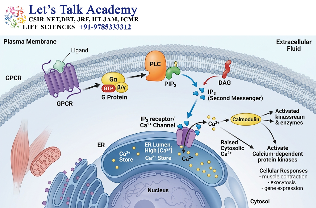
Inositol 1,4,5-trisphosphate (IP₃) is generated from a membrane phospholipid called PIP₂.
The enzyme phospholipase C (PLC) hydrolyzes PIP₂.
PIP₂ → IP₃ + DAG
IP₃ is water-soluble and moves through the cytoplasm.
8.1 IP₃ Function
The major function of IP₃ is to promote the release of Ca²⁺ from the endoplasmic reticulum (ER).
The sequence is:
Ligand → GPCR/RTK → PLC → PIP₂ cleavage → IP₃ → ER Ca²⁺ channel → Ca²⁺ release
IP₃ binds to IP₃ receptors located on the ER membrane.
This opens Ca²⁺ channels and increases cytosolic Ca²⁺ concentration.
9. DAG as a Second Messenger
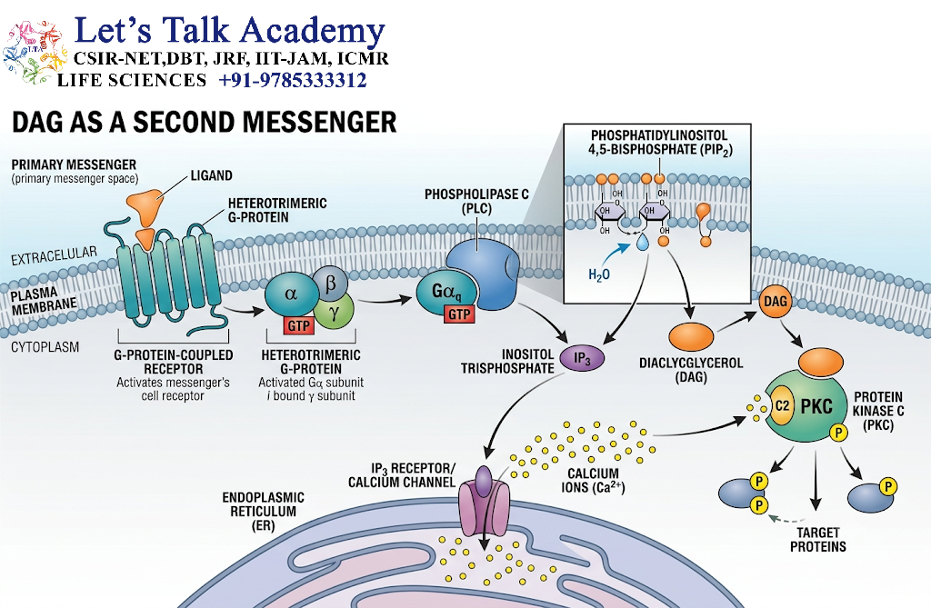
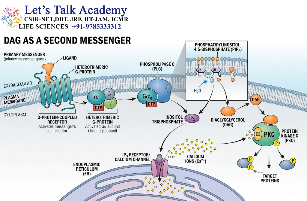
Diacylglycerol (DAG) is generated together with IP₃ when PLC cleaves PIP₂.
PIP₂ → IP₃ + DAG
Unlike IP₃, DAG is lipid-soluble and remains associated with the plasma membrane.
9.1 Activation of Protein Kinase C
DAG contributes to activation of protein kinase C (PKC).
Ca²⁺ can also participate in the activation of several conventional PKC isoforms.
Therefore, the PLC pathway can coordinate both:
- Ca²⁺ signaling
- Protein phosphorylation
9.2 DAG Functions
DAG signaling contributes to:
- Cell proliferation
- Secretion
- Metabolism
- Gene expression
- Cell differentiation
- Immune-cell activation
10. Calcium as a Second Messenger
Calcium ions (Ca²⁺) are among the most important intracellular signaling molecules.
Although Ca²⁺ is an essential structural and metabolic ion, its concentration in the resting cytosol is maintained at a very low level compared with extracellular fluid and intracellular Ca²⁺ stores.
Therefore, a small increase in cytosolic Ca²⁺ can act as a strong signal.
10.1 Sources of Cytosolic Ca²⁺
Major sources include:
- Extracellular fluid
- Endoplasmic reticulum
- Specialized intracellular stores
Ca²⁺ can enter the cytoplasm through ion channels or be released from intracellular stores.
10.2 Ca²⁺ Signaling Through IP₃
A common mechanism is:
Receptor → PLC → IP₃ → IP₃ receptor → ER Ca²⁺ release → Cellular response
10.3 Calcium-Calmodulin System
Ca²⁺ can bind to calmodulin, a calcium-binding regulatory protein.
The complex:
Ca²⁺ + Calmodulin → Ca²⁺-calmodulin complex
can regulate several enzymes and proteins, including certain protein kinases and phosphatases.
10.4 Functions of Ca²⁺ Signaling
Ca²⁺ participates in:
- Muscle contraction
- Neurotransmitter release
- Hormone secretion
- Fertilization
- Gene expression
- Metabolism
- Cell migration
- Cell differentiation
- Apoptosis
11. PLC-IP₃-DAG Signaling Pathway
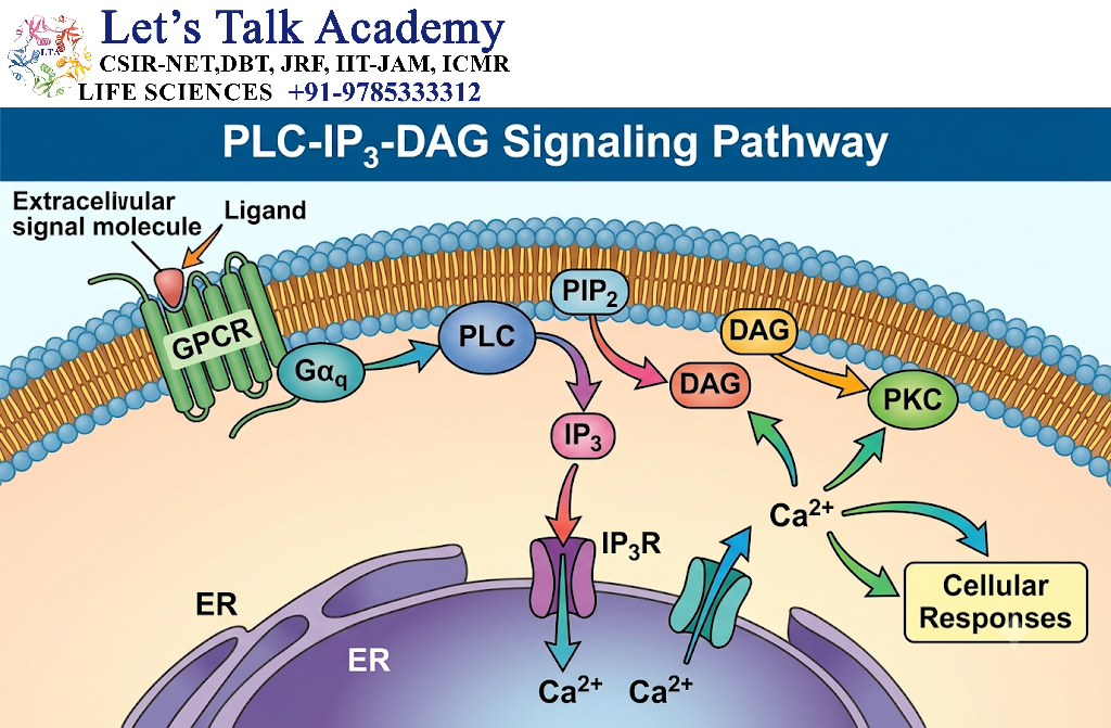
The PLC pathway is one of the most important second-messenger systems.
11.1 General Mechanism
A ligand binds to a receptor and activates PLC.
Depending on the receptor type, PLC may be activated through:
- Gq-coupled GPCRs
- Receptor tyrosine kinases through PLCγ
PLC hydrolyzes PIP₂:
PIP₂ → IP₃ + DAG
Then:
IP₃ → Ca²⁺ release from ER
and:
DAG + Ca²⁺ → PKC activation
The combined effects regulate numerous cellular processes.
11.2 Conceptual Flowchart
Signal molecule
↓
Receptor activation
↓
PLC activation
↓
PIP₂ cleavage
↓
IP₃ + DAG
↓
IP₃ → Ca²⁺ release
↓
DAG + Ca²⁺ → PKC
↓
Protein phosphorylation and cellular response
12. Relationship Between Second Messengers
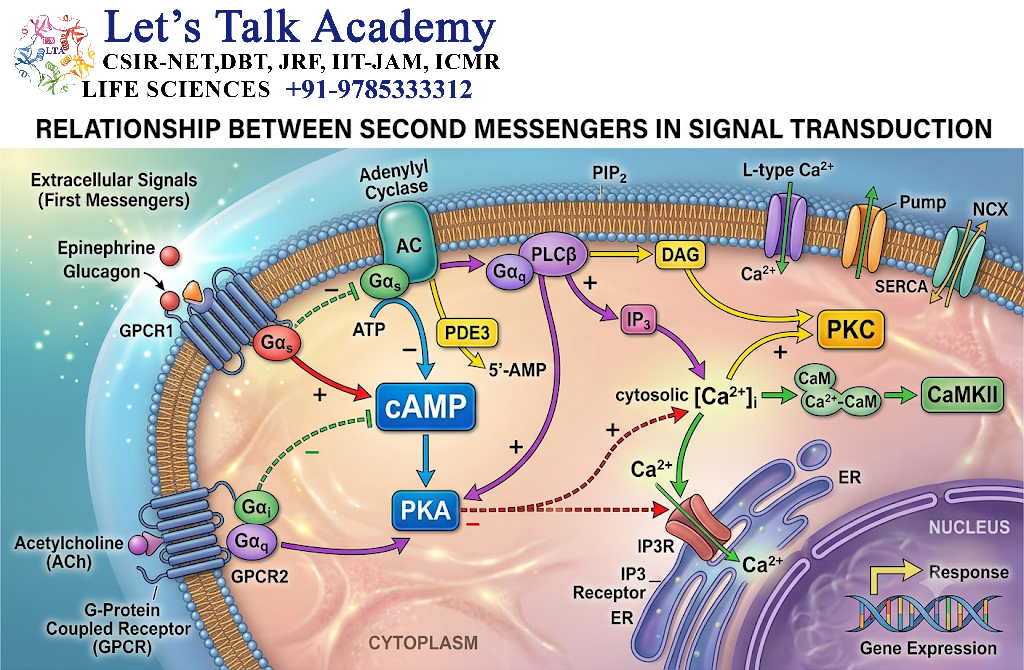
Second messengers rarely function completely independently.
For example, IP₃ and DAG are generated together from PIP₂.
PIP₂ → IP₃ + DAG
IP₃ increases cytosolic Ca²⁺, while DAG remains in the membrane and contributes to PKC activation.
Thus:
IP₃ → Ca²⁺ signaling
DAG → PKC signaling
Ca²⁺ + DAG → coordinated PKC activation
This illustrates how different second-messenger pathways cooperate to produce an integrated cellular response.
13. Signal Amplification by Second Messengers
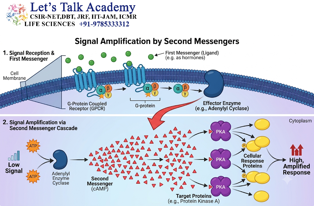
One of the major advantages of second-messenger systems is signal amplification.
A single activated receptor can activate multiple signaling proteins.
For example:
One receptor
↓
Multiple G proteins
↓
Adenylyl cyclase activation
↓
Many cAMP molecules
↓
Multiple PKA molecules
↓
Many target proteins
Thus, a relatively small extracellular signal can produce a large intracellular response.
14. Second Messengers and Protein Kinases
Many second messengers regulate protein kinases.
Important examples include:
| Second Messenger | Major Kinase |
|---|---|
| cAMP | PKA |
| cGMP | PKG |
| DAG/Ca²⁺ | PKC |
| Ca²⁺ | CaM-dependent kinases |
Protein kinases phosphorylate target proteins, commonly on serine, threonine or tyrosine residues depending on the kinase.
Phosphorylation can change:
- Enzyme activity
- Protein localization
- Protein stability
- Protein interactions
- Gene expression
15. Second Messengers and Gene Expression
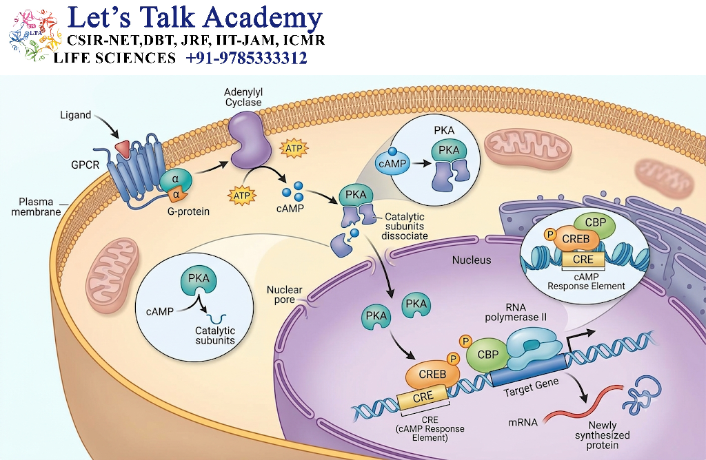
Second messengers can ultimately influence transcription.
For example:
Ligand → GPCR → cAMP → PKA → transcription factor activation → gene expression
A classic example involves CREB (cAMP response element-binding protein).
PKA can activate CREB through phosphorylation, allowing signaling pathways initiated at the cell membrane to influence nuclear gene expression.
16. Termination of Second-Messenger Signals
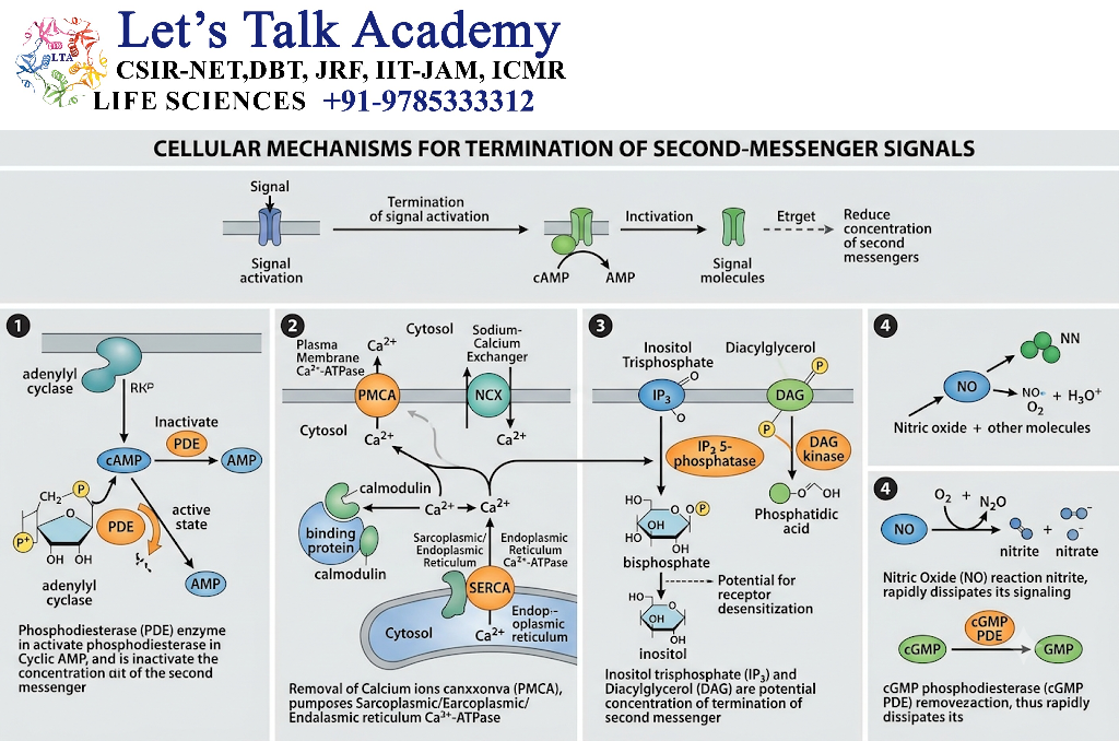
Second-messenger signaling must be carefully controlled.
If a signal remained permanently active, normal cellular regulation could be disrupted.
Major mechanisms include:
16.1 Phosphodiesterases
PDEs degrade cyclic nucleotides.
cAMP → AMP
cGMP → GMP
16.2 Ca²⁺ Removal
Ca²⁺ can be removed from the cytoplasm through:
- Ca²⁺ pumps
- Na⁺/Ca²⁺ exchangers
- Uptake into the ER
- Mitochondrial uptake under appropriate conditions
16.3 IP₃ Metabolism
IP₃ is metabolized through enzymatic pathways, reducing its signaling activity.
16.4 DAG Metabolism
DAG can be metabolized into other lipid molecules, helping terminate signaling.
17. Spatial Regulation of Second Messengers
Second-messenger signaling is not necessarily uniform throughout the cell.
For example, Ca²⁺ may form localized Ca²⁺ microdomains near ion channels.
Similarly, cAMP signaling can occur in specific cellular compartments.
Scaffold and anchoring proteins can position signaling enzymes near their targets.
This spatial organization increases signaling specificity.
18. Temporal Regulation
Second-messenger signals can vary in:
- Duration
- Frequency
- Amplitude
- Timing
For example, Ca²⁺ signaling can occur as:
- Short pulses
- Repeated oscillations
- Sustained increases
Different temporal patterns can produce different cellular responses.
Thus:
Signal amplitude + duration + frequency + location = cellular information
19. Second Messenger Crosstalk
Different signaling pathways can interact with one another.
Examples include:
- cAMP and Ca²⁺
- DAG and Ca²⁺
- cAMP and MAPK
- Ca²⁺ and MAPK
- PI3K-AKT and other kinase pathways
This interaction is known as signal crosstalk.
Crosstalk allows cells to integrate multiple extracellular signals and generate coordinated responses.
20. Second Messengers in Different Cellular Processes
20.1 Metabolism
cAMP signaling regulates metabolic enzymes involved in processes such as glycogen breakdown and lipolysis.
20.2 Muscle Contraction
Ca²⁺ is essential for skeletal, cardiac and smooth-muscle contraction.
20.3 Secretion
Ca²⁺ acts as a major trigger for exocytosis, including neurotransmitter and hormone release.
20.4 Gene Expression
cAMP, Ca²⁺ and other signaling pathways can regulate transcription factors.
20.5 Cell Growth
Second-messenger systems interact with pathways such as MAPK and PI3K-AKT to regulate growth and proliferation.
20.6 Cell Differentiation
Changes in second-messenger signaling can influence differentiation programs in many cell types.
21. Examples of Second-Messenger Signaling
21.1 Adrenaline and cAMP
Adrenaline
↓
β-adrenergic receptor
↓
Gs protein
↓
Adenylyl cyclase
↓
cAMP
↓
PKA
↓
Metabolic response
21.2 Angiotensin II and IP₃/DAG
Angiotensin II
↓
AT₁ receptor
↓
Gq protein
↓
PLC
↓
PIP₂ → IP₃ + DAG
↓
Ca²⁺ release + PKC activation
↓
Cellular response
21.3 Nitric Oxide and cGMP
NO
↓
Soluble guanylyl cyclase
↓
cGMP
↓
PKG
↓
Smooth-muscle relaxation
22. Biological Importance of Second Messengers
Second messengers are essential because they allow cells to convert receptor activation into coordinated intracellular responses.
Their major roles include:
- Signal transmission
- Signal amplification
- Regulation of enzymes
- Protein phosphorylation
- Gene regulation
- Metabolic control
- Muscle contraction
- Secretion
- Cell growth
- Differentiation
- Cell survival
- Communication between signaling pathways
23. Second Messengers and Disease
Abnormal second-messenger signaling can contribute to disease.
23.1 Cancer
Dysregulated signaling involving cAMP, Ca²⁺, PKC, MAPK or PI3K-AKT can contribute to abnormal proliferation and survival.
23.2 Metabolic Disorders
Defects in intracellular signaling can interfere with hormone-regulated metabolism, including insulin-related pathways.
23.3 Cardiovascular Disorders
Altered NO-cGMP signaling can affect vascular tone and cardiovascular regulation.
23.4 Neurological Disorders
Abnormal Ca²⁺ signaling can disturb neuronal function and contribute to cellular injury under pathological conditions.
24. Comparison of Major Second Messengers
| Property | cAMP | cGMP | IP₃ | DAG | Ca²⁺ |
|---|---|---|---|---|---|
| Chemical nature | Cyclic nucleotide | Cyclic nucleotide | Soluble inositol phosphate | Lipid | Ion |
| Main precursor | ATP | GTP | PIP₂ | PIP₂ | ER/extracellular stores |
| Major target | PKA | PKG | IP₃ receptor | PKC | Calmodulin/other proteins |
| Major location | Cytoplasm/compartments | Cytoplasm/compartments | Cytosol | Plasma membrane | Cytosol |
| Major role | Metabolism/gene regulation | Smooth muscle/visual signaling | Ca²⁺ release | PKC activation | Contraction/secretion/signaling |


