
1. Introduction to G-Protein-Coupled Receptor Signaling
Cells constantly receive information from their surroundings. Hormones, neurotransmitters, growth-related signals, odor molecules, and many other chemical substances communicate with cells to regulate their activities. However, most signaling molecules cannot directly pass through the plasma membrane because they are water-soluble or too large to enter the cell.
To respond to these external signals, cells use specialized proteins called cell surface receptors. These receptors recognize signaling molecules outside the cell and convert the information into intracellular signals. One of the largest and most important families of cell surface receptors is the G-protein-coupled receptor (GPCR) family.
G-protein-coupled receptors are membrane proteins that transmit extracellular signals to the interior of the cell through heterotrimeric G proteins. They play essential roles in sensory perception, neurotransmission, hormone action, metabolism, immune responses, cardiovascular regulation, and many other physiological processes.
The defining feature of GPCR signaling is the conversion of an extracellular ligand-binding event into changes in intracellular signaling molecules, ion channel activity, enzyme activity, and gene expression.
For example, when adrenaline binds to a β-adrenergic receptor on a heart cell, it initiates a signaling cascade that increases the rate and force of cardiac contraction. Similarly, the binding of a neurotransmitter to a GPCR in a neuron can alter ion channel activity and influence neuronal excitability.
Understanding GPCR signaling requires knowledge of receptor structure, G-protein activation, nucleotide exchange, second messenger generation, protein kinases, and the mechanisms that terminate signaling.
1.1 Definition of G-Protein-Coupled Receptors
G-protein-coupled receptors (GPCRs) are integral membrane proteins that contain seven transmembrane α-helical segments and communicate extracellular signals to intracellular signaling pathways through G proteins or other receptor-associated signaling proteins.
These receptors are also known as seven-transmembrane receptors (7TM receptors) because their polypeptide chains cross the plasma membrane seven times.
When a signaling molecule binds to a GPCR, the receptor undergoes a conformational change. This structural change enables the receptor to interact with a heterotrimeric G protein located on the cytoplasmic side of the membrane.
The activated receptor promotes the exchange of GDP for GTP on the Gα subunit of the G protein. This initiates downstream signaling through G-protein subunits and their target proteins.
1.2 General Characteristics of GPCRs
The major characteristics of GPCRs include:
-
GPCRs are integral membrane proteins.
-
They contain seven transmembrane α-helices.
-
Their amino-terminal region is generally located on the extracellular side of the plasma membrane.
-
Their carboxyl-terminal region is located in the cytoplasm.
-
They interact with heterotrimeric G proteins.
-
They activate intracellular signaling pathways through ligand-induced conformational changes.
-
They can regulate enzymes, ion channels, transporters, and transcriptional responses.
-
Their signaling can be amplified through enzymatic cascades.
-
They undergo desensitization, internalization, and recycling or degradation.
-
They participate in both rapid cellular responses and long-term changes in gene expression.
GPCRs can respond to a wide variety of chemical and physical stimuli. Depending on the receptor and its cellular environment, activation may produce effects within milliseconds or lead to gene regulatory changes over a much longer period.
1.3 Importance of GPCR Signaling in Cell Communication
GPCR signaling is important because it allows cells to respond rapidly and efficiently to changes in their environment.
Major functions include:
-
Regulation of heart rate and blood vessel diameter.
-
Control of smooth muscle contraction and relaxation.
-
Regulation of glucose and lipid metabolism.
-
Transmission of neuronal signals.
-
Detection of light, odors, and taste molecules.
-
Regulation of immune and inflammatory responses.
-
Control of hormone secretion.
-
Modulation of cell growth and differentiation.
-
Regulation of kidney function and fluid balance.
A single GPCR can activate multiple downstream pathways. Likewise, the same ligand may produce different responses in different tissues because cells express different receptor subtypes, G-protein isoforms, signaling enzymes, and regulatory proteins.
2. Structural Organization of G-Protein-Coupled Receptors
The structure of a GPCR is closely related to its function. The receptor acts as a molecular bridge between the extracellular environment and intracellular signaling machinery.
Most classical GPCRs have a characteristic seven-transmembrane architecture, although their extracellular domains, ligand-binding sites, and intracellular regulatory regions can vary considerably.
2.1 Seven-Transmembrane α-Helical Structure
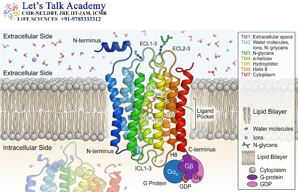
The defining structural feature of a GPCR is its seven transmembrane α-helices, commonly designated TM1 through TM7.
These hydrophobic helices are embedded within the lipid bilayer of the plasma membrane. They form a three-dimensional receptor structure that separates the extracellular and intracellular environments.
The seven helices are connected by:
-
Three extracellular loops.
-
Three intracellular loops.
-
An extracellular amino-terminal domain.
-
An intracellular carboxyl-terminal tail.
The transmembrane helices are not rigid structures. They can move relative to one another when a ligand binds to the receptor. These movements change the shape of the receptor’s intracellular surface and regulate its interactions with signaling proteins.
In many GPCRs, the ligand-binding region is located within the transmembrane helical bundle. However, some GPCRs have large extracellular domains that contribute substantially to ligand recognition.
2.2 Extracellular N-Terminal Domain
The amino-terminal domain of a GPCR is located outside the cell. It contributes to ligand recognition, receptor folding, and interactions with extracellular molecules.
The size and composition of this domain differ among GPCR classes.
For example:
-
Some small-molecule receptors have relatively short extracellular amino-terminal regions.
-
Peptide hormone receptors may have larger extracellular regions.
-
Class C GPCRs, such as metabotropic glutamate receptors, possess large extracellular ligand-binding domains.
The amino-terminal domain may also contain glycosylation sites. The attachment of carbohydrate groups can influence receptor folding, stability, trafficking, and ligand recognition.
2.3 Transmembrane Helices
The seven transmembrane helices form the central structural framework of a GPCR.
These helices are composed primarily of hydrophobic amino acids, allowing them to remain stable within the lipid bilayer. However, certain conserved polar and charged residues within the helices play important roles in ligand binding and receptor activation.
Ligand binding can cause changes in the relative positions of the helices. In many class A GPCRs, activation involves an outward movement of the cytoplasmic portion of transmembrane helix 6, along with coordinated changes in other helices.
This rearrangement creates a binding interface for the activated G protein or other intracellular signaling partners.
2.4 Intracellular Loops and C-Terminal Tail
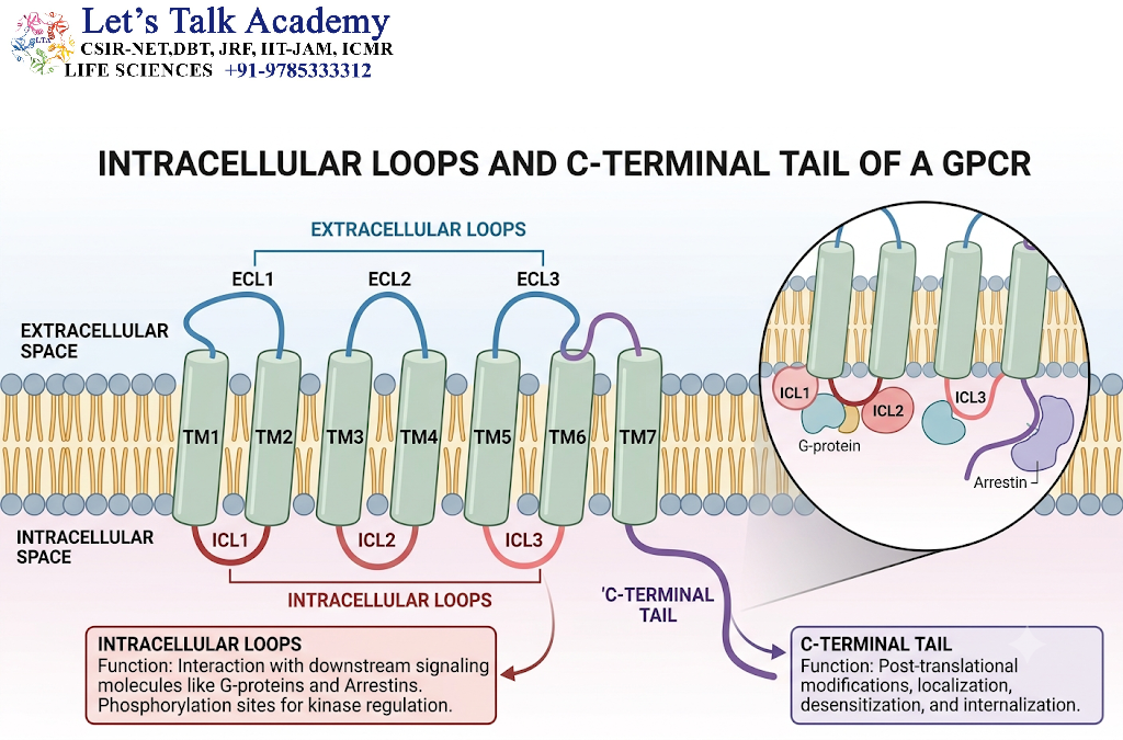
The intracellular loops and carboxyl-terminal tail are important for signal transmission and receptor regulation.
The intracellular loops interact with G proteins, arrestins, and other signaling proteins. Their amino acid sequences influence which intracellular partners can bind to a particular receptor.
The carboxyl-terminal tail often contains serine and threonine residues that can be phosphorylated by GPCR kinases and other protein kinases.
Phosphorylation of these residues may promote the binding of arrestin proteins, which can reduce further G-protein coupling and initiate receptor internalization.
The C-terminal tail may also contain molecular signals that regulate receptor trafficking and recycling.
2.5 Ligand-Binding Sites
GPCRs recognize a wide range of ligands, including:
-
Neurotransmitters.
-
Hormones.
-
Lipid mediators.
-
Peptides.
-
Odorant molecules.
-
Photons, through the retinal chromophore in visual receptors.
-
Metabolites and other extracellular chemical signals.
Ligands can bind to orthosteric or allosteric sites.
Orthosteric sites are the primary binding regions for endogenous ligands. Allosteric sites are distinct regions where other molecules bind and modify receptor activity.
An allosteric ligand may increase receptor activity, decrease receptor activity, or alter the receptor’s response to its primary ligand.
2.6 Structural Classes of GPCRs
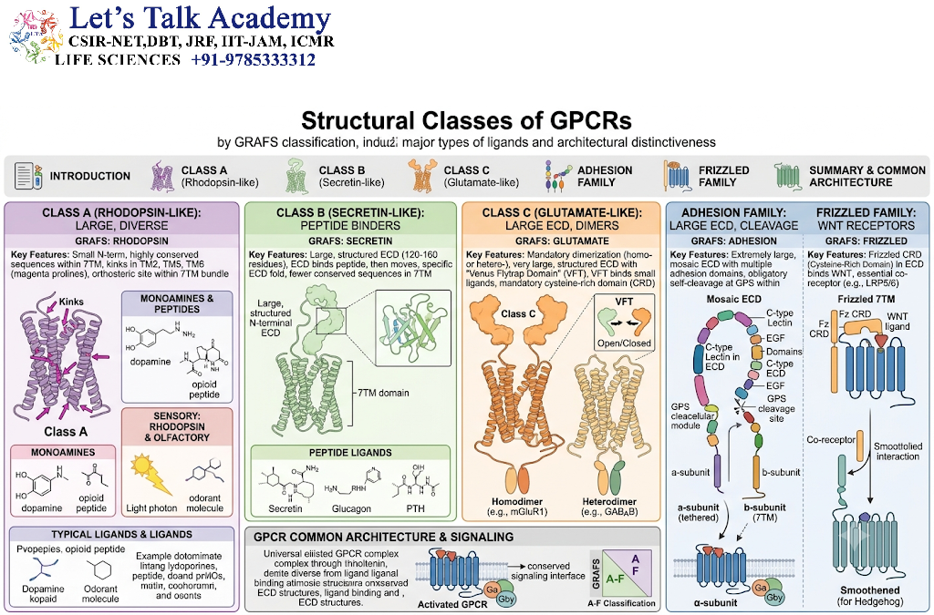
GPCRs are commonly grouped into several structural and evolutionary classes.
2.6.1 Class A: Rhodopsin-Like Receptors
Class A is the largest GPCR class. It includes receptors for many neurotransmitters, hormones, and sensory signals.
Examples include:
-
β-adrenergic receptors.
-
Dopamine receptors.
-
Muscarinic acetylcholine receptors.
-
Rhodopsin.
-
Opioid receptors.
-
Chemokine receptors.
These receptors commonly possess conserved sequence motifs that participate in receptor activation and G-protein coupling.
2.6.2 Class B: Secretin-Family Receptors
Class B GPCRs generally recognize peptide hormones and possess characteristic extracellular ligand-binding domains.
Examples include receptors for:
-
Glucagon.
-
Secretin.
-
Glucagon-like peptide-1 (GLP-1).
-
Parathyroid hormone.
These receptors frequently regulate physiological processes such as metabolism, digestion, and endocrine signaling.
2.6.3 Class C: Metabotropic Receptors
Class C GPCRs typically contain a large extracellular ligand-binding domain.
Examples include:
-
Metabotropic glutamate receptors.
-
GABA-B receptors.
-
Calcium-sensing receptors.
-
Certain taste receptors.
These receptors participate in neurotransmission, calcium homeostasis, and sensory perception.
2.6.4 Adhesion and Other GPCR Families
Adhesion GPCRs possess specialized extracellular regions involved in cell-cell or cell-matrix interactions. Some contain large extracellular domains and undergo distinctive activation mechanisms.
Other GPCR groups include receptors with specialized functions in taste, olfaction, and various physiological processes.
3. Heterotrimeric G Proteins: The Intracellular Signaling Partners
GPCRs transmit signals to the cell interior primarily through heterotrimeric G proteins. These proteins are composed of three subunits: Gα, Gβ, and Gγ.
The term heterotrimeric indicates that the protein complex contains three different types of subunits.
G proteins act as molecular switches. Their activity depends on whether guanine nucleotide GDP or GTP is bound to the Gα subunit.
3.1 Structure of Heterotrimeric G Proteins
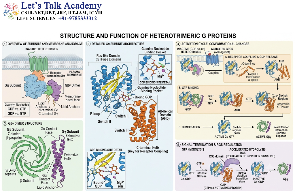
A heterotrimeric G protein consists of:
-
Gα subunit.
-
Gβ subunit.
-
Gγ subunit.
The Gα subunit binds guanine nucleotides and possesses intrinsic GTPase activity. The Gβ and Gγ subunits form a stable complex called the Gβγ dimer.
The Gα subunit is often associated with the inner surface of the plasma membrane through lipid modifications. The Gγ subunit also contains a lipid anchor that helps position the Gβγ complex near the membrane.
This membrane localization allows G proteins to interact efficiently with activated GPCRs and membrane-associated effector proteins.
3.2 Functions of the Gα Subunit
The Gα subunit functions as a guanine nucleotide-dependent molecular switch.
-
When GDP is bound, Gα is generally in an inactive state.
-
When GTP is bound, Gα adopts an active conformation.
-
The active Gα subunit can regulate downstream effectors.
-
Gα hydrolyzes GTP to GDP, helping terminate its activation.
The Gα subunit contains regions that undergo conformational changes when GTP replaces GDP. These changes influence the ability of Gα to bind effector proteins.
3.3 Functions of the Gβγ Dimer
The Gβγ dimer is not simply a structural component. It is also an important signaling unit.
After receptor activation, Gβγ can regulate:
-
Ion channels.
-
Certain phospholipases.
-
Lipid kinases.
-
Protein kinase signaling pathways.
-
Other membrane-associated signaling proteins.
In some pathways, Gβγ directly influences the activity of ion channels and contributes to rapid changes in cellular excitability.
Thus, both Gα and Gβγ can transmit information following GPCR activation.
3.4 GDP and GTP as Molecular Switches
The activity of heterotrimeric G proteins depends on the reversible binding of GDP and GTP.
GDP-bound Gα is associated with the inactive heterotrimer in the classical signaling cycle. When a GPCR becomes activated, it promotes GDP release and GTP binding.
The GTP-bound Gα subunit then changes conformation and regulates its target proteins.
GTP hydrolysis returns Gα to the GDP-bound state, allowing the signaling system to reset.
This cycle is essential because it permits signaling to be controlled in both time and intensity.
4. Mechanism of GPCR Activation
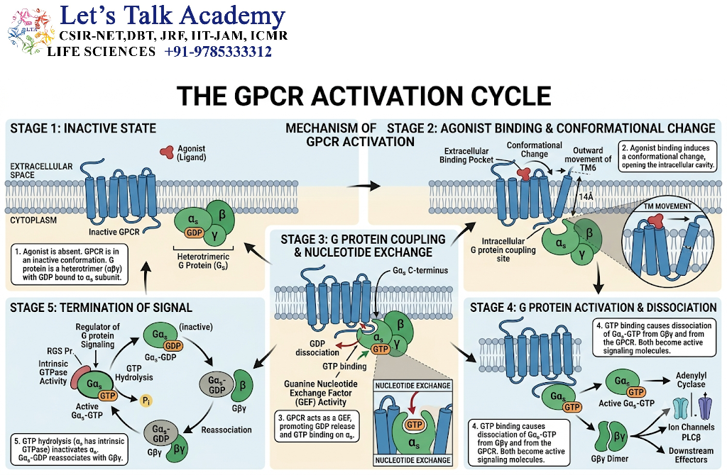
GPCR activation begins when an extracellular ligand binds to its receptor. The receptor then undergoes a conformational change that enables it to interact with a heterotrimeric G protein.
The following stages describe the classical mechanism of GPCR signaling.
4.1 Ligand Binding to the Receptor
The signaling process begins when a ligand binds to a specific site on the extracellular or transmembrane region of the GPCR.
The ligand may be a hormone, neurotransmitter, odorant, or another signaling molecule.
Ligand binding changes the energetic state of the receptor and stabilizes an active receptor conformation. The exact structural change depends on the receptor and the ligand.
Some ligands activate receptors, whereas others stabilize inactive conformations and inhibit receptor signaling.
4.2 Conformational Change in the GPCR
After ligand binding, the receptor undergoes a structural rearrangement.
This rearrangement affects the cytoplasmic regions of the receptor, allowing the receptor to interact with a G protein.
In many class A GPCRs, changes in the transmembrane helices create a binding site for the Gα subunit. The receptor acts as a guanine nucleotide exchange factor for the heterotrimeric G protein.
Unlike many soluble guanine nucleotide exchange factors, an activated GPCR is a membrane receptor that facilitates nucleotide exchange by changing the conformation of the Gα subunit.
4.3 Activation of the Heterotrimeric G Protein
In the inactive state, the Gα subunit is bound to GDP and associated with the Gβγ dimer.
The activated GPCR interacts with the Gα subunit and promotes the release of GDP.
Because the cytoplasm contains GTP, GTP binds to the nucleotide-binding site on Gα.
GTP binding causes the Gα subunit to adopt an active conformation.
The activated Gα-GTP and Gβγ signaling units can then regulate downstream effector proteins.
4.4 Dissociation and Rearrangement of G-Protein Subunits
In the classical model, Gα-GTP separates functionally from the Gβγ dimer after activation.
However, modern studies indicate that the subunits do not always need to undergo complete physical separation. In some cases, changes in their relative orientation allow them to interact with effectors while remaining associated.
Therefore, G-protein activation is better understood as a change in the signaling state and arrangement of the subunits rather than as an obligatory complete dissociation.
4.5 Activation of Downstream Effectors
The active Gα subunit and Gβγ dimer regulate specific downstream effectors.
Examples include:
-
Adenylyl cyclase.
-
Phospholipase C-β.
-
Ion channels.
-
Phosphoinositide 3-kinase and other lipid signaling enzymes in certain contexts.
-
Additional signaling proteins.
The identity of the activated effector depends on the G-protein subtype and the cellular context.
Activation of these effectors leads to the production of second messengers or changes in ion concentrations and initiates downstream cellular responses.
5. Major Families of G-Protein Signaling Pathways
The type of Gα subunit associated with a GPCR largely determines which intracellular signaling pathways are activated. Heterotrimeric G proteins are commonly grouped into four major families: Gαs, Gαi/o, Gαq/11, and Gα12/13.
Each family can regulate different effectors and generate distinct cellular responses. However, GPCR signaling is not always restricted to one G-protein family. A single receptor may interact with multiple G proteins, depending on its structure, ligand, expression level, and cellular environment.
5.1 Gαs-Mediated Signaling Pathway
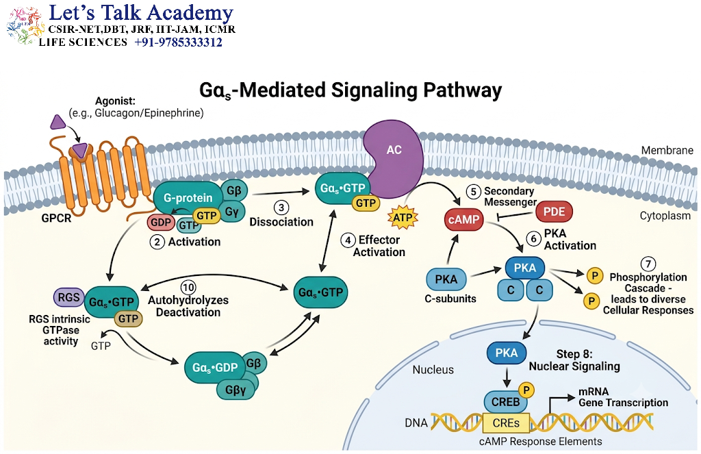
The Gαs pathway primarily stimulates the enzyme adenylyl cyclase, leading to the production of cyclic adenosine monophosphate (cAMP).
5.1.1 Activation of Adenylyl Cyclase
When a ligand activates a GPCR coupled to Gαs, the receptor promotes the exchange of GDP for GTP on the Gαs subunit.
The activated Gαs-GTP interacts with adenylyl cyclase, an enzyme associated with the plasma membrane. Adenylyl cyclase catalyzes the conversion of ATP into cAMP.
The increase in cAMP activates downstream signaling proteins, particularly protein kinase A (PKA).
5.1.2 Activation of Protein Kinase A
Protein kinase A is a serine/threonine protein kinase that is activated by cAMP.
In its inactive state, PKA generally exists as a tetramer containing two regulatory subunits and two catalytic subunits.
When cAMP binds to the regulatory subunits, the catalytic subunits are released and become active.
Active PKA phosphorylates specific target proteins, including enzymes, ion channels, transcription factors, and other regulatory proteins.
The cellular effects of PKA activation depend on the proteins expressed in a particular cell.
5.1.3 Physiological Functions of the Gαs Pathway
The Gαs–cAMP–PKA pathway participates in several processes:
-
Regulation of cardiac contraction.
-
Stimulation of glycogen breakdown in certain tissues.
-
Regulation of lipolysis.
-
Modulation of gene expression.
-
Regulation of hormone secretion.
-
Control of ion channel activity.
For example, activation of β₁-adrenergic receptors in cardiac muscle increases cAMP production and activates PKA. This contributes to increased cardiac contractility and changes in heart rate.
5.1.4 Regulation of Gene Expression by CREB
One important target of PKA is the transcription factor known as CREB (cAMP response element-binding protein).
PKA can phosphorylate CREB at a regulatory site. Phosphorylated CREB interacts with specific DNA sequences called cAMP response elements (CREs) through its DNA-binding domain.
CREB can recruit transcriptional coactivators, including CREB-binding protein (CBP), to regulate the transcription of target genes.
This mechanism connects a rapid membrane receptor signal with longer-term changes in gene expression.
5.2 Gαi/o-Mediated Signaling Pathway
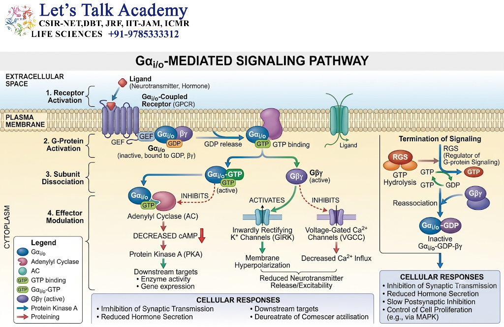
The Gαi/o family commonly inhibits certain isoforms of adenylyl cyclase, thereby reducing cAMP production.
5.2.1 Inhibition of Adenylyl Cyclase
When a GPCR activates Gαi/o, the Gαi/o-GTP subunit can bind to adenylyl cyclase and reduce its catalytic activity.
As a result, the intracellular concentration of cAMP may decrease.
The reduction in cAMP can decrease PKA activity and alter the phosphorylation state of various target proteins.
However, the effects of Gαi/o signaling depend on the adenylyl cyclase isoform and the other signaling proteins present in the cell. Not every adenylyl cyclase responds identically to Gαi/o.
5.2.2 Regulation of Ion Channels by Gβγ
The Gβγ dimer released or rearranged during G-protein activation can directly regulate ion channels.
In certain neurons, Gβγ can activate G-protein-gated inwardly rectifying potassium channels (GIRK channels). Potassium efflux can hyperpolarize the membrane and reduce neuronal excitability.
Gβγ can also inhibit certain voltage-gated calcium channels, reducing calcium entry into the presynaptic terminal.
This mechanism is important in the regulation of neurotransmitter release.
5.2.3 Physiological Significance
Gαi/o-coupled receptors participate in:
-
Inhibition of neurotransmitter release.
-
Modulation of neuronal excitability.
-
Regulation of heart rate.
-
Control of hormone secretion.
-
Regulation of synaptic transmission.
-
Modulation of cellular metabolism.
For example, activation of certain muscarinic acetylcholine receptors in cardiac tissue can activate G-protein-gated potassium channels through Gβγ signaling. This contributes to the slowing of heart rate.
5.3 Gαq/11-Mediated Signaling Pathway
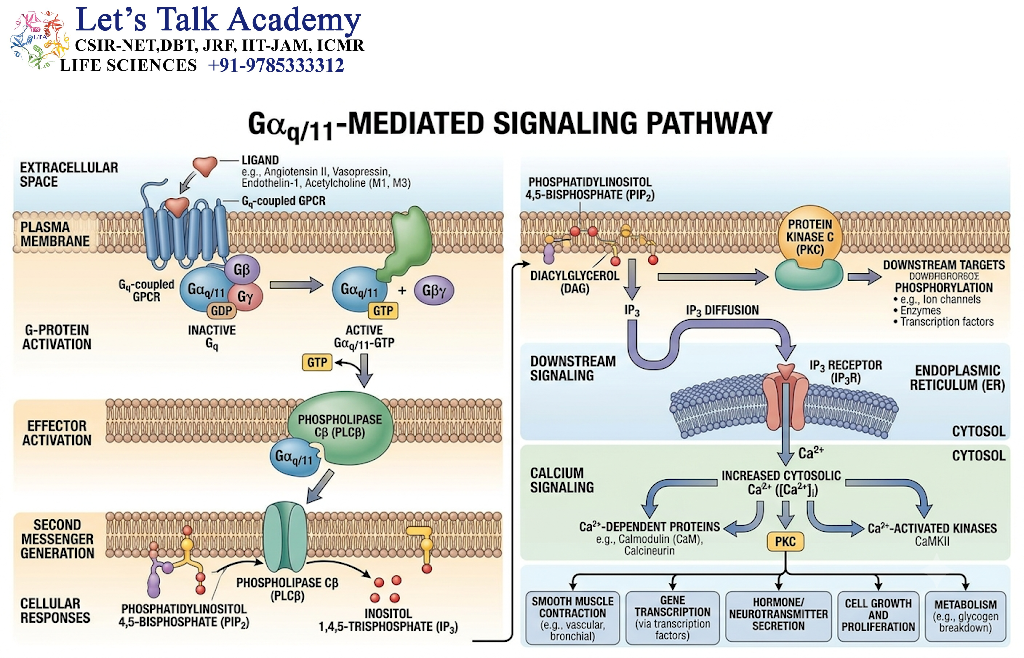
The Gαq/11 family primarily activates phospholipase C-β (PLC-β), leading to the formation of two important second messengers: inositol 1,4,5-trisphosphate (IP₃) and diacylglycerol (DAG).
This pathway is particularly important in calcium signaling, smooth muscle contraction, secretion, and protein kinase C activation.
5.3.1 Activation of Phospholipase C-β
When a ligand activates a GPCR coupled to Gαq/11, the activated Gαq/11-GTP subunit interacts with PLC-β.
PLC-β hydrolyzes the membrane phospholipid phosphatidylinositol 4,5-bisphosphate (PIP₂).
This reaction produces two second messengers with different properties.
-
IP₃ is water-soluble and can diffuse through the cytoplasm.
-
DAG is lipid-soluble and remains associated with the plasma membrane.
5.3.2 IP₃-Mediated Calcium Release
IP₃ travels through the cytoplasm and binds to IP₃ receptors located on the membrane of the endoplasmic reticulum (ER).
The IP₃ receptor functions as a calcium-release channel.
When IP₃ binds to the receptor under appropriate conditions, the channel opens and calcium ions are released from the ER into the cytoplasm.
This causes a rapid increase in cytosolic calcium concentration.
Calcium acts as a second messenger and can regulate several cellular processes.
These include:
-
Muscle contraction.
-
Vesicle fusion.
-
Enzyme activation.
-
Metabolic regulation.
-
Gene expression.
-
Cellular secretion.
5.3.3 DAG-Mediated Activation of Protein Kinase C
DAG remains within the plasma membrane after the hydrolysis of PIP₂.
Together with calcium and other regulatory factors, DAG promotes the activation of conventional protein kinase C (PKC) isoforms.
PKC is a family of serine/threonine protein kinases that phosphorylate specific target proteins.
PKC activation regulates processes such as secretion, contraction, proliferation, and changes in gene expression.
Not all PKC isoforms require calcium. Their regulatory requirements differ according to their structural classification.
5.3.4 Physiological Functions of the Gαq/11 Pathway
The Gαq/11–PLC-β pathway is involved in:
-
Smooth muscle contraction.
-
Secretion of digestive enzymes.
-
Platelet activation.
-
Regulation of vascular tone.
-
Neurotransmitter-mediated signaling.
-
Activation of calcium-dependent enzymes.
-
Regulation of cellular metabolism.
For example, activation of certain α₁-adrenergic receptors in vascular smooth muscle increases intracellular calcium and promotes smooth muscle contraction, which can contribute to vasoconstriction.
5.4 Gα12/13-Mediated Signaling Pathway
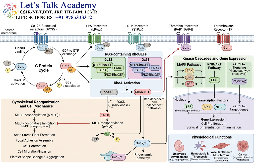
The Gα12/13 family regulates intracellular signaling through proteins known as Rho guanine nucleotide exchange factors (RhoGEFs).
These signaling proteins activate small GTPases of the Rho family, particularly RhoA.
5.4.1 Activation of RhoGEFs
After activation of Gα12/13, the GTP-bound Gα subunit interacts with specific RhoGEFs.
RhoGEFs promote the exchange of GDP for GTP on RhoA.
RhoA-GTP is an active small GTPase that regulates the actin cytoskeleton.
5.4.2 Regulation of the Actin Cytoskeleton
Activated RhoA influences proteins such as Rho-associated protein kinase (ROCK) and other downstream effectors.
The RhoA–ROCK pathway regulates:
-
Actin filament organization.
-
Formation of stress fibers.
-
Cellular contractility.
-
Cell adhesion.
-
Cell migration.
-
Changes in cell shape.
The pathway is important in the regulation of cytoskeletal dynamics and tissue organization.
5.4.3 Physiological Significance
Gα12/13 signaling participates in:
-
Regulation of smooth muscle contractility.
-
Cell migration.
-
Cytoskeletal remodeling.
-
Cell adhesion.
-
Tissue morphogenesis.
-
Regulation of vascular function.
The Gα12/13 pathway demonstrates that GPCRs can regulate the cytoskeleton in addition to controlling classical second messenger systems.
5.5 Comparison of the Major Gα Signaling Families
|
Gα family |
Major effector or target |
Important signaling molecules |
Common cellular effects |
|---|---|---|---|
|
Gαs |
Adenylyl cyclase stimulation |
Increased cAMP, PKA activation |
Metabolism, secretion, gene regulation |
|
Gαi/o |
Inhibition of certain adenylyl cyclases; ion channels through Gβγ |
Reduced cAMP, altered ion flow |
Neuronal modulation, cardiac regulation |
|
Gαq/11 |
PLC-β |
IP₃, DAG, Ca²⁺ |
Contraction, secretion, PKC activation |
|
Gα12/13 |
RhoGEFs |
RhoA-GTP and downstream signals |
Cytoskeletal remodeling, cell shape, migration |
The pathways described above are simplified representations. In living cells, these signaling networks can overlap and influence one another.
6. Second Messengers in GPCR Signaling
Second messengers are intracellular signaling molecules that transmit information from activated receptors and their immediate effectors to downstream targets.
The extracellular signaling molecule is often called the first messenger, whereas intracellular molecules such as cAMP, IP₃, DAG, and calcium function as second messengers.
Second messengers allow a receptor signal to spread rapidly through the cell and activate multiple target proteins.
6.1 Characteristics of Second Messengers
An effective second messenger generally has several properties:
-
It can be produced or released rapidly.
-
Its concentration can change in response to receptor activation.
-
It can interact with specific intracellular target proteins.
-
Its activity can be terminated or reversed.
-
It can amplify an extracellular signal.
-
Its spatial distribution can be regulated within the cell.
Second messengers do not simply turn cellular responses on. Their concentration, duration, location, and frequency of production can determine the type of cellular response.
6.2 Cyclic AMP as a Second Messenger
Cyclic AMP is one of the best-characterized second messengers in GPCR signaling.
It is synthesized from ATP by adenylyl cyclase and broken down by phosphodiesterases (PDEs).
The balance between cAMP synthesis and degradation determines the intracellular cAMP concentration.
6.2.1 Major Targets of cAMP
The principal intracellular targets of cAMP include:
-
Protein kinase A.
-
Exchange proteins directly activated by cAMP (Epac proteins).
-
Certain cyclic nucleotide-gated ion channels.
-
Other cAMP-responsive regulatory proteins.
Epac proteins function as guanine nucleotide exchange factors for particular small GTPases, such as Rap proteins. This provides a PKA-independent route through which cAMP can regulate cell behavior.
6.2.2 Spatial Regulation of cAMP
cAMP does not necessarily diffuse uniformly throughout the entire cell.
Its distribution is influenced by phosphodiesterases, anchoring proteins, local adenylyl cyclase activity, and cellular compartments.
A-kinase anchoring proteins (AKAPs) can organize PKA and other signaling components into localized signaling complexes.
This spatial organization allows cAMP to produce different responses in different regions of the same cell.
6.3 Inositol 1,4,5-Trisphosphate (IP₃)
IP₃ is a water-soluble second messenger produced by the hydrolysis of PIP₂ by PLC-β.
Its primary function in the classical GPCR pathway is to promote the release of calcium ions from intracellular stores.
IP₃ binds to IP₃ receptors on the endoplasmic reticulum. The receptors function as ligand-gated calcium-release channels.
IP₃-mediated calcium release is important for the rapid transmission of signals from the plasma membrane to intracellular organelles.
6.4 Diacylglycerol (DAG)
DAG is a lipid second messenger produced alongside IP₃ during the hydrolysis of PIP₂.
Unlike IP₃, DAG remains associated with the lipid bilayer.
DAG recruits and regulates certain protein kinase C isoforms and other proteins containing lipid-binding domains.
Because DAG is membrane-associated, it helps localize signaling proteins near the plasma membrane.
6.5 Calcium Ions as Second Messengers
Calcium ions are versatile intracellular signaling molecules.
The concentration of free calcium in the cytoplasm is normally much lower than the concentration in the extracellular environment and in certain intracellular stores, such as the endoplasmic reticulum.
The cell maintains calcium gradients through pumps, exchangers, channels, and calcium-binding proteins.
When a GPCR activates PLC-β, IP₃-mediated calcium release can rapidly increase cytosolic calcium levels.
Calcium can bind to regulatory proteins, including calmodulin, and activate calcium-dependent enzymes.
6.5.1 Calcium–Calmodulin Signaling
Calmodulin is a calcium-binding regulatory protein.
When calcium binds to calmodulin, the protein undergoes a conformational change that enables it to interact with various target proteins.
Calcium–calmodulin complexes regulate:
-
Calcium/calmodulin-dependent protein kinases.
-
Certain phosphatases.
-
Metabolic enzymes.
-
Ion channels.
-
Transcriptional regulators.
Calcium signaling is therefore closely linked to enzyme activity, metabolism, muscle contraction, and gene expression.
6.6 Phosphoinositide Lipid Signaling
GPCRs can regulate membrane phospholipids through enzymes such as PLC and, in certain contexts, phosphoinositide 3-kinases.
These lipid signaling pathways can influence protein localization, membrane trafficking, cell growth, and cytoskeletal organization.
It is important to distinguish PLC-mediated PIP₂ hydrolysis from PI3K-mediated phosphorylation of phosphoinositides. Although both pathways involve membrane lipids, they generate different signaling products and recruit different downstream proteins.
7. Signal Amplification in GPCR Signaling
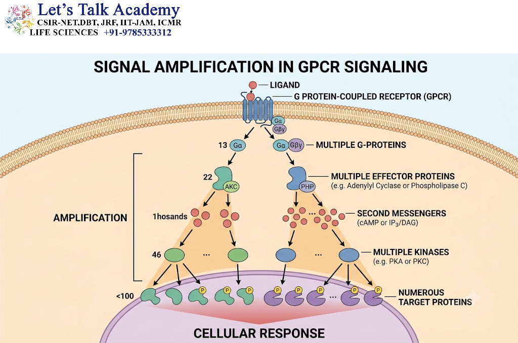
Signal amplification is a fundamental property of many GPCR signaling pathways.
A small number of extracellular ligand molecules can produce a much larger intracellular response because one activated receptor can interact with multiple G proteins over time, and each activated effector can generate numerous second messenger molecules.
7.1 Mechanism of Signal Amplification
Signal amplification can occur at several stages:
-
Ligand binding activates a receptor.
-
One activated receptor can promote activation of multiple G proteins.
-
An activated enzyme can produce many second messenger molecules.
-
A second messenger can activate multiple protein kinase molecules.
-
Each kinase can phosphorylate several target proteins.
-
Downstream signaling can alter the activity of additional regulatory proteins.
This series of events allows a relatively small extracellular signal to produce a substantial cellular response.
7.2 Example of Amplification Through the cAMP Pathway
Consider a GPCR coupled to Gαs.
-
A hormone binds to the receptor.
-
The activated receptor promotes G-protein activation.
-
Gαs-GTP activates adenylyl cyclase.
-
Adenylyl cyclase produces cAMP from ATP.
-
cAMP activates PKA.
-
PKA phosphorylates several target proteins.
-
The target proteins alter metabolism, ion transport, or gene expression.
The magnitude of amplification depends on receptor density, coupling efficiency, enzyme activity, substrate availability, phosphodiesterase activity, and feedback mechanisms.
7.3 Biological Significance of Amplification
Signal amplification is particularly important when the concentration of a signaling molecule is low.
For example, sensory systems often require the detection of very small quantities of external stimuli. Amplification helps convert these weak stimuli into signals that can be detected by the cell.
However, excessive amplification or impaired signal termination can cause abnormal signaling and contribute to disease.
8. Termination and Regulation of GPCR Signaling
GPCR signaling must be carefully controlled. If signaling continues indefinitely, cells may experience excessive activation, altered metabolism, or abnormal physiological responses.
Signal termination occurs through several mechanisms involving GTP hydrolysis, second messenger degradation, receptor phosphorylation, arrestin recruitment, and receptor trafficking.
8.1 GTP Hydrolysis by the Gα Subunit
The Gα subunit possesses intrinsic GTPase activity.
After the Gα subunit activates an effector, it hydrolyzes GTP to GDP and inorganic phosphate.
The GDP-bound Gα subunit returns to an inactive conformation.
It can then reassociate with the Gβγ dimer to form the inactive heterotrimeric G protein.
8.1.1 Role of Regulators of G-Protein Signaling
Proteins known as regulators of G-protein signaling (RGS proteins) accelerate the GTPase activity of specific Gα subunits.
RGS proteins act as GTPase-activating proteins (GAPs) for their target Gα proteins.
By increasing the rate of GTP hydrolysis, they shorten the duration of G-protein signaling.
Different RGS proteins have distinct tissue distributions and selectivities for Gα subunits.
8.2 Degradation of cAMP by Phosphodiesterases
Phosphodiesterases are enzymes that hydrolyze cyclic nucleotides.
In the cAMP pathway, PDEs convert cAMP into AMP, reducing the concentration of the second messenger.
This process limits the duration of PKA activation and other cAMP-dependent responses.
Phosphodiesterases play a major role in shaping the intensity and duration of intracellular cAMP signals.
8.3 Calcium Removal from the Cytoplasm
Calcium signals must be terminated efficiently because prolonged elevation of cytosolic calcium can disrupt cellular homeostasis.
Cells reduce cytosolic calcium through:
-
Calcium ATPases.
-
Sodium–calcium exchangers.
-
Uptake into the endoplasmic reticulum.
-
Uptake into mitochondria under appropriate conditions.
-
Calcium-binding proteins.
The removal of calcium helps restore resting conditions and allows the cell to respond to subsequent signals.
8.4 Receptor Phosphorylation
Activated GPCRs can be phosphorylated by specialized enzymes called G-protein-coupled receptor kinases (GRKs).
GRKs recognize activated receptors and phosphorylate specific serine and threonine residues, particularly within intracellular receptor regions.
Receptor phosphorylation can reduce the ability of the receptor to couple to G proteins and promote the recruitment of arrestin proteins.
This process is an important component of receptor desensitization.
8.5 β-Arrestin-Mediated Desensitization
β-arrestins are regulatory proteins that bind to phosphorylated, activated GPCRs.
Their binding can interfere with further G-protein coupling, reducing receptor signaling through the classical G-protein pathway.
β-arrestins also serve as scaffolding proteins that recruit other signaling proteins.
They can participate in receptor internalization by linking receptors to components of the endocytic machinery.
Importantly, β-arrestins are not merely signal terminators. In some contexts, they can initiate or organize additional signaling pathways.
8.6 Receptor Internalization and Recycling
After activation and arrestin recruitment, many GPCRs are internalized through clathrin-dependent endocytosis.
The internalized receptors enter endosomal compartments, where their activity and fate are regulated.
Depending on the receptor and cellular conditions, internalized GPCRs may:
-
Return to the plasma membrane through recycling.
-
Remain in intracellular compartments.
-
Undergo degradation in lysosomes.
-
Continue signaling from intracellular locations.
Receptor trafficking determines the duration of cellular responsiveness and helps regulate receptor abundance at the cell surface.
8.7 Desensitization, Downregulation, and Resensitization
These three terms describe related but distinct processes.
Desensitization refers to a reduction in receptor responsiveness, often occurring after receptor phosphorylation and arrestin recruitment.
Downregulation refers to a decrease in the number of receptors available at the cell surface or in the cell as a whole, often because of degradation or altered receptor synthesis.
Resensitization refers to the restoration of receptor responsiveness. It may involve receptor dephosphorylation, recycling to the plasma membrane, and reestablishment of productive G-protein coupling.
9. β-Arrestin-Dependent and G-Protein-Independent Signaling
GPCRs were initially studied primarily as receptors that transmit signals through heterotrimeric G proteins. However, research has established that many GPCRs can also activate signaling pathways through other intracellular proteins.
β-arrestins are particularly important in these alternative signaling mechanisms.
9.1 Recruitment of β-Arrestins
Following receptor activation, GRKs can phosphorylate intracellular regions of the GPCR.
β-arrestins recognize specific phosphorylated receptor configurations and bind to the receptor.
The interaction depends on both the receptor’s phosphorylation pattern and its active conformation.
The recruitment of β-arrestin can influence receptor desensitization, trafficking, and signaling through additional protein complexes.
9.2 β-Arrestin as a Signaling Scaffold
β-arrestins can function as scaffolding proteins by bringing signaling proteins into close proximity.
They may organize components of mitogen-activated protein kinase (MAPK) cascades and other signaling networks.
For example, certain GPCRs can promote β-arrestin-dependent regulation of ERK1/2 signaling.
The precise outcome depends on the receptor, ligand, cellular context, and composition of the signaling complex.
9.3 Functional Selectivity or Biased Agonism
Different ligands can stabilize different conformations of the same GPCR.
Some ligands may preferentially promote G-protein signaling, whereas others may favor arrestin recruitment or other receptor-associated pathways.
This phenomenon is known as functional selectivity or biased agonism.
It occurs because GPCRs can adopt multiple active conformations rather than existing only in a single inactive or active state.
9.3.1 Importance of Biased Signaling
Biased signaling is important in pharmacology because it may allow researchers to develop ligands that selectively influence particular signaling pathways.
However, a ligand’s observed signaling bias can also be influenced by receptor expression, signal amplification, assay conditions, and differences in pathway sensitivity.
Therefore, claims of pathway selectivity must be interpreted using carefully controlled experimental comparisons.
10. GPCR Signaling and Mitogen-Activated Protein Kinase Pathways
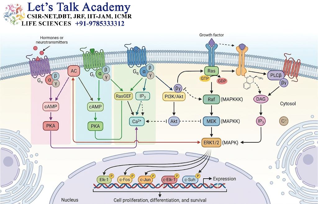
GPCRs can regulate mitogen-activated protein kinase (MAPK) cascades, which control processes such as proliferation, differentiation, survival, and gene expression.
MAPK pathways consist of sequential protein kinases that activate one another through phosphorylation.
10.1 General Organization of a MAPK Cascade
A typical three-tiered MAPK cascade contains:
-
MAP kinase kinase kinase (MAP3K).
-
MAP kinase kinase (MAP2K).
-
MAP kinase (MAPK).
The activation of MAP3K leads to the phosphorylation and activation of MAP2K. MAP2K then phosphorylates and activates MAPK.
Activated MAPKs can phosphorylate transcription factors and other intracellular targets.
10.2 GPCR-Mediated ERK1/2 Activation
Certain GPCRs activate ERK1/2 through G-protein-dependent mechanisms.
For example, Gβγ subunits may activate signaling proteins that ultimately stimulate the Ras–Raf–MEK–ERK cascade.
In other contexts, GPCRs can influence receptor tyrosine kinases, such as the epidermal growth factor receptor, through processes known as transactivation.
β-arrestin-associated signaling complexes can also contribute to ERK1/2 regulation.
The precise mechanism depends on the receptor and cell type.
10.3 Biological Significance
GPCR regulation of MAPK signaling can influence:
-
Cell proliferation.
-
Cell differentiation.
-
Cell survival.
-
Gene expression.
-
Cellular migration.
-
Tissue remodeling.
Abnormal GPCR–MAPK communication may contribute to pathological changes when signaling becomes excessive, prolonged, or improperly regulated.
11. Physiological Functions of GPCR Signaling
GPCRs participate in nearly every major physiological system. Their ability to recognize diverse extracellular signals makes them important regulators of communication between cells, tissues, and organs.
11.1 GPCRs in the Nervous System
Many neurotransmitters act through GPCRs.
Examples include:
-
Dopamine.
-
Serotonin.
-
Noradrenaline.
-
Acetylcholine.
-
GABA.
-
Glutamate, through metabotropic glutamate receptors.
-
Opioid peptides.
GPCRs can regulate neuronal excitability, neurotransmitter release, synaptic plasticity, and sensory processing.
Unlike ionotropic receptors, which directly form ion channels, metabotropic GPCRs generally act through intracellular signaling pathways and can produce slower but longer-lasting effects.
11.2 GPCRs in the Cardiovascular System
GPCRs regulate cardiac activity and vascular function.
β₁-adrenergic receptors activate Gαs-dependent signaling in cardiac muscle and contribute to increased cardiac contractility.
α₁-adrenergic receptors can activate Gαq/11 signaling in vascular smooth muscle, promoting calcium-dependent contraction.
Other GPCRs regulate blood vessel relaxation, platelet activation, and cardiovascular homeostasis.
11.3 GPCRs in the Endocrine System
Many hormones exert their effects through GPCRs.
Examples include receptors for:
-
Glucagon.
-
Vasopressin.
-
Parathyroid hormone.
-
Thyroid-stimulating hormone.
-
Adrenocorticotropic hormone.
-
Luteinizing hormone.
-
Follicle-stimulating hormone.
These receptors regulate metabolism, reproduction, growth, water balance, and endocrine communication.
11.4 GPCRs in Sensory Perception
GPCRs are essential for several sensory systems.
11.4.1 Vision
Rhodopsin is a GPCR located in the membranes of rod photoreceptor cells.
When light is absorbed by the retinal chromophore associated with rhodopsin, the receptor changes conformation and activates the heterotrimeric G protein transducin.
Transducin activates a cyclic GMP phosphodiesterase, leading to a reduction in cyclic GMP concentration.
The reduction in cyclic GMP causes cyclic nucleotide-gated ion channels to close, producing a change in the electrical state of the photoreceptor.
This is a major step in visual signal transduction.
11.4.2 Smell
Olfactory receptors are GPCRs expressed in olfactory sensory neurons.
Odorant molecules activate these receptors, which can stimulate Gαolf-dependent signaling.
The pathway commonly involves adenylyl cyclase, cAMP production, and cyclic nucleotide-gated ion channels.
The resulting electrical signals contribute to odor detection.
11.4.3 Taste
Certain taste receptors are GPCRs.
For example, sweet, bitter, and umami taste detection involves specialized GPCRs that activate intracellular signaling pathways in taste receptor cells.
These pathways can regulate intracellular calcium signaling and neurotransmitter release.
11.5 GPCRs in the Immune System
Chemokine receptors are GPCRs that guide the movement of immune cells.
Chemokines bind to their receptors and activate signaling pathways that regulate:
-
Cell migration.
-
Leukocyte recruitment.
-
Immune-cell positioning.
-
Inflammatory responses.
-
Immune-cell activation.
GPCR signaling is therefore essential for the coordination of immune responses.
12. GPCR Signaling in Plants and Other Organisms
Although GPCR signaling is extensively studied in animals, G-protein-mediated signaling also occurs in plants and other organisms.
However, the composition of the receptor systems and the mechanisms of signal transduction can differ substantially between species.
12.1 G-Protein Signaling in Plants
Plants possess heterotrimeric G proteins that participate in the regulation of growth, development, stress responses, and environmental adaptation.
In some plant species, the canonical G-protein cycle can occur through mechanisms that differ from the classical animal GPCR model.
For example, plant-specific proteins such as G-protein regulators can influence the activation state of heterotrimeric G proteins.
Some plant G proteins also exhibit relatively rapid intrinsic nucleotide cycling compared with many animal G proteins.
12.2 Functions of Plant G-Protein Signaling
Plant G-protein signaling has been associated with:
-
Regulation of seed germination.
-
Control of plant growth.
-
Regulation of stomatal behavior.
-
Responses to environmental stress.
-
Hormone-related signaling.
-
Regulation of cell proliferation and development.
The exact mechanisms vary among plant lineages, and not all plant G-protein signaling pathways require a conventional seven-transmembrane GPCR.
13. Experimental Approaches to Study GPCR Signaling
Researchers use a variety of experimental methods to investigate the structure, activation, regulation, and physiological effects of GPCRs.
Understanding these methods is important because GPCR signaling involves dynamic molecular interactions that cannot always be explained by static protein structures alone.
13.1 Ligand-Binding Assays
Ligand-binding assays are used to study interactions between a ligand and its receptor.
Common approaches include:
-
Radioligand-binding assays.
-
Fluorescence-based binding assays.
-
Fluorescence polarization assays.
-
Surface-based binding methods.
These methods can help determine receptor affinity, ligand selectivity, and receptor density.
13.2 cAMP Detection Assays
Researchers can measure changes in intracellular cAMP using biochemical or genetically encoded sensors.
These assays help determine whether a receptor stimulates or inhibits adenylyl cyclase signaling.
They can also reveal the timing and magnitude of cAMP responses.
13.3 Calcium Imaging
Calcium imaging is used to observe changes in intracellular calcium concentration.
Fluorescent calcium indicators and genetically encoded calcium sensors can help visualize calcium signals in individual cells.
This approach is particularly useful for studying GPCRs that activate PLC-β and release calcium from intracellular stores.
13.4 β-Arrestin Recruitment Assays
β-arrestin recruitment can be studied using techniques such as:
-
Bioluminescence resonance energy transfer (BRET).
-
Fluorescence resonance energy transfer (FRET).
-
Protein complementation assays.
-
Microscopy-based approaches.
These methods can help determine whether a ligand promotes receptor–arrestin interactions.
13.5 Receptor Internalization Studies
Fluorescence microscopy and biochemical assays can be used to investigate receptor trafficking.
Researchers may examine:
-
Receptor movement from the plasma membrane.
-
Endosomal localization.
-
Recycling to the cell surface.
-
Receptor degradation.
These studies provide information about receptor regulation and the duration of signaling.
13.6 Structural Biology
Structural biology methods, including cryo-electron microscopy and X-ray crystallography, have contributed significantly to understanding GPCR activation.
They can reveal receptor conformations, ligand-binding sites, G-protein interactions, and the structural basis of signaling selectivity.
However, structural snapshots represent particular molecular states and may not capture the full range of conformations present in living cells.
14. Pharmacological Regulation of GPCR Signaling
GPCRs are important targets for pharmacological research because they regulate a wide range of physiological processes. Many therapeutic compounds act by increasing, decreasing, or modifying GPCR activity.
The effect of a drug depends on its interaction with the receptor, its ability to stabilize particular receptor conformations, and the signaling pathways activated in the target tissue.
14.1 Agonists
An agonist is a ligand that binds to a receptor and stabilizes a receptor conformation capable of producing a biological response.
Agonists may mimic the action of naturally occurring signaling molecules.
For example, certain β-adrenergic receptor agonists stimulate signaling pathways involved in smooth muscle relaxation.
The magnitude of an agonist-induced response depends on its efficacy, receptor density, coupling efficiency, and the signaling environment.
14.2 Antagonists
An antagonist binds to a receptor but does not activate it in the same way as an agonist. It can prevent or reduce the effects of an agonist.
For example, β-adrenergic receptor antagonists can block the effects of adrenaline and noradrenaline at particular receptor subtypes.
Antagonists may be competitive or noncompetitive, depending on how they interfere with receptor activation.
14.3 Partial Agonists
A partial agonist activates a receptor but produces a lower maximal response than a full agonist under the same experimental conditions.
A partial agonist may act as an activator in one situation and reduce the response to a full agonist in another situation by competing for receptor occupancy.
The observed effect depends on receptor expression and the signaling conditions of the cell.
14.4 Inverse Agonists
Some GPCRs exhibit a degree of constitutive activity, meaning that they can signal even in the absence of an externally added agonist.
An inverse agonist binds to the receptor and stabilizes a conformation associated with reduced basal activity.
This distinguishes inverse agonists from neutral antagonists, which primarily block receptor activation without necessarily reducing constitutive activity.
14.5 Allosteric Modulators
Allosteric modulators bind to sites that are different from the primary ligand-binding site.
They can alter the receptor’s response to an orthosteric ligand.
Allosteric modulators may be:
-
Positive allosteric modulators, which enhance receptor responses.
-
Negative allosteric modulators, which reduce receptor responses.
-
Neutral allosteric ligands, which bind without substantially altering receptor function.
Allosteric regulation provides an additional mechanism for controlling GPCR activity.
15. GPCRs and Human Diseases
GPCR signaling must be regulated precisely to maintain normal cellular function. Mutations, abnormal receptor expression, excessive stimulation, or defects in downstream signaling can disrupt physiological processes.
GPCR-related abnormalities have been associated with endocrine disorders, sensory defects, neurological conditions, cardiovascular problems, and other diseases.
15.1 Mutations in GPCRs
Mutations can affect GPCR structure, ligand binding, trafficking, G-protein coupling, or receptor regulation.
Depending on the mutation, a receptor may become:
-
Unable to bind its ligand.
-
Unable to reach the plasma membrane.
-
Constitutively active.
-
Less responsive to stimulation.
-
Abnormally responsive to specific ligands.
The consequences depend on the receptor and the tissues in which it is expressed.
15.2 Constitutive Receptor Activity
A constitutively active receptor signals even when its endogenous ligand is absent or present at very low concentrations.
This may result from mutations that stabilize an active receptor conformation.
Persistent signaling can disturb normal cellular regulation.
Constitutive activity has been studied in several GPCR systems, including receptors involved in endocrine regulation.
15.3 Defects in Receptor Trafficking
A receptor must be properly folded and transported to the plasma membrane to respond to extracellular ligands.
Mutations that interfere with receptor folding or intracellular trafficking may cause the receptor to accumulate inside the cell instead of reaching the cell surface.
This can reduce normal receptor signaling, even if the receptor retains its ability to bind a ligand.
15.4 Abnormal G-Protein Signaling
Changes in G-protein activity can alter downstream signaling pathways.
For example, activating mutations in certain G-protein subunits can produce persistent stimulation of downstream effectors.
In other cases, loss-of-function mutations may impair signal transmission.
Abnormal G-protein signaling has been implicated in certain endocrine disorders and cancers.
15.5 GPCRs as Therapeutic Targets
GPCRs are among the most extensively studied targets in drug discovery.
Therapeutic strategies include:
-
Receptor activation using agonists.
-
Receptor inhibition using antagonists.
-
Modulation of receptor activity using allosteric ligands.
-
Development of ligands with pathway-selective signaling properties.
-
Regulation of receptor trafficking and desensitization.
The development of a safe therapeutic agent requires consideration of receptor subtype selectivity, tissue distribution, downstream signaling, and possible adverse effects.
16. Advanced Concepts in GPCR Signaling
GPCR signaling is a dynamic process involving multiple receptor conformations, interacting signaling proteins, and spatially organized molecular complexes.
Modern research has expanded the classical receptor–G protein–effector model to include signaling from intracellular compartments, receptor oligomerization, and context-dependent pathway regulation.
16.1 Receptor Conformational Dynamics
GPCRs are flexible proteins that can adopt multiple conformations.
Different ligands may stabilize different receptor states, which can influence:
-
G-protein coupling.
-
β-arrestin recruitment.
-
Ligand affinity.
-
Receptor phosphorylation.
-
Intracellular trafficking.
A receptor should therefore not be considered a simple molecular switch with only two possible states.
Instead, GPCR activation is better understood as a shift in the distribution of multiple conformations.
16.2 Receptor Oligomerization
Some GPCRs can form dimers or higher-order oligomers.
These complexes may influence receptor folding, trafficking, ligand recognition, and signaling.
However, the functional importance of receptor oligomerization varies among receptor families and experimental systems.
The formation of an oligomer does not automatically imply that the receptor complex has a unique physiological function.
16.3 Compartmentalized Signaling
GPCR signaling can occur at different cellular locations.
Although the plasma membrane is a major site of receptor activation, some receptors continue to signal after internalization into endosomes.
For example, certain receptor–arrestin complexes may support signaling from intracellular compartments.
Compartmentalized signaling allows cells to regulate the location, duration, and specificity of intracellular responses.
16.4 Crosstalk Between GPCRs and Other Receptors
GPCRs can interact functionally with other receptor systems.
Examples include signaling interactions involving:
-
Receptor tyrosine kinases.
-
Ion channels.
-
Integrins.
-
Cytokine receptors.
-
Other GPCRs.
Such interactions can involve direct protein interactions, shared signaling molecules, or changes in the activity of downstream enzymes.
Receptor crosstalk allows cells to integrate signals from multiple extracellular stimuli.
16.5 Feedback Regulation
GPCR signaling pathways often contain feedback mechanisms.
For example, a downstream kinase may phosphorylate signaling proteins and alter their activity. A second messenger may regulate the enzyme responsible for its own synthesis or degradation.
Feedback can be:
-
Positive, increasing or prolonging a response.
-
Negative, reducing signaling intensity or duration.
Feedback regulation helps maintain cellular stability and prevents inappropriate responses.
17. Integrated Example: β-Adrenergic Receptor Signaling
The β-adrenergic receptor provides a useful example of how an extracellular hormone can regulate intracellular metabolism and cellular activity.
β-adrenergic receptors are GPCRs activated by catecholamines such as adrenaline.
17.1 Step-by-Step Mechanism
Step 1: Ligand Binding
Adrenaline binds to a β-adrenergic receptor on the plasma membrane of a target cell.
Step 2: Receptor Activation
The receptor undergoes a conformational change that allows it to interact with a heterotrimeric G protein.
Step 3: G-Protein Activation
The receptor promotes GDP release from the Gαs subunit.
GTP then binds to Gαs, producing the active Gαs-GTP state.
Step 4: Adenylyl Cyclase Stimulation
Gαs-GTP activates adenylyl cyclase.
The enzyme converts ATP into cAMP.
Step 5: PKA Activation
The increased cAMP concentration activates protein kinase A.
Step 6: Protein Phosphorylation
PKA phosphorylates specific target proteins.
In cardiac muscle, these changes can influence calcium handling and contribute to increased contractility.
In certain metabolic tissues, PKA can regulate enzymes involved in glycogen breakdown and lipid metabolism.
Step 7: Signal Termination
Gαs hydrolyzes GTP to GDP.
Phosphodiesterases degrade cAMP, and receptor phosphorylation and arrestin recruitment may reduce further receptor activation.
This example demonstrates how GPCR activation can connect an extracellular signal to multiple intracellular effects.


