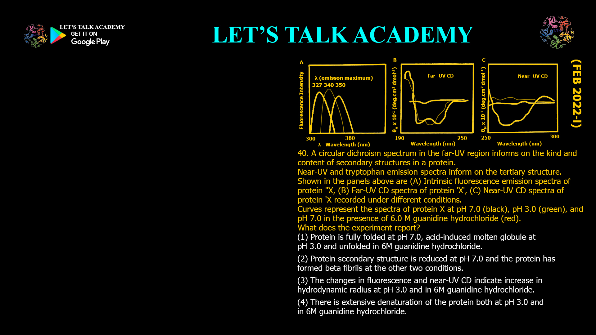
40. A circular dichroism spectrum in the far-UV region informs on the kind and content of secondary structures in a protein. Near-UV and tryptophan emission spectra inform on the tertiary structure. Shown in the panels above are (A) intrinsic fluorescence emission spectra of protein ‘X’, (B) far-UV CD spectra of protein ‘X’, (C) near-UV CD spectra of protein ‘X’ recorded under different conditions. Curves represent the spectra of protein X at pH 7.0 (black), pH 3.0 (green), and pH 7.0 in the presence of 6.0 M guanidine hydrochloride (red).
What does the experiment report?
(A) Protein is fully folded at pH 7.0, acid-induced molten globule at pH 3.0, and unfolded in 6 M guanidine hydrochloride.
(B) Protein secondary structure is reduced at pH 7.0 and the protein has formed β-fibrils at the other two conditions.
(C) The changes in fluorescence and near-UV CD indicate increase in hydrodynamic radius at pH 3.0 and in 6 M guanidine hydrochloride.
(D) There is extensive denaturation of the protein both at pH 3.0 and in 6 M guanidine hydrochloride.
Interpreting Fluorescence, Far-UV Circular Dichroism, and Near-UV Circular Dichroism Spectra for Protein Folding
Correct Answer
Option (1): Protein is fully folded at pH 7.0, acid-induced molten globule at pH 3.0 and unfolded in 6 M guanidine hydrochloride.
Explanation
This question combines three complementary spectroscopic techniques to determine the conformational state of a protein under different environmental conditions. Each technique reports a different level of structural organization. Intrinsic tryptophan fluorescence reflects the local environment surrounding aromatic residues and therefore provides information about tertiary structure. Far-UV Circular Dichroism reports the secondary structure by monitoring the peptide backbone, whereas Near-UV Circular Dichroism reflects the packing of aromatic side chains and therefore provides information about tertiary structure.
At pH 7.0, the fluorescence spectrum exhibits the shortest emission wavelength, approximately 327 nm. Tryptophan residues emit at shorter wavelengths when they are buried within a hydrophobic core. The Far-UV CD spectrum displays strong characteristic signals corresponding to well-defined secondary structure, while the Near-UV CD spectrum also shows pronounced signals arising from a rigid and well-organized tertiary structure. Together, these observations indicate that the protein is present in its fully folded native conformation.
At pH 3.0, the fluorescence emission maximum shifts to approximately 340 nm, indicating partial exposure of tryptophan residues to the solvent. However, the Far-UV CD spectrum remains very similar to that observed at pH 7.0, demonstrating that most of the secondary structure is still preserved. In contrast, the Near-UV CD signal decreases markedly, showing that the ordered packing of aromatic side chains has largely been disrupted. This combination of preserved secondary structure with a largely disrupted tertiary structure is the defining characteristic of a molten globule state.
In the presence of 6 M guanidine hydrochloride, the fluorescence emission maximum shifts further to approximately 350 nm, indicating that the tryptophan residues are now fully exposed to the aqueous environment. Simultaneously, both the Far-UV CD and Near-UV CD signals are almost completely lost. The disappearance of the Far-UV CD signal indicates destruction of the secondary structure, while the absence of the Near-UV CD signal demonstrates complete loss of tertiary structure. These observations are characteristic of a fully unfolded protein.
Why Option (1) is Correct
This option accurately explains all three experimental observations. The native protein at pH 7.0 possesses both secondary and tertiary structure. Lowering the pH to 3.0 preserves most of the secondary structure but disrupts the tertiary structure, producing an acid-induced molten globule. Guanidine hydrochloride acts as a strong chaotropic agent that unfolds the protein completely, leading to the loss of both secondary and tertiary structures.
Why Option (2) is Incorrect
The Far-UV CD spectrum at pH 7.0 clearly demonstrates that the protein possesses well-defined secondary structure rather than reduced secondary structure. Furthermore, there is no spectroscopic evidence supporting the formation of β-fibrils. Protein fibrils typically exhibit characteristic β-sheet-rich CD spectra that are not observed in the figure.
Why Option (3) is Incorrect
Although unfolding and molten globule formation generally increase the hydrodynamic radius of a protein, fluorescence spectroscopy and Circular Dichroism do not directly measure hydrodynamic radius. These techniques provide information about protein structure rather than molecular size. Determination of hydrodynamic radius requires methods such as dynamic light scattering or analytical ultracentrifugation.
Why Option (4) is Incorrect
This option incorrectly states that extensive denaturation occurs at both pH 3.0 and in guanidine hydrochloride. The Far-UV CD spectrum at pH 3.0 clearly shows that the secondary structure remains largely intact. Therefore, the protein is only partially unfolded at acidic pH and exists as a molten globule rather than a completely denatured molecule.
Interpretation of Intrinsic Fluorescence
Tryptophan fluorescence is highly sensitive to the polarity of its surrounding environment. Buried tryptophan residues located within the hydrophobic core of a folded protein emit at shorter wavelengths, whereas exposure to water during unfolding causes the emission maximum to shift toward longer wavelengths. This progressive red shift from 327 nm to 340 nm and finally to 350 nm reflects increasing exposure of the tryptophan residues as the protein loses its native structure.
Role of Far-UV and Near-UV Circular Dichroism
Far-UV Circular Dichroism primarily monitors the conformation of the peptide backbone and is therefore used to estimate the amount of α-helices, β-sheets, and random coils. Near-UV Circular Dichroism originates from aromatic amino acid side chains and reflects their fixed spatial arrangement within the folded protein. A protein may retain substantial secondary structure while losing its tertiary structure, producing a spectrum characteristic of the molten globule state. Complete disappearance of both Far-UV and Near-UV CD signals indicates extensive unfolding of the protein.
Characteristics of the Molten Globule State
The molten globule is an intermediate conformational state in which most of the native secondary structure is retained, but the tightly packed tertiary structure is disrupted. Aromatic side chains become more mobile, hydrophobic regions become partially exposed, and tryptophan fluorescence shifts toward longer wavelengths. This state commonly appears during protein folding, acid-induced unfolding, or mild denaturation and is distinguished from the fully unfolded state by the preservation of significant secondary structure.
Conclusion
The combined fluorescence, Far-UV CD, and Near-UV CD spectra demonstrate that protein X exists in three distinct conformational states. At pH 7.0 the protein is fully folded, at pH 3.0 it adopts an acid-induced molten globule conformation that retains secondary structure but loses much of its tertiary organization, and in 6 M guanidine hydrochloride it becomes completely unfolded. Therefore, the correct answer is Option (1).


