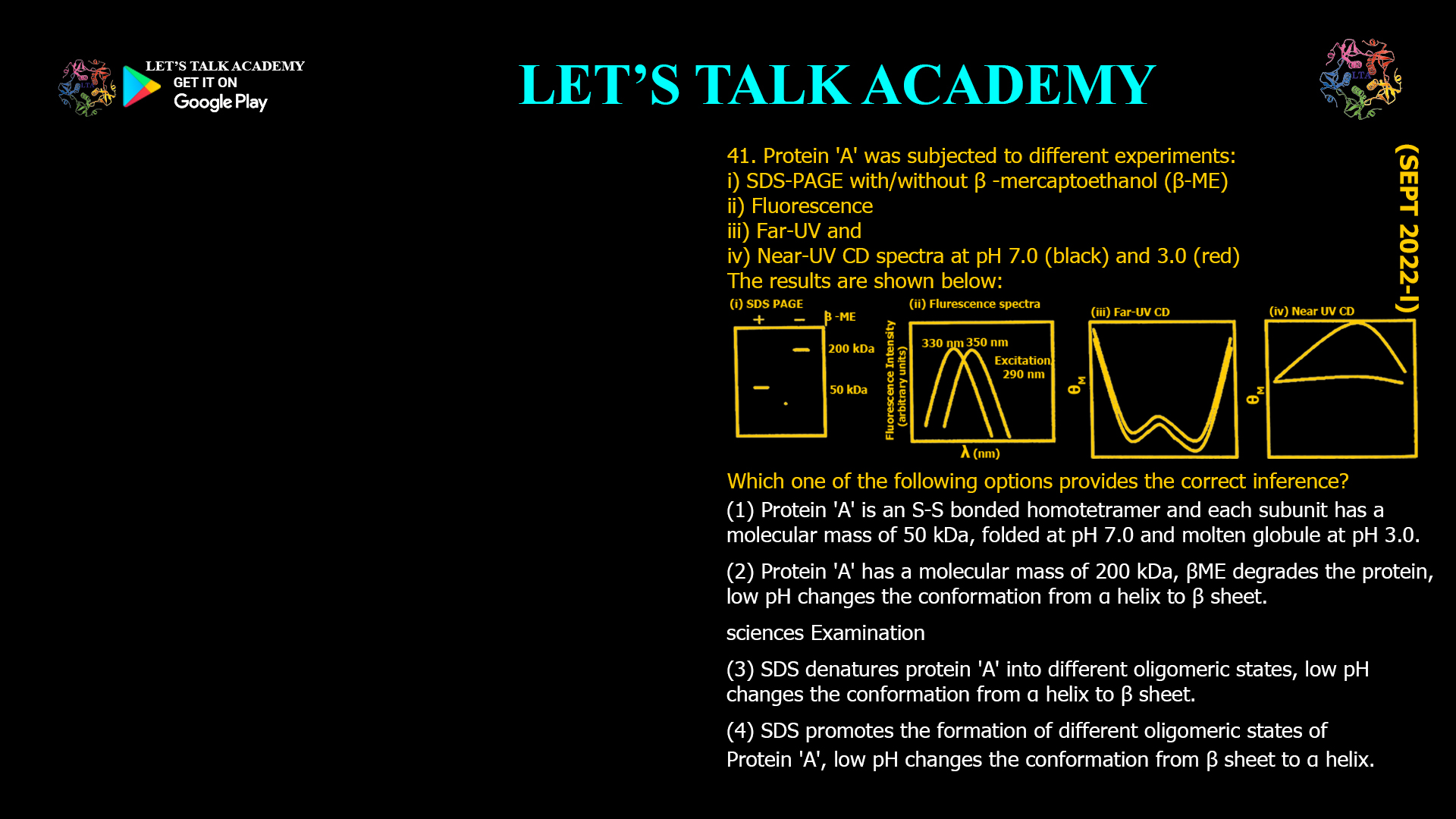
41. Protein ‘A’ was subjected to different experiments:
(i) SDS-PAGE with/without β-mercaptoethanol (β-ME)
(ii) Fluorescence
(iii) Far-UV CD
(iv) Near-UV CD spectra at pH 7.0 (black) and 3.0 (red)
The results are shown below.
Which one of the following options provides the correct inference?
(A) Protein ‘A’ is an S-S bonded homotetramer and each subunit has a molecular mass of 50 kDa, folded at pH 7.0 and molten globule at pH 3.0.
(B) Protein ‘A’ has a molecular mass of 200 kDa, β-ME degrades the protein, low pH changes the conformation from α-helix to β-sheet.
(C) SDS denatures protein ‘A’ into different oligomeric states, low pH changes the conformation from α-helix to β-sheet.
(D) SDS promotes the formation of different oligomeric states of Protein ‘A’, low pH changes the conformation from β-sheet to α-helix.
Interpreting SDS-PAGE, Fluorescence, and Circular Dichroism Data to Determine Protein Structure
Correct Answer
Option (1): Protein ‘A’ is an S-S bonded homotetramer and each subunit has a molecular mass of 50 kDa, folded at pH 7.0 and molten globule at pH 3.0.
Explanation
This question combines information obtained from four independent biochemical techniques. Each experiment provides evidence about a different aspect of protein structure. SDS-PAGE reveals the oligomeric organization of the protein, fluorescence spectroscopy reports changes in the local environment of aromatic residues, Far-UV Circular Dichroism provides information about secondary structure, and Near-UV Circular Dichroism reflects the integrity of tertiary structure. The correct interpretation requires combining all of these observations rather than analyzing each experiment separately.
Interpretation of the SDS-PAGE Experiment
In the absence of β-mercaptoethanol, the protein migrates as a single band corresponding to approximately 200 kDa. When β-mercaptoethanol is added, the protein migrates as a band of approximately 50 kDa. β-Mercaptoethanol specifically reduces disulfide bonds but does not hydrolyze peptide bonds or degrade proteins. Therefore, the appearance of a 50 kDa band after reduction demonstrates that the native protein consists of four identical 50 kDa subunits held together by intermolecular disulfide bonds. This identifies Protein A as a disulfide-linked homotetramer.
Interpretation of the Fluorescence Spectrum
The fluorescence emission maximum shifts from approximately 330 nm at pH 7.0 to approximately 350 nm at pH 3.0. Tryptophan residues emit at shorter wavelengths when buried inside the hydrophobic core of a folded protein. A shift toward longer wavelengths indicates increased solvent exposure caused by partial unfolding. The observed red shift therefore shows that lowering the pH disrupts the tertiary structure surrounding the aromatic residues.
Interpretation of the Far-UV Circular Dichroism Spectrum
The Far-UV CD spectra recorded at pH 7.0 and pH 3.0 are very similar. Since this spectral region reflects the conformation of the peptide backbone, the nearly identical spectra indicate that the protein retains most of its secondary structural elements despite acid treatment. There is no evidence for conversion of α-helices into β-sheets or vice versa.
Interpretation of the Near-UV Circular Dichroism Spectrum
The Near-UV CD spectrum shows a strong signal at pH 7.0 but becomes almost flat at pH 3.0. Near-UV CD originates from aromatic amino acid side chains that are held in fixed orientations within a folded tertiary structure. Loss of this signal indicates disruption of tertiary packing while the secondary structure remains largely preserved. This pattern is characteristic of a molten globule state.
Why Option (1) is Correct
This option explains every experimental observation consistently. The SDS-PAGE experiment demonstrates that the native protein is a disulfide-linked homotetramer composed of four identical 50 kDa subunits. The fluorescence spectrum indicates partial unfolding at low pH, the Far-UV CD spectrum shows that the secondary structure is retained, and the Near-UV CD spectrum demonstrates loss of tertiary structure. Together, these features define a molten globule conformation at pH 3.0 while the protein remains fully folded at pH 7.0.
Why Option (2) is Incorrect
β-Mercaptoethanol does not degrade proteins. Its function is to reduce disulfide bonds, allowing covalently linked subunits to separate. Furthermore, the Far-UV CD spectra do not indicate conversion from an α-helical structure to a β-sheet-rich structure. Since both statements are incorrect, this option is not supported by the experimental evidence.
Why Option (3) is Incorrect
SDS denatures proteins and disrupts non-covalent interactions, but it does not generate different oligomeric states. The different bands observed arise because β-mercaptoethanol reduces disulfide bonds, not because SDS produces new oligomers. In addition, the Far-UV CD spectra provide no evidence for an α-helix to β-sheet transition.
Why Option (4) is Incorrect
SDS does not promote oligomer formation; instead, it dissociates protein complexes by disrupting hydrophobic interactions. Moreover, the CD spectra do not support a transition from β-sheet to α-helix at low pH. The secondary structure remains largely unchanged, whereas the tertiary structure is disrupted.
Relationship Between Far-UV and Near-UV Circular Dichroism
Far-UV Circular Dichroism primarily reflects the conformation of the peptide backbone and therefore provides information about α-helices, β-sheets, and random coils. Near-UV Circular Dichroism arises from aromatic amino acid side chains and disulfide bonds arranged within a rigid three-dimensional structure. A protein may retain its secondary structure while losing its tertiary organization, producing little change in the Far-UV CD spectrum but a substantial reduction in the Near-UV CD signal. This combination of observations is one of the defining features of a molten globule.
Fluorescence Changes During Partial Protein Unfolding
Tryptophan fluorescence is extremely sensitive to its local environment. When the protein is folded, tryptophan residues are often buried within hydrophobic regions and emit at shorter wavelengths. Partial unfolding exposes these residues to water, producing a red shift in the emission maximum. The shift from approximately 330 nm to 350 nm therefore indicates disruption of the tertiary structure without necessarily affecting the overall secondary structure.
Conclusion
The combined results from SDS-PAGE, fluorescence spectroscopy, Far-UV CD, and Near-UV CD demonstrate that Protein A is a disulfide-linked homotetramer composed of four 50 kDa subunits. At pH 7.0 the protein is fully folded, whereas at pH 3.0 it retains its secondary structure but loses most of its tertiary organization, producing a molten globule state. Therefore, the correct answer is Option (1).


