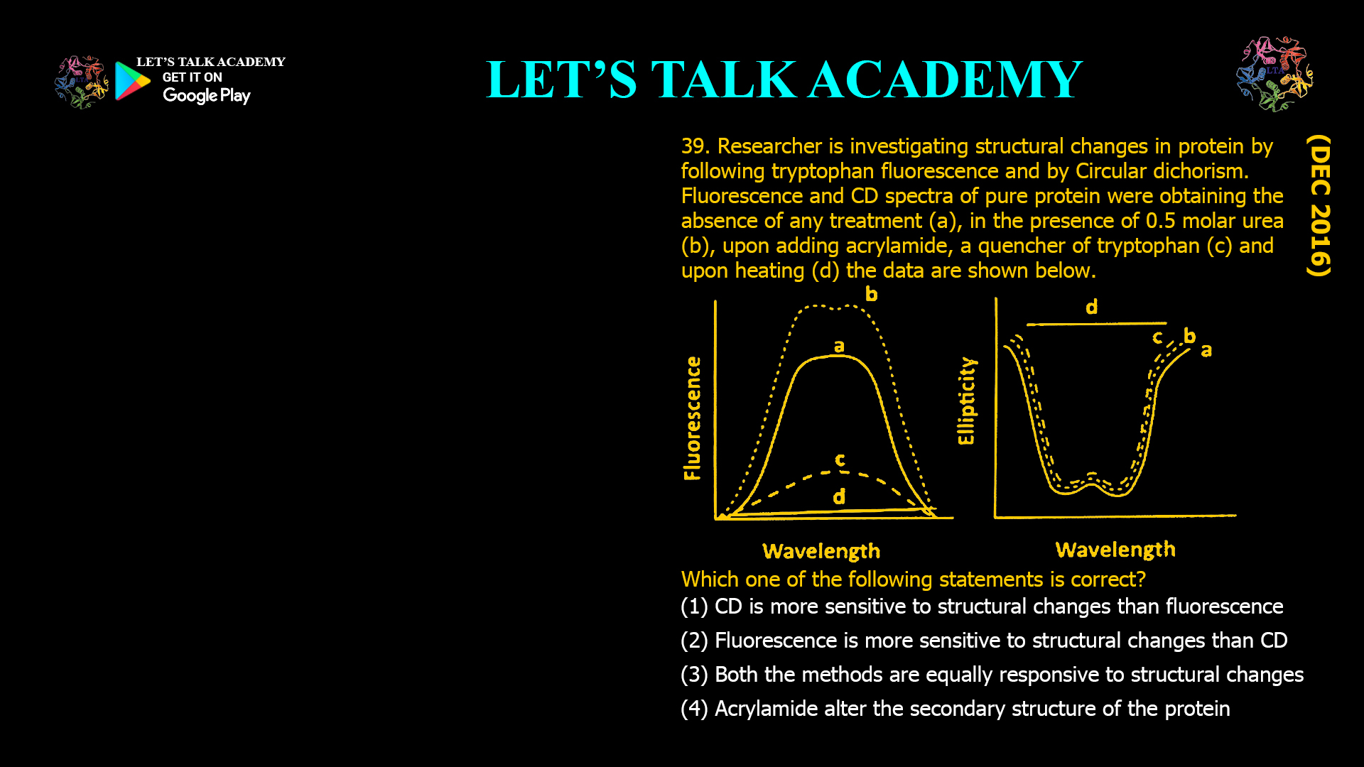
39. A researcher is investigating structural changes in a protein by following tryptophan fluorescence and circular dichroism. Fluorescence and CD spectra of the pure protein were obtained in the absence of any treatment (A), in the presence of 0.5 M urea (B), upon adding acrylamide, a quencher of tryptophan (C), and upon heating (D). The data are shown below.
Which one of the following statements is correct?
(A) CD is more sensitive to structural changes than fluorescence.
(B) Fluorescence is more sensitive to structural changes than CD.
(C) Both the methods are equally responsive to structural changes.
(D) Acrylamide alters the secondary structure of the protein.
Fluorescence versus Circular Dichroism for Monitoring Protein Structural Changes
Correct Answer
Option (2): Fluorescence is more sensitive to structural changes than Circular Dichroism.
Explanation
Fluorescence spectroscopy and Circular Dichroism (CD) spectroscopy are two complementary techniques commonly used to investigate protein structure. Although both methods can detect conformational changes, they monitor different structural features. Fluorescence spectroscopy primarily reports changes in the local environment surrounding aromatic fluorophores such as tryptophan, whereas Circular Dichroism mainly provides information about the secondary structure of the protein, including α-helices and β-sheets.
In the fluorescence spectra shown, even a mild treatment with 0.5 M urea produces a noticeable increase in fluorescence intensity. This indicates that a small conformational change has altered the environment around the tryptophan residue. Partial unfolding exposes the fluorophore to a different environment and reduces interactions that previously quenched its fluorescence. Such local structural perturbations are detected immediately by fluorescence spectroscopy.
In contrast, the corresponding CD spectrum changes only slightly after treatment with 0.5 M urea. This indicates that although the tertiary structure surrounding the tryptophan residue has been perturbed, the overall secondary structure of the protein remains largely intact. Since CD primarily detects changes in the peptide backbone, it is less sensitive to subtle local rearrangements that do not significantly alter α-helices or β-sheets.
The addition of acrylamide causes a dramatic decrease in fluorescence intensity because acrylamide is an efficient dynamic quencher of tryptophan fluorescence. It interacts with the excited tryptophan residue and promotes non-radiative relaxation, thereby reducing fluorescence emission. However, the CD spectrum remains almost unchanged because acrylamide does not disrupt the secondary structure of the protein. This demonstrates that fluorescence can respond strongly to changes that have little or no effect on the protein backbone.
Heating produces the largest changes in both spectra. Elevated temperature unfolds the protein, altering both the local environment of tryptophan residues and the overall secondary structure. Consequently, fluorescence intensity changes substantially, while the characteristic CD bands associated with ordered secondary structures diminish, indicating extensive denaturation.
Why Option (1) is Incorrect
The experimental data show that fluorescence detects structural perturbations produced by low concentrations of urea even when the CD spectrum changes very little. This demonstrates that fluorescence responds to subtle conformational changes before significant alterations in secondary structure occur. Therefore, CD is not more sensitive than fluorescence for detecting early structural changes.
Why Option (2) is Correct
Fluorescence spectroscopy is highly sensitive to the immediate environment of tryptophan residues. Even minor changes in protein folding, solvent accessibility, or interactions with nearby amino acids produce measurable changes in fluorescence intensity and emission characteristics. The figure clearly shows that fluorescence changes considerably after mild perturbation, whereas the CD spectrum remains nearly unchanged. Therefore, fluorescence is the more sensitive technique for detecting early structural changes.
Why Option (3) is Incorrect
If both techniques were equally responsive, comparable spectral changes would be observed under all experimental conditions. The figure shows that fluorescence undergoes much larger changes than CD after treatment with urea and acrylamide. This difference demonstrates that the two techniques differ in their sensitivity and the structural information they provide.
Why Option (4) is Incorrect
Acrylamide functions as a fluorescence quencher rather than a protein denaturant. It decreases fluorescence intensity through collisional quenching of excited tryptophan residues but does not significantly alter the secondary structure of the protein. The nearly unchanged CD spectrum confirms that the α-helices and β-sheets remain essentially intact after acrylamide treatment.
Why Fluorescence Detects Structural Changes Earlier
Tryptophan fluorescence depends strongly on its local molecular environment. Small changes in solvent exposure, hydrogen bonding, hydrophobic interactions, or proximity to quenching groups immediately affect fluorescence intensity and emission wavelength. Because these local changes often occur before the protein undergoes significant secondary structural rearrangement, fluorescence spectroscopy is particularly effective for detecting early stages of protein unfolding.
Role of Circular Dichroism in Protein Analysis
Circular Dichroism spectroscopy measures the differential absorption of left- and right-circularly polarized light by chiral molecules. In proteins, the far-UV region primarily reflects the conformation of the peptide backbone and is therefore used to estimate the proportion of α-helices, β-sheets, and random coils. Significant changes in the CD spectrum generally occur only after substantial alteration of the protein’s secondary structure.
Effect of Acrylamide on Tryptophan Fluorescence
Acrylamide is widely used as a neutral collisional quencher for studying the accessibility of tryptophan residues. If a tryptophan residue is exposed to the solvent, acrylamide can diffuse close to the fluorophore and efficiently quench its fluorescence. Buried tryptophan residues are less accessible and therefore exhibit weaker quenching. This property allows fluorescence quenching experiments to provide valuable information about protein folding and solvent accessibility without disrupting the protein structure.
Conclusion
The experimental results demonstrate that fluorescence spectroscopy detects subtle conformational changes that produce little or no change in the secondary structure measured by Circular Dichroism. Mild unfolding caused by urea significantly alters fluorescence before noticeable changes appear in the CD spectrum, while acrylamide selectively quenches fluorescence without affecting secondary structure. Therefore, the correct answer is Option (2): Fluorescence is more sensitive to structural changes than Circular Dichroism.


