
Composition, Structure and Function of Biomolecules: Nucleic Acids
1. Introduction to Nucleic Acids
Nucleic acids are among the most important biological macromolecules because they are responsible for storing, transmitting, expressing, and regulating genetic information. The two major classes of nucleic acids are deoxyribonucleic acid (DNA) and ribonucleic acid (RNA).
DNA primarily serves as the long-term repository of genetic information, whereas RNA performs a much wider variety of functions, including information transfer, protein synthesis, regulation of gene expression, catalysis, and genome organization in many organisms. Nucleic acids are therefore not simply genetic molecules; they are central components of virtually every major biological process.
At the molecular level, nucleic acids are polymers composed of repeating units called nucleotides. Each nucleotide consists of a nitrogenous base, a pentose sugar, and one or more phosphate groups. Nucleotides are connected through phosphodiester bonds to form long polynucleotide chains.
Nucleic acids are particularly important because questions can be asked not only from their basic composition but also from their chemical bonding, polarity, structural organization, base pairing, DNA forms, RNA structures, thermodynamic stability, replication, transcription, and structure–function relationships.
2. Chemical Composition of Nucleic Acids
Nucleic acids are composed mainly of three fundamental chemical components:
- Nitrogenous base
- Pentose sugar
- Phosphate group
The combination of a nitrogenous base with a pentose sugar forms a nucleoside. When one or more phosphate groups are added to a nucleoside, the resulting molecule is called a nucleotide.
Therefore:
Nitrogenous base + Pentose sugar → Nucleoside
Nucleoside + Phosphate → Nucleotide
Many nucleotides linked together → Polynucleotide / Nucleic acid
3. Nitrogenous Bases
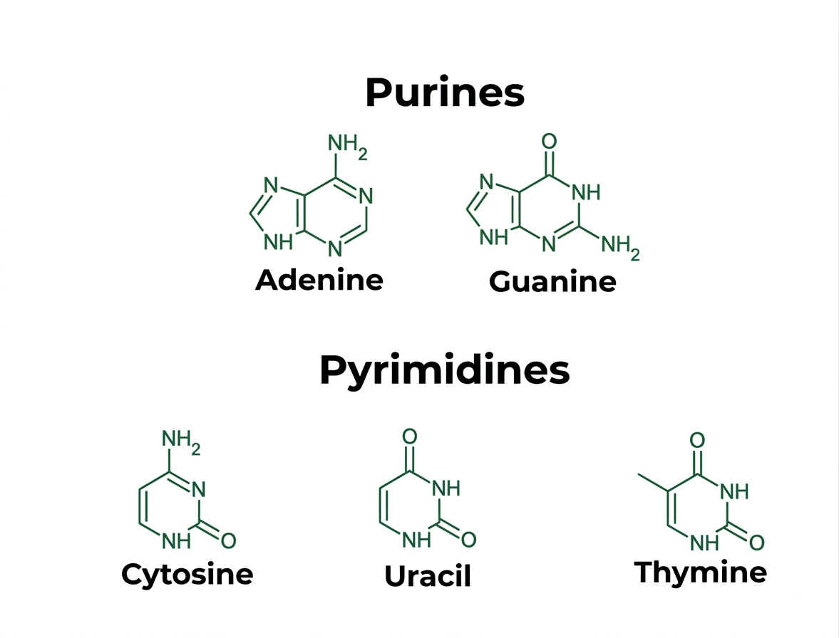
Nitrogenous bases are heterocyclic organic compounds containing nitrogen atoms. They are divided into two major groups:
3.1 Purines
Purines are nitrogen-containing bases with a two-ring structure.
The two purines found in nucleic acids are:
- Adenine (A)
- Guanine (G)
Purines contain a fused six-membered and five-membered ring system.
3.2 Pyrimidines
Pyrimidines contain a single six-membered ring.
The pyrimidine bases are:
- Cytosine (C)
- Thymine (T)
- Uracil (U)
DNA contains adenine, guanine, cytosine, and thymine, whereas RNA generally contains adenine, guanine, cytosine, and uracil.
Purines: A and G
Pyrimidines: C, T, and U
A useful conceptual distinction is that the number of rings is different:
Purine → Two rings
Pyrimidine → One ring
4. Pentose Sugars in Nucleic Acids
The sugar component of nucleic acids is a five-carbon sugar called a pentose.
The sugar differs between DNA and RNA.
4.1 Deoxyribose in DNA
DNA contains 2-deoxyribose.
At the 2′ carbon, DNA contains a hydrogen atom instead of the hydroxyl group present in ribose.
Thus:
DNA sugar → 2-deoxyribose
4.2 Ribose in RNA
RNA contains ribose.
Ribose possesses a hydroxyl group at the 2′ carbon.
Thus:
RNA sugar → Ribose
This apparently small chemical difference has major biological consequences. The 2′-OH group of RNA makes RNA more chemically reactive and contributes to its structural flexibility and susceptibility to alkaline hydrolysis compared with DNA.
DNA versus RNA Sugar
Feature |
DNA |
RNA |
|---|---|---|
| Sugar | 2-Deoxyribose | Ribose |
| 2′ position | H | OH |
| Relative chemical stability | Higher | Lower |
| Main biological role | Genetic information storage | Information transfer, translation, regulation, catalysis |
5. Nucleotides
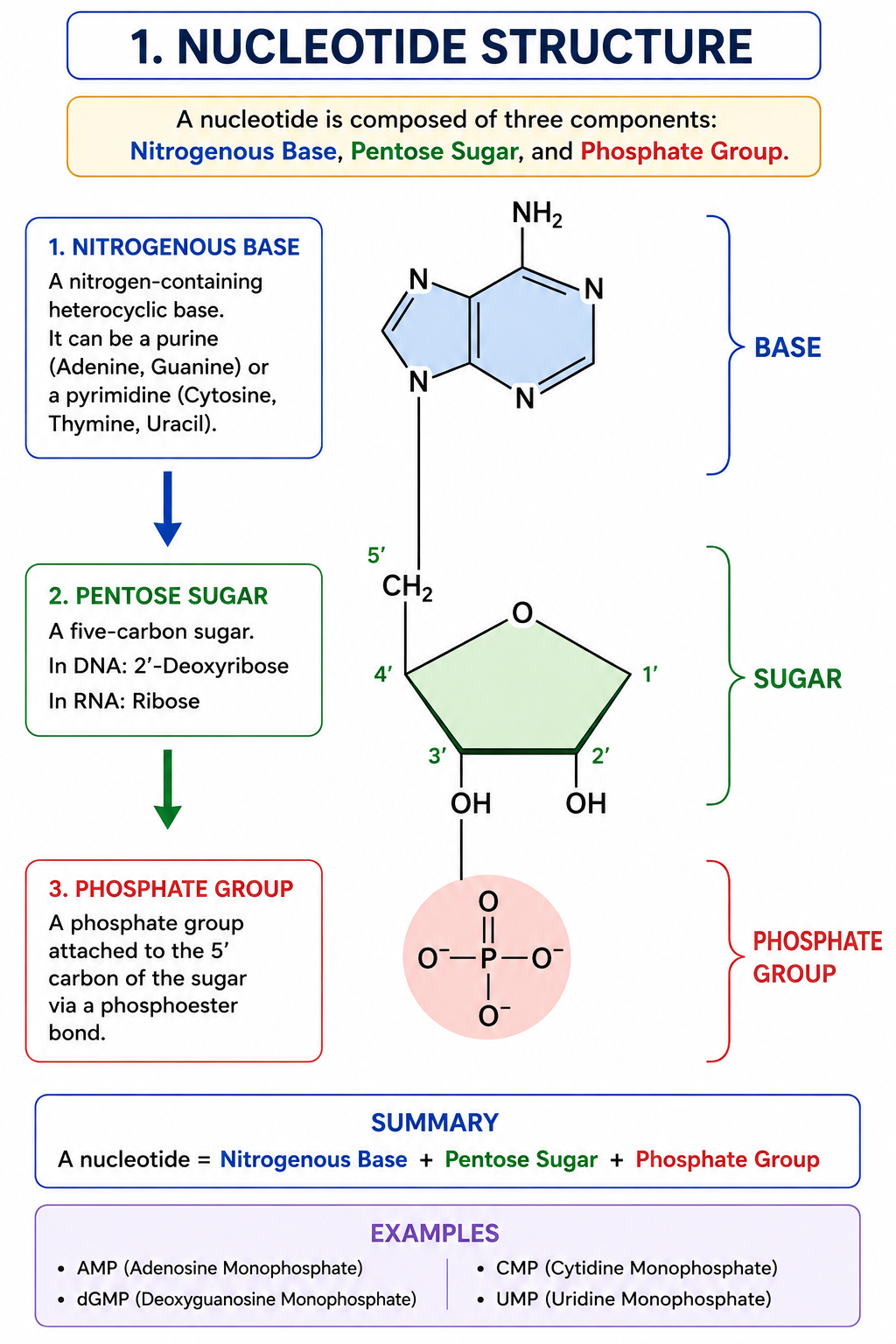
A nucleotide consists of:
Nitrogenous base + Pentose sugar + Phosphate group
Nucleotides are the fundamental monomeric units from which DNA and RNA are constructed.
Examples include:
- AMP — adenosine monophosphate
- ADP — adenosine diphosphate
- ATP — adenosine triphosphate
- GMP — guanosine monophosphate
- GDP — guanosine diphosphate
- GTP — guanosine triphosphate
- CMP — cytidine monophosphate
- UMP — uridine monophosphate
Nucleotides are not only structural units of nucleic acids. Several nucleotides also function independently as energy carriers, signaling molecules, enzyme cofactors, and metabolic intermediates.
For example:
ATP → cellular energy transfer
GTP → protein synthesis and signaling
cAMP → intracellular second messenger
NAD⁺ and FAD → electron-transfer cofactors containing nucleotide-derived components
Thus, nucleotide biology extends far beyond DNA and RNA.
6. Nucleosides
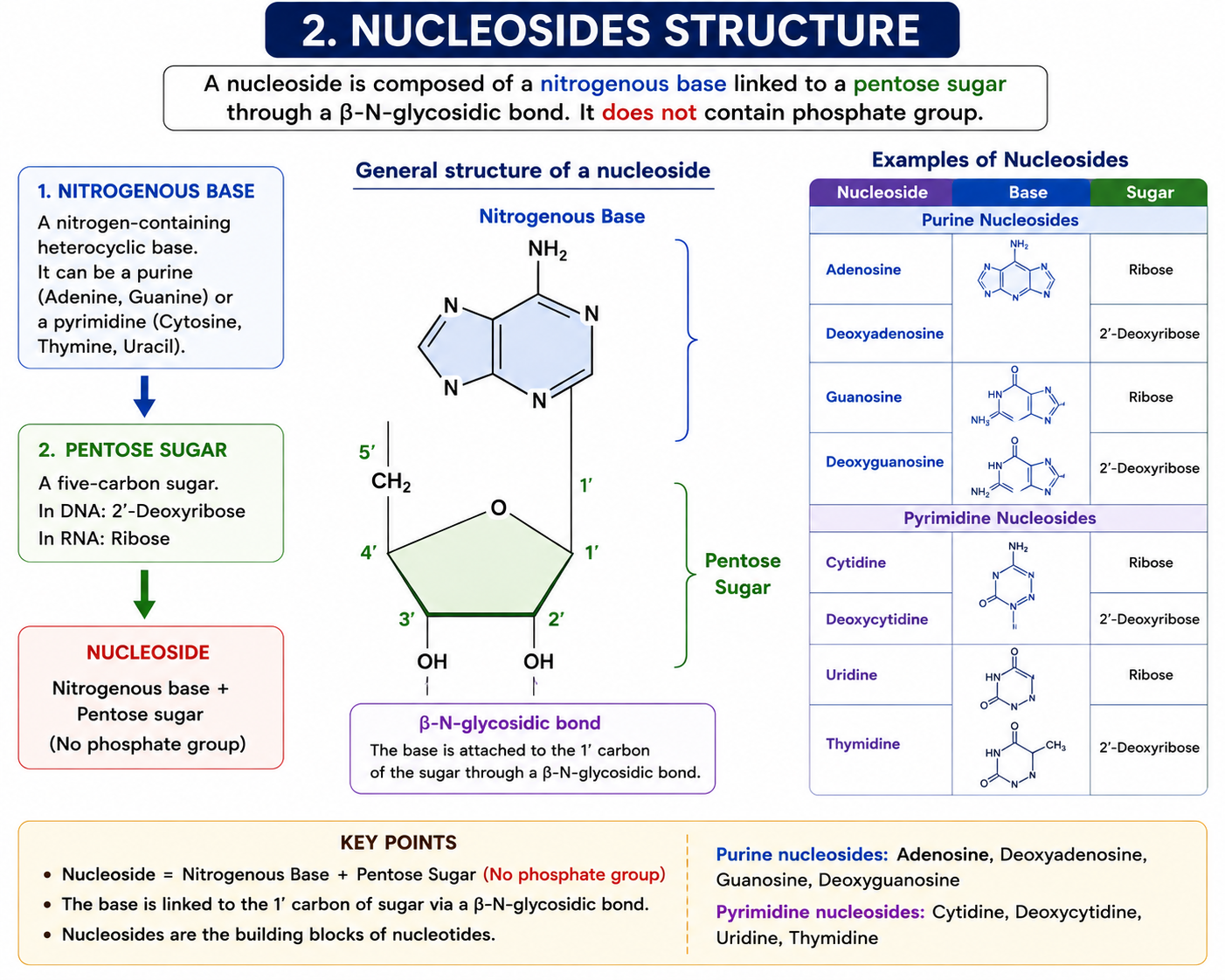
A nucleoside is formed when a nitrogenous base is covalently attached to a pentose sugar.
There is no phosphate group in a nucleoside.
The nitrogenous base is connected to the sugar through an N-glycosidic bond.
Important Nucleosides
Base |
DNA nucleoside |
RNA nucleoside |
|---|---|---|
| Adenine | Deoxyadenosine | Adenosine |
| Guanine | Deoxyguanosine | Guanosine |
| Cytosine | Deoxycytidine | Cytidine |
| Thymine | Deoxythymidine | — |
| Uracil | — | Uridine |
The bond between the nitrogenous base and sugar is an important structural feature of nucleic acids. In purines, the glycosidic linkage involves N9, whereas in pyrimidines it involves N1.
7. Phosphate Groups and Their Importance
Phosphate groups contribute significantly to the chemical properties of nucleic acids.
The phosphate groups form part of the sugar-phosphate backbone and give nucleic acids a strong negative charge under physiological conditions.
This negative charge is important because nucleic acids interact with positively charged molecules such as:
- Histones
- Basic DNA-binding proteins
- Metal ions
- Positively charged amino acid residues
The phosphate groups also participate in the formation of phosphodiester bonds, which connect adjacent nucleotides in a nucleic acid chain.
8. Phosphodiester Bond
Nucleotides within a polynucleotide chain are connected through 3′–5′ phosphodiester bonds.
A phosphodiester bond is formed between:
- the 3′-OH group of one sugar, and
- the 5′-phosphate group of the adjacent nucleotide.
Therefore, the backbone can be represented as:
5′ → Sugar → Phosphate → Sugar → Phosphate → Sugar → 3′
The phosphodiester linkage creates the continuous sugar-phosphate backbone of DNA and RNA.
The phosphodiester bond is a covalent bond and therefore forms the strong structural backbone of nucleic acids.
In contrast, complementary bases between DNA strands are primarily stabilized by hydrogen bonding and base-stacking interactions.
9. Polarity of Nucleic Acid Strands
A nucleic acid strand has a definite directionality.
One end is called the 5′ end, while the other is called the 3′ end.
The 5′ end generally contains a free phosphate group, whereas the 3′ end contains a free hydroxyl group.
Therefore, a DNA or RNA sequence is conventionally written:
5′ → 3′
For example:
5′-ATGCGTAC-3′
The complementary strand would run in the opposite direction:
3′-TACGCATG-5′
This opposite orientation is called antiparallel arrangement.
Directionality is extremely important for understanding:
- DNA replication
- Transcription
- Translation
- Polymerase activity
- Primer extension
- PCR
- Sequencing
10. Structure of DNA
DNA is generally composed of two complementary polynucleotide strands arranged in a double-helical structure.
The classical Watson–Crick model explains how DNA can simultaneously store genetic information and permit accurate replication.
The major structural characteristics of DNA include:
- Two polynucleotide strands
- Antiparallel orientation
- Sugar-phosphate backbone
- Nitrogenous bases directed toward the interior
- Complementary base pairing
- Hydrogen bonding
- Base stacking
- Helical organization
The sequence of nucleotides along DNA represents biological information.
11. Watson–Crick Base Pairing
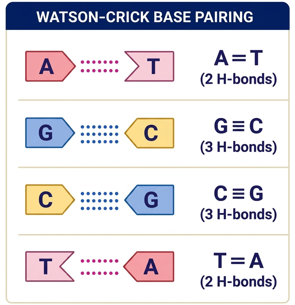
In conventional DNA double-stranded structures:
Adenine pairs with Thymine
Guanine pairs with Cytosine
The interactions are:
A = T → 2 hydrogen bonds
G ≡ C → 3 hydrogen bonds
This complementary base pairing allows one DNA strand to act as a template for synthesis of the other strand.
Why Is Base Pairing Important?
Base pairing provides:
- Accurate information storage
- Template-directed DNA replication
- DNA repair
- Genetic transmission
- Sequence recognition
- Specific nucleic acid hybridization
12. Chargaff’s Rules
For double-stranded DNA, Chargaff’s observations can be expressed as:
A = T
G = C
Therefore:
A + G = T + C
This also means:
Purines = Pyrimidines
However, the exact percentage of A, T, G, and C can differ between species.
Numerical Concept
If a double-stranded DNA molecule contains:
30% adenine
Then:
T = 30%
The remaining 40% consists of G + C.
Since G = C:
G = 20%
C = 20%
13. Stability of DNA Double Helix
The DNA double helix is stabilized by several types of interactions.
13.1 Hydrogen Bonding
Hydrogen bonds occur between complementary bases.
A-T contains two hydrogen bonds, while G-C contains three.
However, DNA stability cannot be explained only by counting hydrogen bonds.
13.2 Base Stacking
Adjacent aromatic nitrogenous bases stack over one another inside the helix.
Hydrophobic effects, van der Waals interactions, and other favorable interactions associated with base stacking make an important contribution to helix stability.
Therefore, a common examination trap is to assume that hydrogen bonding alone determines DNA stability.
14. DNA Melting and Melting Temperature
The two strands of DNA can separate when sufficient thermal energy is supplied.
This process is called DNA melting, denaturation, or strand separation.
The temperature at which approximately half of a DNA population is denatured is called the melting temperature (Tm).
Several factors influence Tm, including:
- GC content
- DNA length
- Ionic strength
- Sequence composition
- Mismatches
- Experimental conditions
DNA with relatively higher GC content generally has a higher melting temperature under comparable conditions because GC-rich sequences tend to have stronger overall base-stacking and pairing interactions.
Do not interpret the statement:
“GC-rich DNA is more stable because G-C has three hydrogen bonds”
as a complete explanation.
The stability of nucleic acid duplexes results from multiple interactions, with base stacking making a major contribution.
15. Major Structural Forms of DNA
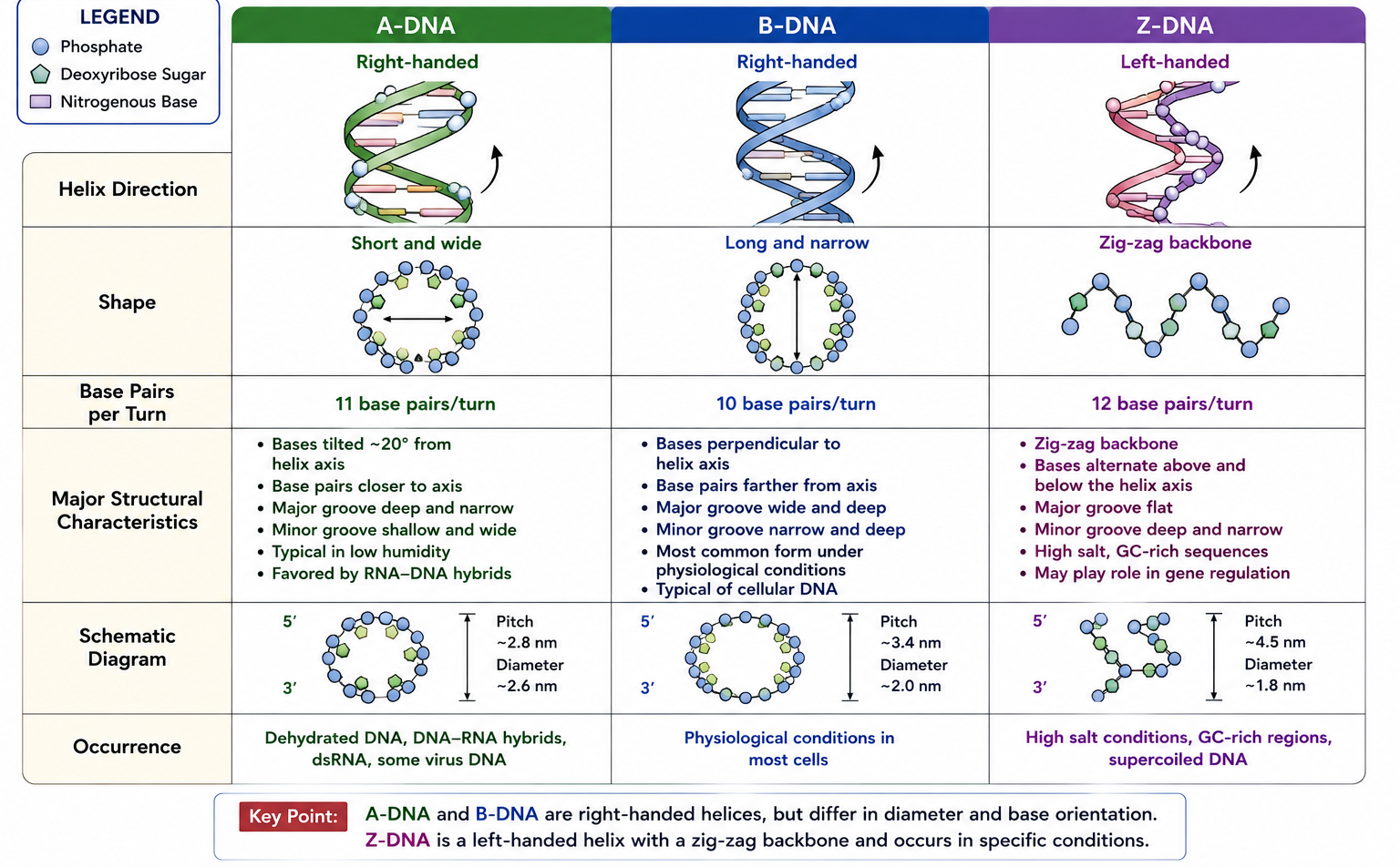
DNA can adopt different helical conformations depending on sequence, hydration, ionic conditions, and interaction with proteins.
The major forms studied in molecular biology are:
- A-DNA
- B-DNA
- Z-DNA
15.1 B-DNA
B-DNA is the classical Watson–Crick form and is the predominant conformation under many physiological conditions.
It is:
- Right-handed
- Approximately 2 nm in diameter
- Approximately 10.5 base pairs per turn
- Characterized by major and minor grooves
The grooves provide surfaces through which DNA-binding proteins can recognize specific sequences and structural features.
15.2 A-DNA
A-DNA is also a right-handed helix.
It is shorter and wider than B-DNA and is favored under conditions of reduced hydration.
RNA-DNA hybrid duplexes and double-stranded RNA frequently adopt A-form-like geometry.
15.3 Z-DNA
Z-DNA is unusual because it is a left-handed helix.
It has a characteristic zig-zag appearance of its sugar-phosphate backbone.
Z-DNA formation is favored by particular sequence and environmental conditions, especially certain alternating purine-pyrimidine sequences.
High-Yield Comparison
Feature |
A-DNA |
B-DNA |
Z-DNA |
|---|---|---|---|
| Helix | Right-handed | Right-handed | Left-handed |
| General shape | Short and broad | Long and relatively narrow | Zig-zag backbone |
| Common association | Dehydrated DNA/RNA-containing duplexes | Physiological DNA | Specific sequence/conditions |
| Importance | Structural variation | Major cellular form | Regulatory/structural interest |
16. Major and Minor Grooves
The DNA double helix contains two grooves:
- Major groove
- Minor groove
The grooves arise because the glycosidic bonds connecting bases to sugars are not positioned directly opposite one another.
Major Groove
The major groove is wider and exposes more chemical information from the base pairs.
Many DNA-binding proteins recognize specific DNA sequences through interactions in the major groove.
Minor Groove
The minor groove is narrower and can also be recognized by DNA-binding proteins and small molecules.
A protein does not always need to separate the DNA strands to recognize a sequence. DNA-binding proteins can often identify specific base-pair patterns by interacting with the exposed chemical groups within the grooves.
17. DNA as Genetic Material
The biological importance of DNA arises from its ability to:
- Store genetic information
- Replicate accurately
- Transmit information
- Undergo mutation
- Serve as a template for RNA synthesis
- Control protein production indirectly
- Participate in genome organization
The nucleotide sequence functions like a molecular information system.
The sequence of bases provides the information, while the physical and chemical properties of the DNA molecule allow that information to be stored and copied.
18. DNA Replication and Structural Complementarity
DNA replication depends fundamentally on complementary base pairing.
Each parental strand serves as a template for synthesis of a new complementary strand.
Thus:
Parental DNA
↓
Strand separation
Each strand acts as a template
↓
Complementary nucleotide incorporation
↓
Two daughter DNA molecules
This is called semiconservative replication, because each daughter DNA molecule contains one parental strand and one newly synthesized strand.
The complementary structure of DNA therefore provides a direct molecular explanation for accurate information transmission.
19. RNA: Composition and Structure
RNA stands for ribonucleic acid.
Like DNA, RNA is a polymer of nucleotides. However, RNA contains:
- Ribose sugar
- Adenine
- Guanine
- Cytosine
- Uracil
The most important chemical difference from DNA is the presence of the 2′-OH group in ribose and the replacement of thymine by uracil.
RNA is usually single-stranded, although single RNA molecules can fold extensively and form double-stranded regions through intramolecular base pairing.
RNA structure can therefore be highly complex despite being based on a single chain.
20. Levels of RNA Structure
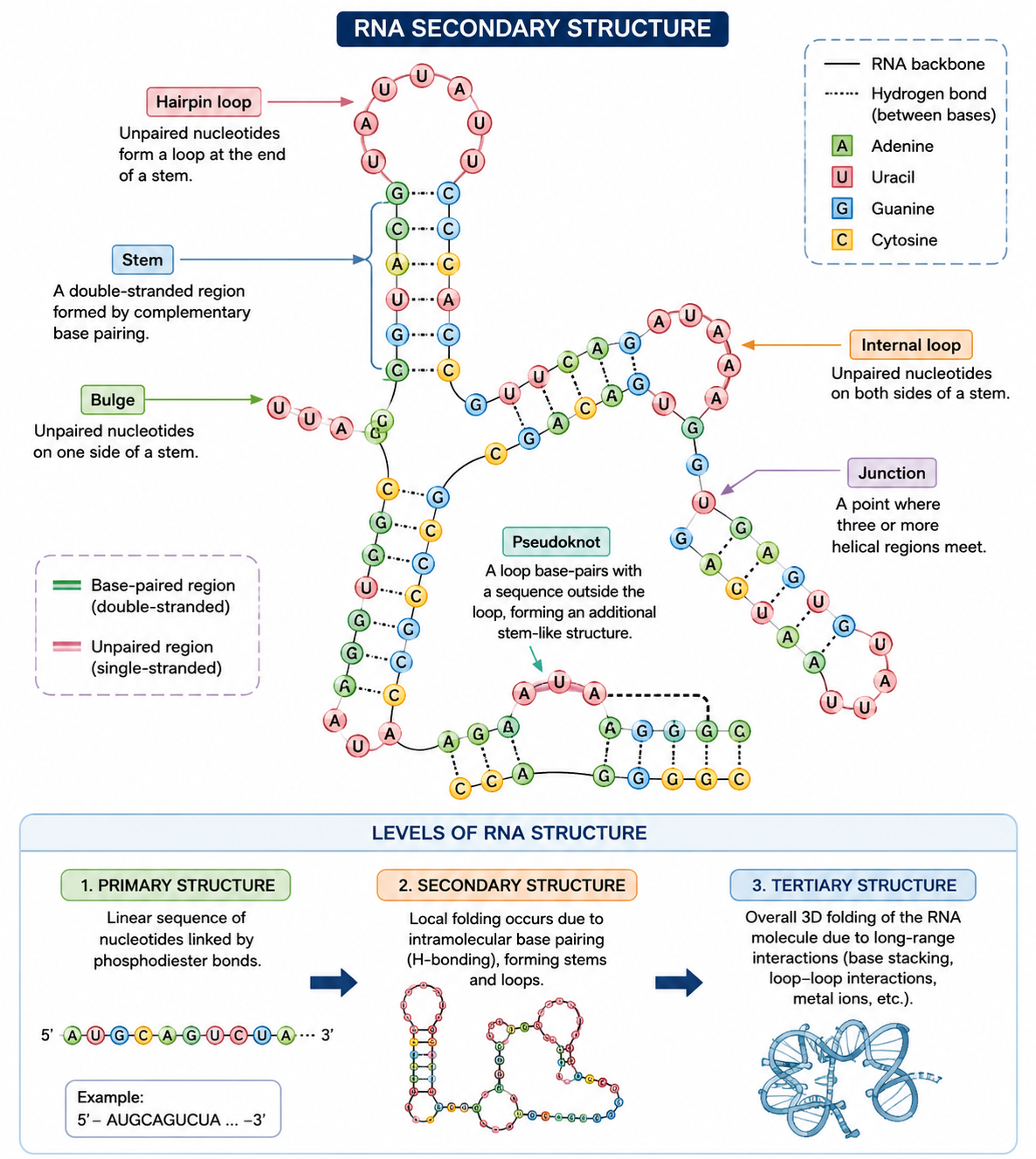
RNA can be discussed at several structural levels.
20.1 Primary Structure
Primary structure refers to the linear sequence of ribonucleotides.
For example:
5′-AUGCUAGCUA-3′
The nucleotides are connected by 3′–5′ phosphodiester bonds.
20.2 Secondary Structure
Secondary structure develops when complementary regions within an RNA molecule base-pair.
Common features include:
- Stem
- Hairpin loop
- Bulge
- Internal loop
- Junction
- Pseudoknot
RNA secondary structure is particularly important for understanding tRNA, rRNA, ribozymes, and regulatory RNAs.
20.3 Tertiary Structure
Tertiary structure refers to the complete three-dimensional folding of RNA.
It is stabilized by:
- Hydrogen bonding
- Base stacking
- Noncanonical base pairing
- Ionic interactions
- Metal-ion coordination
- Long-range interactions
RNA can therefore fold into complex three-dimensional structures capable of molecular recognition and catalysis.
21. Major Types of RNA
RNA molecules are functionally diverse.
The three classical RNA types are:
- Messenger RNA (mRNA)
- Transfer RNA (tRNA)
- Ribosomal RNA (rRNA)
However, modern molecular biology recognizes many additional RNA classes.
These include:
- snRNA
- snoRNA
- miRNA
- siRNA
- piRNA
- lncRNA
- circRNA
- antisense RNA
- CRISPR-associated RNAs
22. Messenger RNA (mRNA)
mRNA carries genetic information from DNA to the ribosome for protein synthesis.
The nucleotide sequence of mRNA contains codons that specify the amino acid sequence of a protein.
The basic information flow can be represented as:
DNA
↓
Transcription
↓
mRNA
↓
Translation
↓
Protein
mRNA therefore acts as an intermediary between genetic information stored in DNA and protein synthesis.
23. Transfer RNA (tRNA)
tRNA functions as an adaptor molecule during translation.
Each tRNA contains:
- An anticodon region
- An amino acid attachment site
- A characteristic folded structure
The anticodon recognizes a complementary codon in mRNA, while the amino acid attachment site carries the appropriate amino acid.
Thus:
mRNA codon ↔ tRNA anticodon
The amino acid carried by the tRNA is then incorporated into the growing polypeptide chain.
24. Ribosomal RNA (rRNA)
rRNA forms a major structural and functional component of ribosomes.
It is not merely a structural scaffold. rRNA participates directly in the molecular events of translation.
The ribosome therefore represents an important example of how RNA can possess both structural and catalytic functions.
This is one reason RNA is considered much more functionally versatile than the traditional concept of RNA as merely a messenger molecule.
25. Catalytic RNA and Ribozymes
Some RNA molecules possess catalytic activity.
Catalytic RNAs are called ribozymes.
Examples of RNA-associated catalytic functions include reactions involved in:
- RNA processing
- Peptide bond formation
- RNA cleavage
- RNA splicing
The catalytic role of RNA provides important evidence that RNA can function as both an information-containing molecule and a catalyst.
This concept is also central to discussions of the RNA world hypothesis, which proposes that early biological systems may have relied heavily on RNA for both information storage and catalysis.
26. DNA versus RNA
Feature |
DNA |
RNA |
|---|---|---|
| Full name | Deoxyribonucleic acid | Ribonucleic acid |
| Sugar | 2-Deoxyribose | Ribose |
| 2′ group | H | OH |
| Purines | A, G | A, G |
| Pyrimidines | C, T | C, U |
| Typical structure | Double-stranded | Usually single-stranded |
| Main role | Long-term genetic information storage | Information transfer, translation, regulation, catalysis |
| Chemical stability | Relatively higher | Relatively lower |
| Base characteristic | Thymine | Uracil |
The difference in chemical composition explains many of the functional differences between DNA and RNA.
27. Why Is DNA More Stable Than RNA?
DNA is chemically more stable than RNA largely because DNA lacks the 2′-OH group present in RNA.
The 2′-OH group of RNA can participate in intramolecular reactions that facilitate backbone cleavage, particularly under alkaline conditions.
DNA therefore provides a more chemically stable molecule for long-term storage of genetic information.
RNA, in contrast, is more chemically reactive and structurally versatile, properties that are advantageous for temporary information transfer, molecular regulation, and catalysis.
28. Nucleotides as Energy and Signaling Molecules
Nucleotides have functions beyond their role as components of DNA and RNA.
ATP
ATP is the major cellular energy-transfer molecule.
It participates in:
- Biosynthetic reactions
- Active transport
- Mechanical work
- Phosphorylation reactions
GTP
GTP is important in:
- Protein synthesis
- Signal transduction
- Microtubule dynamics
cAMP
Cyclic AMP is an important intracellular second messenger.
It mediates responses to extracellular signals through pathways involving protein kinases and other signaling proteins.
Thus, nucleotide chemistry is deeply integrated into cellular metabolism and signaling.
29. Nucleic Acid–Protein Interactions
Nucleic acids interact extensively with proteins.
DNA interacts with:
- Histones
- DNA polymerases
- RNA polymerases
- Transcription factors
- DNA repair proteins
- Chromatin-remodeling proteins
RNA interacts with:
- Ribosomal proteins
- RNA polymerases
- Splicing factors
- RNA-binding proteins
- Translational machinery
These interactions allow nucleic acids to be packaged, replicated, transcribed, processed, transported, and regulated.
30. DNA Packaging and Chromatin
In eukaryotic cells, DNA is extremely long and must be efficiently organized within the nucleus.
DNA associates with histone proteins to form nucleosomes, which are fundamental units of chromatin organization.
The general hierarchy can be represented as:
DNA
↓
DNA + Histone proteins
↓
Nucleosome
↓
Chromatin
↓
Higher-order chromosome organization
This packaging is not merely mechanical. Chromatin organization also influences DNA accessibility and gene expression.
31. Denaturation and Renaturation of Nucleic Acids
Denaturation
Denaturation refers to the disruption of interactions holding complementary nucleic acid strands together.
In DNA, this results in separation of the two strands.
Denaturation can be induced by:
- Heat
- Extreme pH
- Certain chemical agents
Importantly, denaturation generally disrupts noncovalent interactions without breaking the covalent phosphodiester backbone.
Renaturation
When suitable conditions are restored, complementary strands can reassociate.
This process is called:
Renaturation or annealing
These properties are fundamental to techniques such as:
- PCR
- Southern blotting
- Northern blotting
- DNA hybridization
- Probe-based detection
- Nucleic acid sequencing
32. Nucleic Acid Hybridization
Hybridization occurs when complementary nucleic acid strands associate through base pairing.
For example:
DNA strand + complementary DNA strand → DNA duplex
or:
RNA strand + complementary DNA strand → RNA-DNA hybrid
Hybridization is highly useful in molecular biology because sequence complementarity can be exploited for detecting specific nucleic acid sequences.
Important applications include:
- PCR
- Southern blotting
- Northern blotting
- Fluorescence in situ hybridization
- Microarrays
- Molecular diagnostics
- DNA sequencing
33. Biological Functions of Nucleic Acids
Nucleic acids perform several major biological functions.
33.1 Genetic Information Storage
DNA stores hereditary information in its nucleotide sequence.
33.2 Genetic Information Transmission
DNA is replicated and transmitted to daughter cells and, in organisms, across generations.
33.3 Gene Expression
DNA serves as the template for RNA synthesis.
33.4 Protein Synthesis
mRNA, tRNA, and rRNA work together to facilitate protein synthesis.
33.5 Regulation of Gene Expression
Several RNA molecules regulate transcription, mRNA stability, translation, and chromatin organization.
33.6 Catalysis
Certain RNA molecules function as ribozymes.
33.7 Cellular Signaling
Nucleotide derivatives such as cAMP and cGMP function as intracellular signaling molecules.
33.8 Energy Transfer
ATP and GTP participate in energy-dependent cellular processes.
34. Central Dogma and Nucleic Acids
The classical central dogma describes the major directional flow of genetic information:
DNA → RNA → Protein
The first step is transcription, in which RNA is synthesized using DNA as a template.
The second major step is translation, in which information in mRNA is used to determine the amino acid sequence of a protein.
However, molecular biology contains important variations and extensions, including:
RNA → DNA through reverse transcription.
This occurs in retroviruses and is also a fundamental mechanism in several cellular processes.
Therefore, the central dogma should be understood as a framework for information transfer rather than as an absolute statement that information can never move from RNA to DNA.
35. Nucleic Acids and Genetic Information
The information stored in DNA is determined by the order of its nucleotides.
A sequence such as:
5′-ATGCGTACG…-3′
contains information based on the arrangement of A, T, G, and C.
This sequence can be copied, transcribed, mutated, repaired, and recombined.
The enormous information-storage capacity of DNA arises because even a four-letter molecular alphabet can generate an extraordinarily large number of possible sequences.
36. Mutations in Nucleic Acids
A mutation is a change in the nucleotide sequence.
Mutations may include:
- Substitution
- Insertion
- Deletion
- Duplication
- Inversion
- Repeat expansion
- Larger genomic rearrangements
Mutations can be:
- Neutral
- Beneficial
- Harmful
depending on their location, molecular consequence, environmental context, and effects on gene function.
Mutations are also an important source of genetic variation and therefore contribute to evolution.
37. Nucleic Acid Structure–Function Relationship
A central concept in biomolecular science is that structure determines function.
For DNA:
Stable double-stranded structure → reliable information storage
Complementary base pairing → accurate replication
Grooves → sequence-specific protein recognition
Helical organization → compact molecular structure
For RNA:
2′-OH group → increased chemical reactivity
Intramolecular base pairing → secondary structure
Three-dimensional folding → molecular recognition and catalysis
Thus, the chemical structure of nucleic acids directly explains their biological functions.


