
1. Introduction
Morphogenesis is the biological process through which cells, tissues, and organs acquire their characteristic shape and spatial organization during development. A developing embryo contains cells that must determine not only what type of cell they should become, but also where they are located and how they should be arranged.
One of the important mechanisms responsible for establishing positional information during development is the formation of morphogenetic gradients.
A morphogenetic gradient is a spatial distribution of a signaling molecule in which its concentration varies across a developing tissue. Cells positioned at different locations experience different concentrations of the signaling molecule and may therefore activate different sets of genes. In this way, a relatively simple chemical gradient can provide positional information to a large population of cells.
The concept can be summarized as:
Localized signal production → diffusion or transport → concentration gradient → concentration-dependent cellular response → differential gene expression → tissue pattern formation
Morphogenetic gradients are particularly important in embryonic development, where they help establish body axes, specify cell fates, regulate tissue boundaries, and coordinate the formation of organs.
2. Concept of Morphogenetic Gradients
2.1 Definition
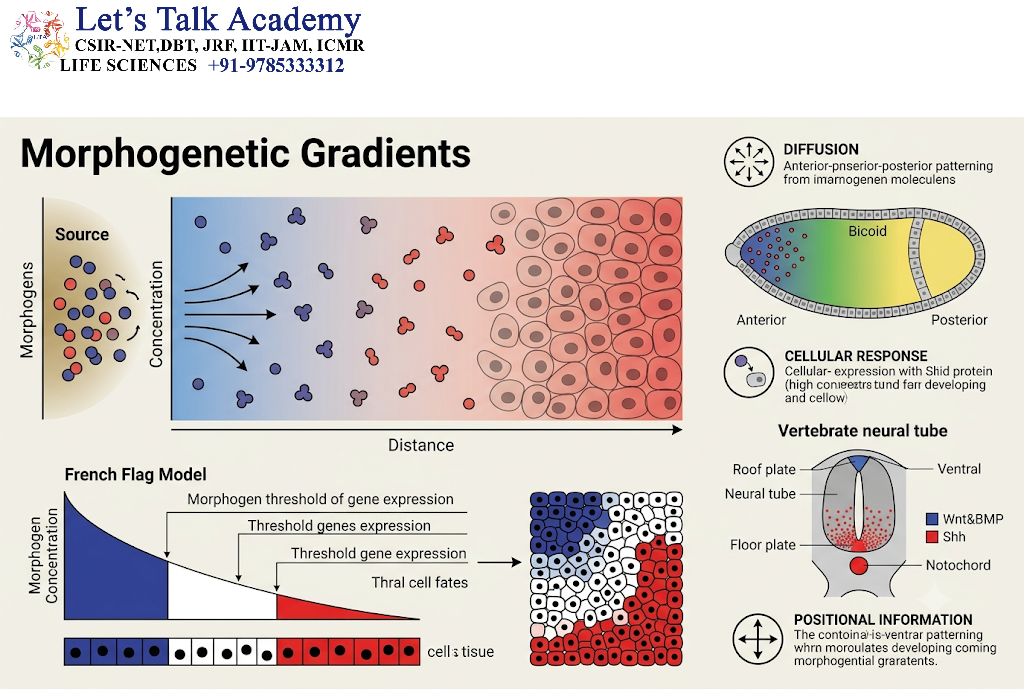
A morphogenetic gradient is a spatial concentration gradient of a signaling molecule that provides positional information to cells within a developing tissue.
The signaling molecule involved is often referred to as a morphogen when it fulfills the functional criteria associated with morphogen activity.
A classical morphogen has three important characteristics:
- It is produced from a localized source.
- It spreads through the surrounding tissue and establishes a concentration gradient.
- Different concentrations can induce different cellular responses.
Thus, cells can interpret their position according to the concentration of the morphogen to which they are exposed.
2.2 Positional Information
The fundamental idea behind a morphogen gradient is that cells need information about their location.
For example, consider a developing tissue extending from one side to another:
Source
↓
High morphogen concentration
↓
Intermediate concentration
↓
Low concentration
Cells near the source receive a strong signal, whereas cells farther away receive progressively weaker signals.
If cells respond to different concentration thresholds, the same morphogen can specify multiple cell types.
3. Morphogens and Morphogenetic Gradients
3.1 What Is a Morphogen?
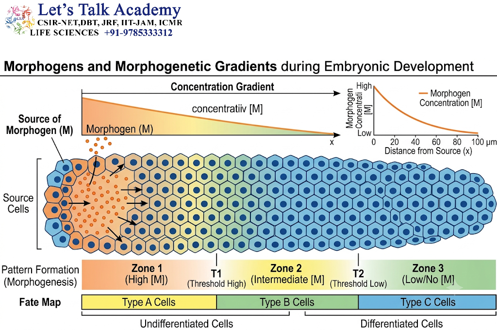
A morphogen is a signaling molecule that can provide positional information through its spatial distribution and concentration-dependent effects.
Morphogens may belong to different molecular classes, including:
- Secreted proteins
- Growth factors
- Lipid-modified signaling proteins
- Other extracellular signaling molecules
Well-known developmental signaling systems associated with morphogen activity include:
- Hedgehog signaling
- BMP signaling
- Wnt signaling
- Retinoic acid signaling
However, not every signaling molecule that forms a gradient necessarily functions as a classical morphogen.
3.2 Morphogen Versus General Signaling Molecule
A signaling molecule can act locally without functioning as a morphogen.
The important distinction is that morphogen activity involves spatial information and differential cellular responses related to signal concentration.
Therefore:
Signal molecule ≠ automatically a morphogen
A molecule is considered morphogenetic when its distribution and interpretation contribute to positional patterning.
4. Formation of a Morphogenetic Gradient
4.1 Localized Production
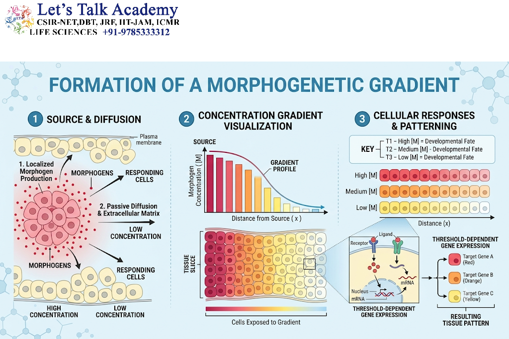
The first requirement is usually a localized source of the signaling molecule.
A group of cells may produce and release the morphogen.
For example:
Source cells → morphogen production → release into extracellular space
The source establishes the initial spatial asymmetry.
4.2 Transport
Once released, the morphogen must reach cells at different distances from the source.
Transport can involve:
- Diffusion
- Extracellular transport
- Cell-mediated transport
- Receptor-mediated movement
- Vesicular transport
- Tissue-specific transport mechanisms
The exact mechanism differs among developmental systems.
4.3 Removal and Degradation
A stable gradient requires not only production and transport but also removal.
Morphogens may be removed through:
- Receptor-mediated internalization
- Enzymatic degradation
- Extracellular degradation
- Intracellular degradation
- Binding to extracellular components
Therefore, gradient formation can be understood as a balance between:
Production + transport − removal
4.4 Establishment of the Concentration Gradient
As a result of localized production and spatial transport, morphogen concentration decreases with increasing distance from the source.
A simplified representation is:
High concentration → intermediate concentration → low concentration
This spatial difference allows cells in different positions to receive different levels of signaling.
5. Concentration-Dependent Cellular Responses
5.1 Threshold Model
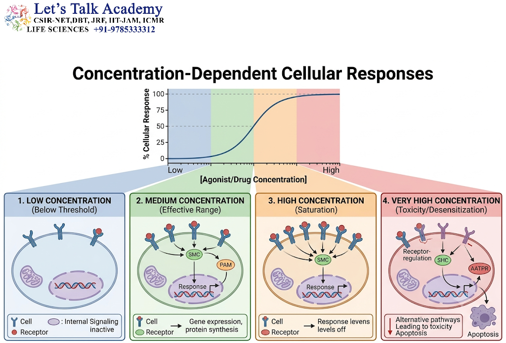
One of the most important features of morphogen signaling is that cells can respond differently at different concentration thresholds.
For example:
High concentration → Cell fate A
Intermediate concentration → Cell fate B
Low concentration → Cell fate C
Below signaling threshold → Cell fate D
Thus, a single gradient can establish several developmental domains.
5.2 Thresholds and Gene Expression
Cells contain receptors and intracellular signaling machinery that detect morphogen concentration.
Different concentrations can produce different levels or durations of intracellular signaling.
These differences can alter transcription factor activity and ultimately change gene expression.
The general sequence is:
Morphogen concentration
↓
Receptor activation
↓
Intracellular signaling
↓
Transcription factor activity
↓
Target-gene expression
↓
Cellular differentiation
6. Morphogen Interpretation by Cells
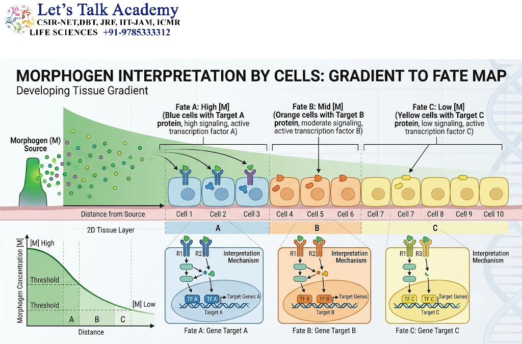
6.1 Receptor Binding
A morphogen generally interacts with receptors on or within responding cells.
The number and activity of receptors can influence cellular sensitivity to the signal.
6.2 Signal Transduction
After receptor activation, intracellular signaling pathways transmit information toward the nucleus.
These pathways may involve:
- Protein kinases
- Phosphatases
- Transcription factors
- Secondary messengers
- Protein–protein interactions
6.3 Gene Regulatory Networks
Ultimately, morphogen signaling modifies gene expression.
The response is therefore not simply a direct result of the extracellular concentration.
It depends on:
- Receptor abundance
- Intracellular signaling components
- Transcription-factor networks
- Chromatin state
- Previous signaling history
- Interactions with other pathways
Consequently, two cells exposed to the same morphogen concentration may not always produce identical responses.
7. Morphogen Gradients and Cell Fate
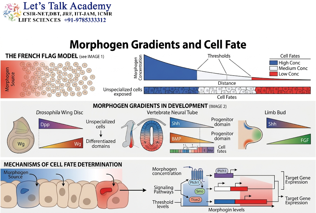
7.1 Cell-Fate Specification
Cell fate refers to the developmental pathway that a cell is expected to follow.
Morphogen gradients can divide a tissue into regions with different developmental identities.
For example:
High signal → neural subtype A
Intermediate signal → neural subtype B
Low signal → neural subtype C
The exact outcomes depend on the developmental system and signaling pathway.
7.2 Spatial Pattern Formation
Morphogen gradients provide a mechanism for converting molecular differences into spatial patterns.
This is one of the fundamental principles of developmental biology:
Chemical gradient → positional information → differential gene expression → spatial pattern
8. Sonic Hedgehog as a Morphogenetic Signaling System
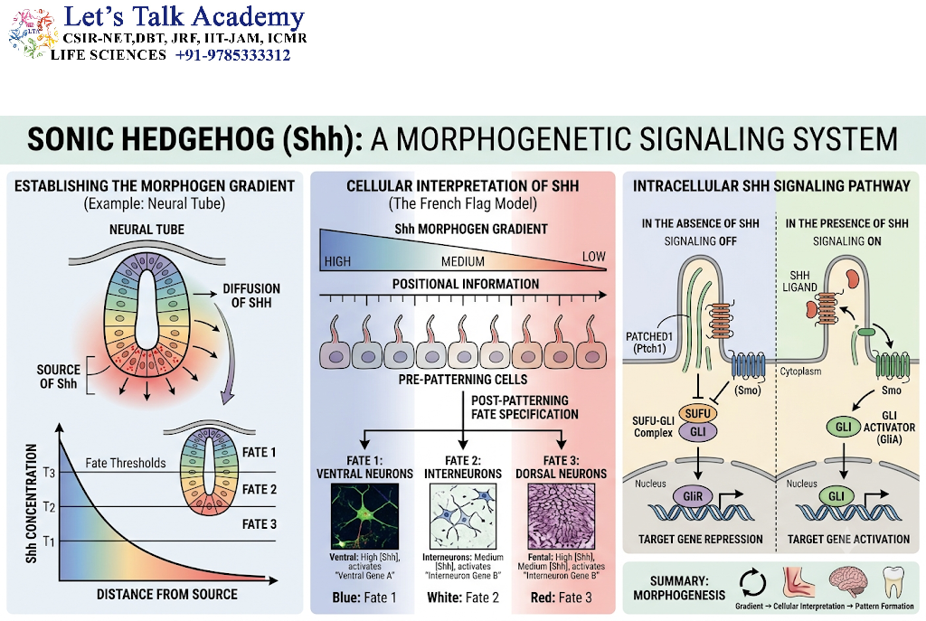
8.1 Hedgehog Signaling
The Hedgehog family of signaling proteins plays important roles in embryonic development.
In vertebrates, Sonic hedgehog (SHH) is particularly important in:
- Neural development
- Limb development
- Patterning of the ventral neural tube
- Organization of several embryonic structures
8.2 SHH Gradient in Neural Development
In the developing neural tube, SHH is produced in ventral regions, including the notochord and floor plate.
SHH signaling forms a spatial pattern across the neural tissue.
Cells exposed to different levels of SHH signaling activate different transcriptional programs.
This contributes to the specification of distinct neuronal progenitor domains.
8.3 Concentration and Cellular Identity
The classical model can be represented as:
High SHH signaling
↓
Ventral cell identities
Intermediate SHH signaling
↓
Intermediate progenitor identities
Lower SHH signaling
↓
More dorsal cell identities
This is a classic example of how a spatial signaling system can contribute to positional specification.
9. BMP Gradients
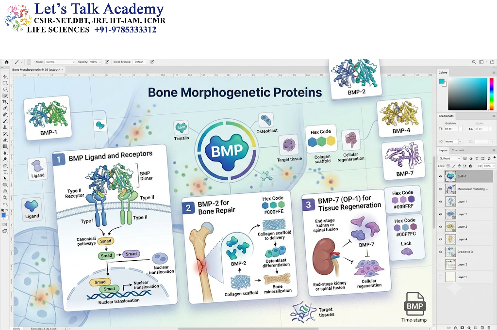
9.1 Bone Morphogenetic Proteins
BMPs belong to the transforming growth factor-beta superfamily.
They participate in:
- Embryonic patterning
- Tissue differentiation
- Bone formation
- Neural development
- Organ development
9.2 BMP Signaling
BMPs bind to receptor complexes and activate intracellular SMAD proteins.
A simplified pathway is:
BMP → receptor activation → SMAD phosphorylation → SMAD complex formation → nuclear entry → gene regulation
The intensity and context of BMP signaling can influence different developmental outcomes.
9.3 BMP and Tissue Patterning
BMP gradients or BMP-related signaling domains contribute to spatial patterning in several embryonic tissues.
The outcome depends on:
- BMP concentration
- Antagonists
- Receptor availability
- Tissue context
- Interactions with other pathways
10. Wnt Signaling Gradients
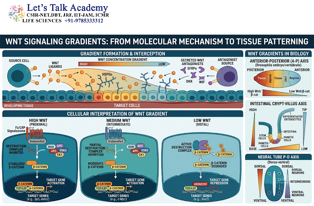
10.1 Wnt Proteins
Wnt proteins are important developmental signaling molecules.
They participate in:
- Body-axis formation
- Cell proliferation
- Cell differentiation
- Stem-cell maintenance
- Tissue regeneration
10.2 Wnt Gradient and Positional Information
Wnt signaling can form spatially organized signaling domains.
Cells at different positions may experience different levels of Wnt activity.
This contributes to regional specification during development.
10.3 Canonical Wnt Signaling
In the canonical pathway:
Wnt → Frizzled/LRP receptors → β-catenin stabilization → nuclear accumulation → transcriptional regulation
The cellular response depends on the level and duration of pathway activation.
11. Retinoic Acid Gradients
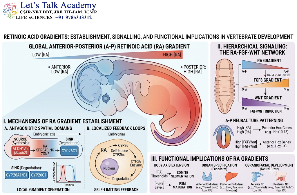
11.1 Retinoic Acid as a Morphogen-Like Signal
Retinoic acid is a small lipid-soluble signaling molecule derived from vitamin A metabolism.
It is particularly important in embryonic patterning.
Retinoic acid can influence gene expression by interacting with nuclear receptors.
11.2 Hox Gene Regulation
One important role of retinoic acid is the regulation of Hox genes, which contribute to positional identity along the body axis.
The general concept is:
Retinoic acid distribution → nuclear receptor activation → Hox gene regulation → positional identity
11.3 Importance of Retinoic Acid Concentration
Both insufficient and excessive retinoic acid signaling can disturb normal embryonic development.
This illustrates an important principle:
Development requires precisely regulated signaling rather than simply “more” or “less” signal.
12. Gradient Interpretation and Thresholds
12.1 Multiple Thresholds
A single gradient can produce multiple cell populations if cells respond at different thresholds.
For example:
| Morphogen concentration | Developmental response |
|---|---|
| Very high | Fate A |
| High | Fate B |
| Intermediate | Fate C |
| Low | Fate D |
| Very low | Fate E |
This creates multiple domains from one continuous concentration gradient.
12.2 Threshold Crossing
Suppose a morphogen concentration decreases gradually across a tissue.
At a particular position, concentration may cross a threshold required for activation of gene X.
At another position, it may cross a lower threshold required for gene Y.
Thus, gene-expression boundaries can correspond to specific concentration thresholds.
13. Temporal Regulation of Morphogen Signaling
Morphogen interpretation depends not only on concentration but also on time.
13.1 Duration of Exposure
Cells may respond differently to:
- Short exposure
- Prolonged exposure
- Repeated signaling
- Sustained signaling
Therefore:
Concentration + duration = biological response
13.2 Signal Adaptation
Cells may become less responsive to prolonged stimulation.
This can occur through:
- Receptor downregulation
- Inhibitory proteins
- Negative-feedback loops
- Changes in intracellular signaling
These mechanisms help prevent excessive signaling.
14. Regulation of Morphogen Gradients
Morphogen gradients are tightly regulated.
14.1 Production
The amount and location of morphogen production influence the gradient.
14.2 Diffusion and Transport
The rate at which the molecule moves through tissue affects its spatial distribution.
14.3 Receptor Binding
Receptors can capture morphogens and influence their effective range.
14.4 Degradation
Enzymatic degradation limits signal duration and range.
14.5 Extracellular Matrix
Extracellular matrix components can bind signaling molecules and influence their distribution.
14.6 Antagonists
Developmental signaling pathways often have extracellular antagonists that bind ligands or interfere with receptor activation.
For example, BMP signaling can be regulated by extracellular antagonists such as:
- Noggin
- Chordin
- Follistatin
These regulators help shape functional signaling gradients.
15. Morphogen Gradients and Reaction–Diffusion Mechanisms
15.1 Basic Concept
Developmental patterns can emerge from interactions between molecular production, diffusion, activation, inhibition, and degradation.
A simplified reaction–diffusion system contains:
Activator → production or propagation
and
Inhibitor → restriction of signaling
Such systems can generate spatial patterns.
15.2 Turing-Type Pattern Formation
Theoretical developmental biology has proposed that interacting activator–inhibitor systems can spontaneously generate spatial patterns.
The important principle is:
Local activation + broader inhibition → spatial pattern formation
This provides a theoretical framework for understanding how biological patterns can emerge from molecular interactions.
However, not every developmental gradient is generated by a classical Turing mechanism.
16. Morphogen Gradients and Embryonic Axis Formation
16.1 Anterior–Posterior Axis
Developmental signaling systems help establish positional information along the anterior–posterior axis.
Important signaling pathways include:
- Wnt
- FGF
- Retinoic acid
- Hox gene networks
16.2 Dorsal–Ventral Axis
BMP and related signaling systems contribute to dorsal–ventral patterning in several developmental contexts.
16.3 Left–Right Patterning
Left–right asymmetry involves specialized signaling systems, including Nodal-related signaling.
Thus, gradients and spatial signaling networks contribute to the organization of the three-dimensional body plan.
17. Morphogen Gradients During Limb Development
17.1 Sonic Hedgehog and the Limb
The developing limb provides a classic example of spatial patterning.
A region known as the zone of polarizing activity (ZPA) produces SHH during limb development.
SHH signaling contributes to anterior–posterior patterning of the limb.
17.2 Positional Information
Different levels and durations of SHH signaling contribute to differences in digit development.
The classical model emphasizes the relationship among:
- Signal concentration
- Exposure time
- Cellular response
Modern research has shown that morphogen interpretation can be more complex than a simple concentration-only model.
18. Morphogen Gradients and Organ Development
Morphogenetic signaling is important during the formation of:
- Brain
- Spinal cord
- Limbs
- Kidney
- Lung
- Liver
- Heart
- Pancreas
- Digestive organs
Different combinations of signaling pathways establish distinct developmental domains.
Thus, organogenesis involves multiple overlapping signaling gradients rather than a single universal gradient.
19. Interaction Between Morphogen Gradients
Cells are rarely exposed to only one developmental signal.
A cell may simultaneously receive:
- Wnt
- BMP
- FGF
- Hedgehog
- Retinoic acid
- Notch
- TGF-β
The final developmental response depends on the integration of these signals.
A simplified model is:
Signal A + Signal B + Signal C
↓
Integrated intracellular response
↓
Gene regulatory network
↓
Cell fate
Therefore, morphogen gradients operate within signaling networks rather than as isolated systems.
20. Morphogen Gradients and Gene Regulatory Networks
20.1 From Gradient to Gene Expression
A morphogen does not directly create a mature tissue.
Instead, it initiates changes in gene expression.
The sequence is:
Morphogen gradient
↓
Receptor activation
↓
Signal transduction
↓
Transcription-factor regulation
↓
Target-gene activation or repression
↓
Cellular differentiation
↓
Tissue pattern
20.2 Feedback Regulation
Target genes can themselves regulate components of the signaling pathway.
This creates feedback loops.
For example:
Morphogen → transcription factor → signaling inhibitor
The inhibitor then reduces further signaling.
Such feedback helps sharpen boundaries and stabilize developmental patterns.
21. Morphogen Gradients and Tissue Boundaries
A developing tissue must establish precise boundaries between different cell populations.
Morphogen thresholds can contribute to these boundaries.
For example:
High signal region | Intermediate signal region | Low signal region
Each region can activate a different combination of transcription factors.
The resulting gene-expression boundaries can then become more stable through feedback and cell–cell interactions.
22. Morphogen Gradients and Positional Memory
Once cells receive positional information, they may maintain their identity even after the original signal changes.
This can occur through:
- Stable transcription-factor networks
- Epigenetic mechanisms
- Positive feedback
- Cell–cell signaling
Thus, a transient developmental signal can produce a relatively stable cellular identity.
23. Morphogen Gradients in Regeneration
Morphogen-related signaling is not limited to embryonic development.
Similar pathways can function during tissue regeneration.
Examples include signaling involving:
- Wnt
- Hedgehog
- BMP
- FGF
During regeneration, these pathways can influence:
- Stem-cell proliferation
- Cell differentiation
- Tissue patterning
- Repair
However, regeneration and embryonic development are not identical processes.
24. Abnormal Morphogen Signaling and Disease
Abnormal developmental signaling can contribute to disease.
24.1 Cancer
Developmental pathways such as:
- Wnt
- Hedgehog
- Notch
- TGF-β
can become dysregulated in cancer.
Persistent activation of developmental signaling can contribute to:
- Abnormal proliferation
- Cell survival
- Stem-like cellular states
- Invasion
- Tumor progression
24.2 Developmental Disorders
Abnormal morphogen signaling during embryogenesis can result in defects in:
- Body-axis formation
- Limb development
- Neural development
- Organ formation
Therefore, precise control of developmental gradients is essential for normal development.
25. Mathematical Representation of a Morphogen Gradient
A simplified morphogen concentration profile can often be represented using an exponential decay model:
C(x)=C0e−x/λC(x)=C_0e^{-x/\lambda}
where:
- C(x)C(x) = morphogen concentration at position xx
- C0C_0 = concentration near the source
- xx = distance from the source
- λ\lambda = characteristic length scale of the gradient
This equation illustrates a basic principle:
As distance from the source increases, morphogen concentration decreases.
The mathematical description of real biological gradients can be considerably more complicated because production, diffusion, degradation, receptor binding, tissue geometry, and active transport may all contribute.
26. Factors Affecting Gradient Shape
The shape of a morphogen gradient depends on several variables.
26.1 Production Rate
Higher production can increase signal availability.
26.2 Diffusion Rate
Greater mobility can increase the spatial range.
26.3 Degradation Rate
Rapid degradation can shorten the range.
26.4 Tissue Geometry
The physical dimensions and architecture of a tissue affect gradient distribution.
26.5 Receptor Density
High receptor density can alter ligand availability and cellular sensitivity.
26.6 Extracellular Binding
Binding to extracellular matrix components can modify movement and retention.
26.7 Feedback Regulation
Positive and negative feedback can reshape the signaling profile.
27. Important Features of Morphogenetic Gradients
The major features can be summarized as:
- Localized source
- Spatial distribution
- Concentration-dependent signaling
- Threshold responses
- Differential gene expression
- Cell-fate specification
- Tissue pattern formation
- Feedback regulation
- Temporal regulation
- Interaction with other signaling pathways
28. Conceptual Example
Consider a developing tissue containing three groups of cells.
A signaling molecule is released from the left side.
Source → → → tissue
The concentration profile is:
High → Medium → Low
The cells respond according to thresholds:
High concentration
→ Gene A activated
→ Cell type A
Intermediate concentration
→ Gene B activated
→ Cell type B
Low concentration
→ Gene C activated
→ Cell type C
Thus, a continuous gradient produces discrete developmental domains.
This is one of the central ideas behind morphogen-based positional information.
29. Morphogenetic Gradient Versus Concentration Gradient
These terms should not be treated as completely synonymous.
A concentration gradient simply means that the concentration of a substance varies spatially.
A morphogenetic gradient refers to a biologically meaningful signaling gradient that contributes to spatial patterning and developmental organization.
Therefore:
Every morphogenetic gradient is a concentration gradient, but not every concentration gradient is a morphogenetic gradient.
30. Morphogen Gradient Versus Morphogenesis
These terms are also different.
Morphogen gradient:
A spatial signaling mechanism providing positional information.
Morphogenesis:
The broader developmental process through which tissues and organs acquire their shape and organization.
A morphogen gradient can contribute to morphogenesis, but morphogenesis also involves:
- Cell migration
- Cell proliferation
- Cell adhesion
- Cell differentiation
- Cell death
- Cytoskeletal remodeling
- Tissue mechanics
- Extracellular matrix remodeling
31. Experimental Approaches for Studying Morphogen Gradients
Morphogen gradients can be studied using several experimental approaches.
31.1 Fluorescence Microscopy
Fluorescently labeled proteins or antibodies can be used to visualize spatial distribution.
31.2 Reporter Genes
A signaling-responsive promoter can be linked to a fluorescent reporter.
This allows researchers to visualize pathway activity.
31.3 Genetic Manipulation
Gene knockout, knockdown, overexpression, and mutation can be used to determine pathway function.
31.4 Quantitative Imaging
Quantitative microscopy can measure:
- Signal intensity
- Spatial distribution
- Gradient length
- Temporal dynamics
31.5 Single-Cell Analysis
Single-cell approaches can reveal differences in signaling responses among individual cells.
32. Importance of Morphogenetic Gradients in Developmental Biology
Morphogenetic gradients provide a powerful mechanism for organizing developing tissues.
They help answer fundamental developmental questions:
- How do cells know where they are?
- How do different cell types arise in different positions?
- How are tissue boundaries established?
- How are body axes organized?
- How can one signaling source influence a large tissue?
The central answer is that cells can interpret spatial and temporal differences in signaling activity and convert them into distinct gene-expression programs.


