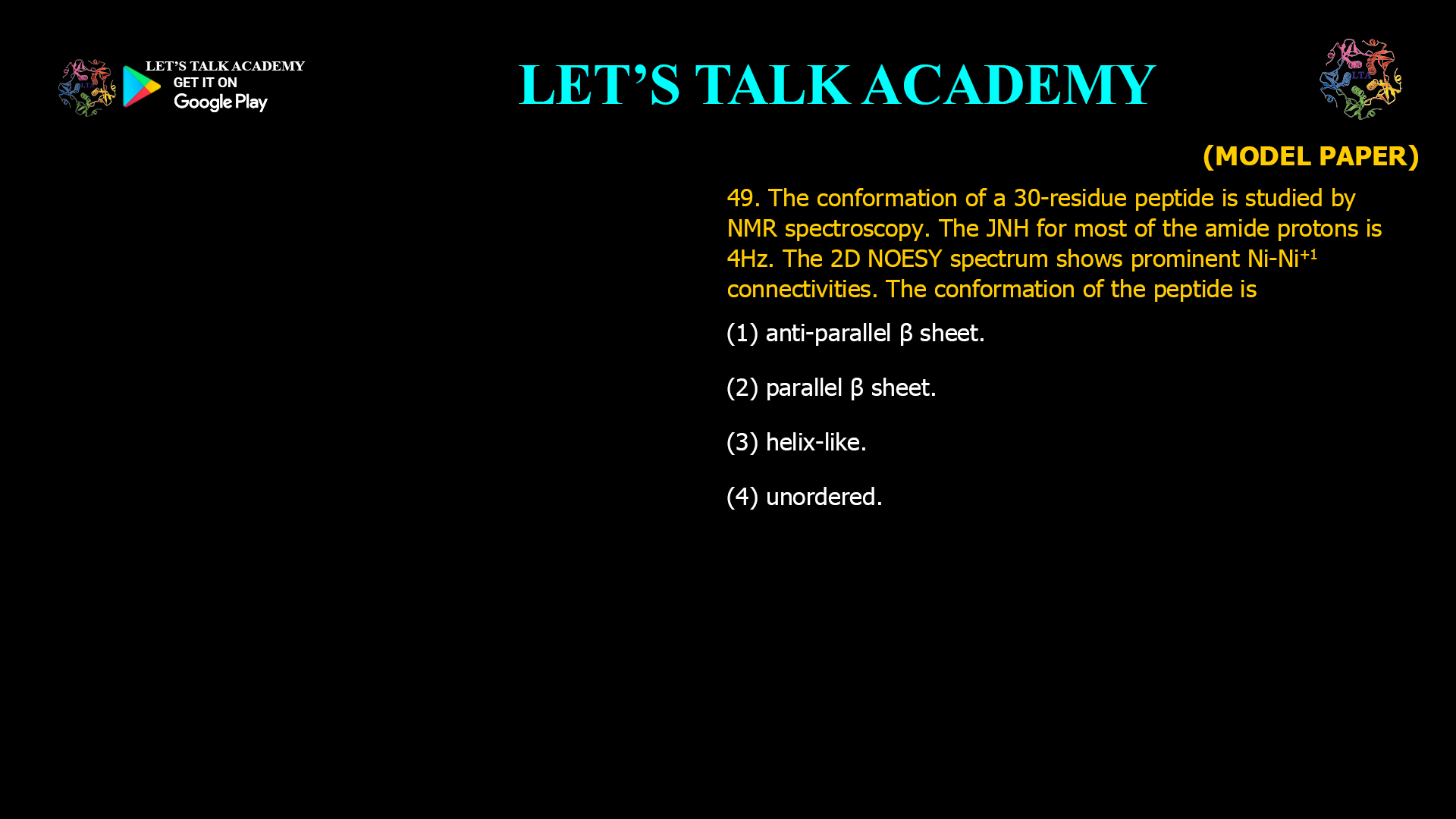
49. The conformation of a 30-residue peptide is studied by NMR spectroscopy. The JNH for most of the amide protons is 4 Hz. The 2D NOESY spectrum shows prominent Nᵢ–Nᵢ₊₁ connectivities. The conformation of the peptide is
(A) Anti-parallel β sheet.
(B) Parallel β sheet.
(C) Helix-like.
(D) Unordered.
Identifying Helical Peptide Conformation Using J Coupling and NOESY NMR Spectroscopy
Correct Answer
Option (3): Helix-like.
Explanation
Nuclear Magnetic Resonance (NMR) spectroscopy is one of the most powerful techniques for determining the secondary structure of peptides in solution. Structural information is obtained from several experimental parameters, among which vicinal scalar coupling constants and Nuclear Overhauser Effect (NOE) connectivities are particularly important. In this question, both the measured 3JHN-Hα coupling constant and the NOESY spectrum independently indicate the same type of secondary structure.
The vicinal coupling constant 3JHN-Hα depends on the dihedral angle between the amide proton and the α-proton according to the Karplus relationship. In α-helices, the backbone adopts relatively small dihedral angles, producing coupling constants typically between 3 and 5 Hz. In contrast, β-sheet conformations possess larger dihedral angles and generally exhibit coupling constants between 8 and 10 Hz. Since the measured value for most residues is approximately 4 Hz, the peptide backbone is consistent with a helical conformation.
The NOESY spectrum provides additional confirmation. A Nuclear Overhauser Effect arises when two protons are separated by approximately 5 Å or less in space. Helical peptides display strong sequential Ni–Ni+1 NOE cross-peaks because adjacent backbone amide protons are brought into close proximity by the regular helical geometry. Continuous stretches of these sequential connectivities are a characteristic feature of α-helical structures.
The agreement between the scalar coupling data and the NOESY connectivities provides strong evidence that the peptide adopts a predominantly helical arrangement. Because the question asks for the overall conformation rather than a perfectly ideal α-helix, the most appropriate description is helix-like.
Why Option (1) is Incorrect
Anti-parallel β-sheets typically exhibit 3JHN-Hα coupling constants of approximately 8–10 Hz because of their extended backbone geometry. Their NOESY spectra are dominated by inter-strand NOE contacts rather than continuous sequential Ni–Ni+1 connectivities. Therefore, the experimental observations are inconsistent with an anti-parallel β-sheet.
Why Option (2) is Incorrect
Parallel β-sheets also possess extended backbone conformations that produce relatively large vicinal coupling constants. Furthermore, their characteristic NOESY spectra contain long-range inter-strand contacts rather than the strong sequential NH-NH connectivities described in the question. Thus, the data do not support a parallel β-sheet structure.
Why Option (3) is Correct
The measured coupling constant of approximately 4 Hz falls within the range expected for a helical backbone, and the prominent sequential Ni–Ni+1 NOEs are characteristic of helical peptides. Since both independent NMR parameters indicate the same structural arrangement, the peptide is correctly described as adopting a helix-like conformation.
Why Option (4) is Incorrect
Unordered peptides generally exhibit poor chemical shift dispersion, weak or irregular NOE patterns, and coupling constants that vary widely from residue to residue. The presence of consistent 4 Hz coupling constants together with strong sequential NOE connectivities demonstrates that the peptide possesses a well-defined secondary structure rather than a random coil.
Role of the Karplus Relationship in NMR
The Karplus relationship describes how vicinal proton-proton scalar coupling constants vary with the dihedral angle separating the coupled nuclei. Because each secondary structural element possesses characteristic backbone dihedral angles, coupling constants provide valuable information about peptide conformation. Smaller coupling constants are typically associated with helical geometries, whereas larger values indicate extended conformations such as β-sheets.
Importance of NOESY in Secondary Structure Determination
NOESY spectroscopy detects through-space interactions between nearby nuclei. Since different secondary structures position backbone protons differently in three-dimensional space, each structural motif generates a characteristic NOE pattern. Helical peptides produce strong sequential and medium-range NOEs, whereas β-sheets generate numerous long-range inter-strand contacts. Analysis of these connectivities allows reliable identification of the underlying secondary structure.
Combining Scalar Coupling and NOE Data
Reliable structural assignment in NMR is achieved by integrating multiple independent experimental observations. Scalar coupling constants provide information about backbone dihedral angles, while NOESY spectra reveal spatial relationships between atoms. When both measurements independently support the same structural model, the confidence in the conformational assignment increases substantially.
Conclusion
The 3JHN-Hα coupling constant of approximately 4 Hz indicates a helical backbone geometry, and the prominent sequential Ni–Ni+1 NOESY connectivities further support this interpretation. Together, these observations demonstrate that the peptide adopts a helix-like conformation. Therefore, the correct answer is Option (3).


