
Composition, Structure and Function of Biomolecules:Proteins
Proteins are one of the most important classes of biological macromolecules and are fundamental to almost every aspect of cellular life. They participate in catalysis, transport, movement, signalling, defence, structural organization, gene regulation and maintenance of cellular homeostasis. A cell cannot function normally without a precisely regulated set of proteins.
The word protein is derived from the Greek word proteios, meaning “of primary importance.” This name is appropriate because proteins perform an exceptionally wide variety of biological functions. While nucleic acids store and transmit genetic information, proteins are the major molecular machines that execute many of the instructions encoded by genes.
Proteins are polymers made up primarily of amino acids joined together by peptide bonds. The properties of a protein are not determined merely by the number of amino acids present but also by their sequence, chemical properties, three-dimensional arrangement and interactions with other molecules.
A strong conceptual understanding requires connecting amino-acid chemistry → peptide bond → primary structure → secondary structure → tertiary structure → quaternary structure → folding → stability → biological function.
1. Introduction to Proteins
Proteins are high-molecular-weight biological macromolecules composed of one or more polypeptide chains. Each polypeptide chain consists of amino-acid residues linked through peptide bonds.
Most proteins found in living organisms are constructed from a common set of 20 standard amino acids. The amino acids differ primarily in their side chains, commonly represented as the R group. These side chains determine whether an amino acid is hydrophobic, polar, acidic, basic, aromatic, flexible, rigid or chemically reactive.
The sequence of amino acids in a protein is determined by genetic information. During gene expression, information encoded in DNA is transcribed into RNA and subsequently translated into a polypeptide sequence. However, the newly synthesized polypeptide must generally fold into a specific three-dimensional conformation before it can perform its biological function.
Therefore, protein biology can be understood at several interconnected levels:
Amino-acid composition → sequence → folding → three-dimensional structure → molecular function.
A change in even a single amino acid can sometimes have little effect, while in other cases it can dramatically alter protein structure and function. This is particularly important in understanding mutations, genetic diseases and molecular evolution.
2. Composition of Proteins
Proteins are mainly composed of:
- Carbon (C)
- Hydrogen (H)
- Oxygen (O)
- Nitrogen (N)
Many proteins also contain significant amounts of sulfur (S) because of the amino acids cysteine and methionine.
Some proteins may additionally contain other elements such as phosphorus, iron, zinc, copper, magnesium or selenium. These elements may occur as part of prosthetic groups, cofactors or metal-binding sites.
The elemental composition of a protein is therefore not always limited to C, H, O and N.
2.1 Amino Acids as the Building Blocks of Proteins
Amino acids are the fundamental structural units of proteins.
A typical protein amino acid contains a central α-carbon, to which four groups are attached:
- An amino group (–NH₂)
- A carboxyl group (–COOH)
- A hydrogen atom (–H)
- A variable side chain (–R)
The general structure can therefore be represented as:
NH₂–CH(R)–COOH
The identity and chemical properties of the amino acid depend mainly on its R group.
In aqueous biological environments, amino acids generally exist in ionized forms. At physiological pH, the amino group commonly exists as –NH₃⁺, whereas the carboxyl group commonly exists as –COO⁻.
Thus, amino acids frequently exist as zwitterions, molecules containing both positive and negative charges but having no net charge under particular conditions.
3. Classification of Amino Acids
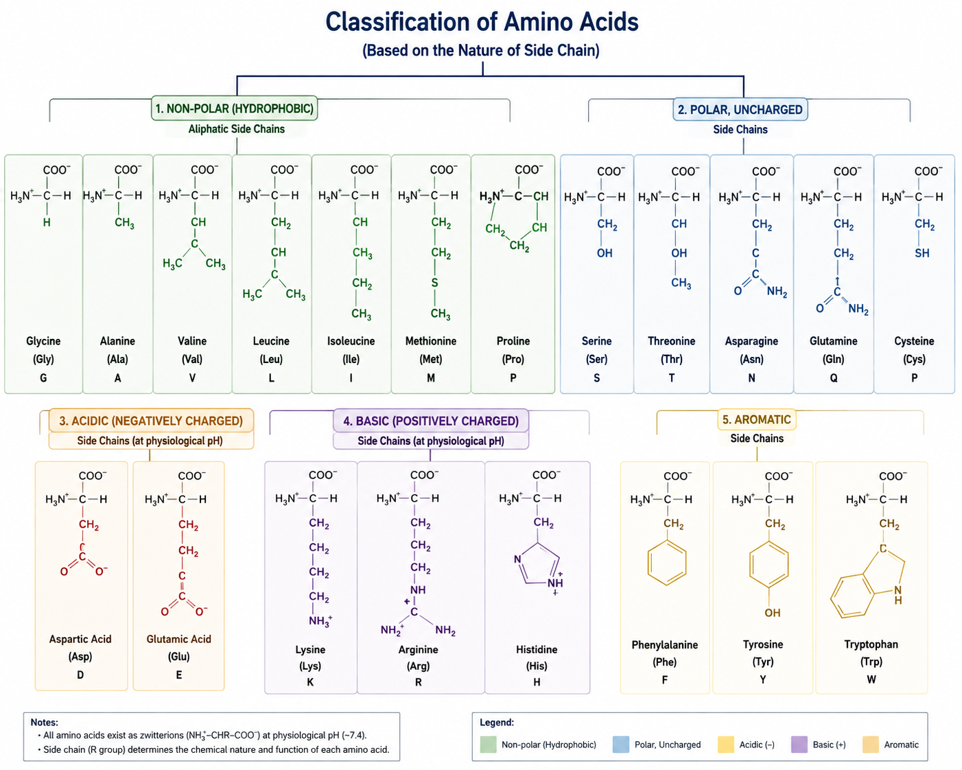
Amino acids can be classified in several ways. Classification according to side-chain properties is particularly important.
3.1 Classification Based on Side-Chain Polarity
Nonpolar or Hydrophobic Amino Acids
These amino acids contain side chains that are generally hydrophobic and tend to avoid contact with water.
Important examples include:
- Glycine
- Alanine
- Valine
- Leucine
- Isoleucine
- Methionine
- Proline
Hydrophobic amino acids frequently become buried inside globular proteins, where they contribute significantly to the hydrophobic core.
Polar Uncharged Amino Acids
These amino acids possess polar side chains but generally do not carry a net electrical charge at physiological pH.
Important examples include:
- Serine
- Threonine
- Asparagine
- Glutamine
- Cysteine
- Tyrosine
These residues frequently participate in hydrogen bonding and can occur on protein surfaces or within active sites.
Acidic Amino Acids
The major acidic amino acids are:
- Aspartate
- Glutamate
Their side chains contain additional carboxyl groups and are generally negatively charged at physiological pH.
Basic Amino Acids
The major basic amino acids are:
- Lysine
- Arginine
- Histidine
Their side chains contain nitrogen-containing groups capable of accepting protons.
Histidine is particularly important because its side-chain pKa is close enough to physiological pH that it can participate effectively in proton-transfer reactions. This makes histidine common in enzyme active sites.
4. Essential and Non-Essential Amino Acids
Amino acids can also be classified nutritionally according to whether they must be obtained from the diet.
4.1 Essential Amino Acids
Essential amino acids cannot be synthesized in sufficient amounts by humans and therefore must be obtained through dietary sources.
The commonly recognized essential amino acids include:
Histidine, Isoleucine, Leucine, Lysine, Methionine, Phenylalanine, Threonine, Tryptophan and Valine.
4.2 Non-Essential Amino Acids
Non-essential amino acids can generally be synthesized by the human body from metabolic intermediates.
Examples include:
- Alanine
- Aspartate
- Glutamate
- Serine
However, the terms essential and non-essential are nutritional classifications, not statements about whether an amino acid is biologically important. Every amino acid used in protein synthesis has an important biological role.
5. Special Amino Acids
Some amino acids have structural or functional properties that make them especially important in protein chemistry.
5.1 Glycine
Glycine has the smallest side chain, consisting only of a hydrogen atom.
Because of its small size, glycine provides considerable conformational flexibility to polypeptide chains.
It is frequently found in turns and regions where tight backbone conformations are required.
5.2 Proline
Proline is structurally unusual because its side chain forms a ring with the amino nitrogen.
This restricts rotation around the backbone and makes proline relatively rigid.
Proline can disrupt regular α-helical structures and is frequently associated with bends or turns in polypeptide chains.
5.3 Cysteine
Cysteine contains a sulfhydryl or thiol group:
–SH
Two cysteine residues can undergo oxidation to form a covalent disulfide bond:
Cys–S–S–Cys
Disulfide bonds can contribute significantly to the stability of many extracellular proteins.
5.4 Methionine
Methionine contains sulfur in a thioether group.
It is particularly important because the AUG codon specifies methionine and commonly serves as the translation initiation codon.
5.5 Tyrosine
Tyrosine contains an aromatic ring with a hydroxyl group.
Its hydroxyl group can undergo phosphorylation, making tyrosine important in cell signalling.
6. Peptide Bond
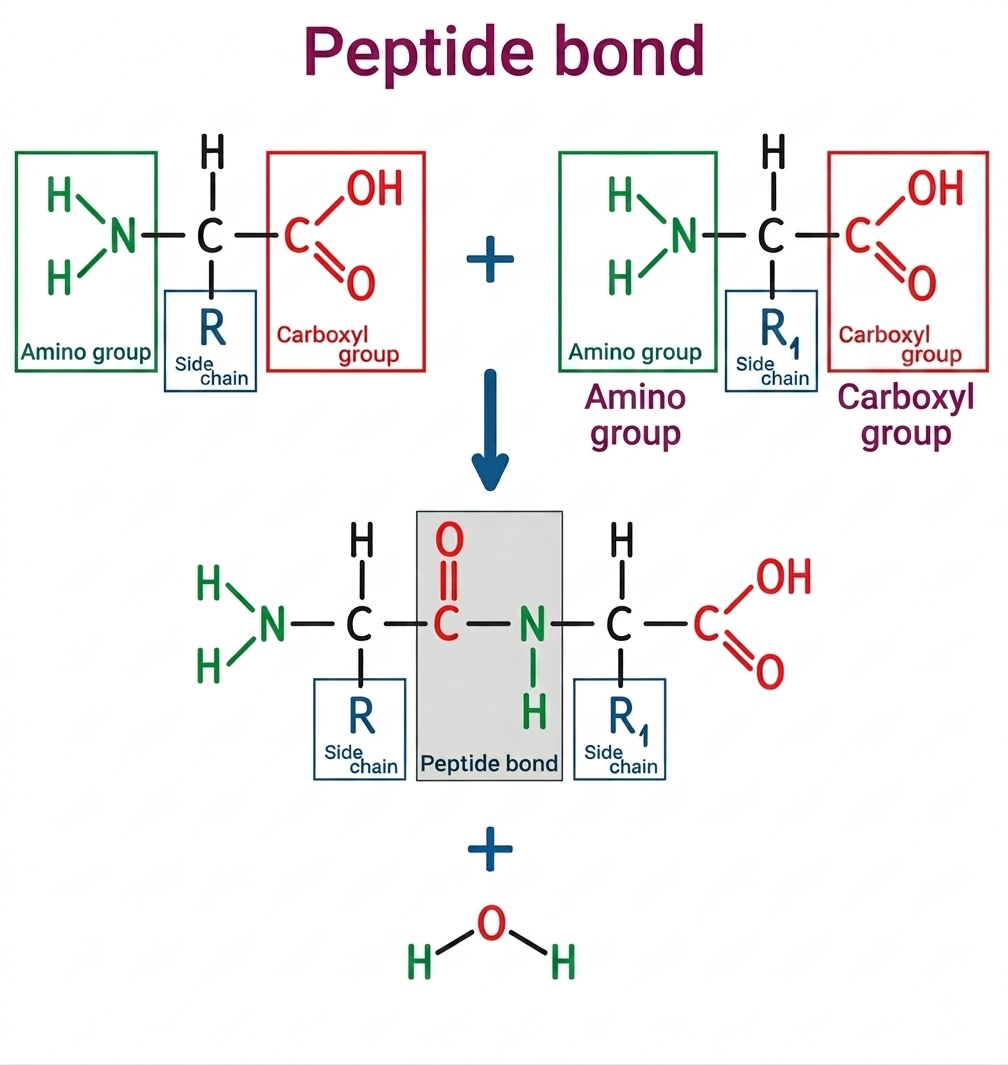
Amino acids are connected to each other through peptide bonds.
A peptide bond is formed between the carboxyl group of one amino acid and the amino group of another amino acid.
This reaction involves the removal of a molecule of water.
The general reaction is:
Amino acid 1 + Amino acid 2 → Dipeptide + H₂O
The bond formed between the carbonyl carbon and the nitrogen atom is the peptide bond.
Proteins are therefore formed through repeated peptide-bond formation between amino acids.
6.1 Important Properties of the Peptide Bond
The peptide bond has partial double-bond character because of resonance.
As a consequence:
- Rotation around the peptide C–N bond is restricted.
- The peptide group is approximately planar.
- The peptide bond generally adopts the trans configuration.
- The peptide backbone has restricted conformational freedom.
These properties are extremely important because they determine how a polypeptide chain can fold.
7. Peptides, Polypeptides and Proteins
The terms peptide, polypeptide and protein are related but are not always interchangeable.
A short chain of amino acids is generally called a peptide.
A longer chain is called a polypeptide.
A biologically functional macromolecule may consist of one or more polypeptide chains and is generally referred to as a protein.
A protein may therefore contain:
- A single polypeptide chain
- Two or more interacting polypeptide chains
- Additional non-protein components
The biological function depends on the final structural organization rather than simply on the length of the chain.
8. Directionality of a Polypeptide Chain
A polypeptide chain has directionality.
One end contains a free amino group and is called the:
N-terminus
The other end contains a free carboxyl group and is called the:
C-terminus
Protein sequences are conventionally written from:
N-terminus → C-terminus
This is an important CSIR-NET concept.
For example:
Ala–Gly–Val–Lys
means that alanine is present at the N-terminal end and lysine at the C-terminal end.
9. Levels of Protein Structure
Protein structure is commonly discussed at four levels:
- Primary structure
- Secondary structure
- Tertiary structure
- Quaternary structure
These levels are not independent. Rather, they represent increasing levels of structural organization.
The primary sequence influences folding, folding determines three-dimensional architecture, and the final architecture determines biological function.
10. Primary Structure of Proteins
The primary structure of a protein refers to the precise linear sequence of amino acids in its polypeptide chain.
For example:
Met–Ala–Gly–Ser–Val–Lys–Leu
represents a particular amino-acid sequence.
The amino acids are connected by peptide bonds.
Primary structure is extremely important because the sequence contains the information necessary for the protein to acquire its characteristic three-dimensional structure under appropriate conditions.
10.1 Importance of Primary Structure
A change in the amino-acid sequence can influence:
- Protein folding
- Stability
- Active-site architecture
- Protein-protein interactions
- Cellular localization
- Biological activity
A classic example is sickle-cell disease, in which a single amino-acid substitution in the β-globin chain changes a glutamate residue to valine.
This seemingly small change alters the physicochemical behavior of hemoglobin and contributes to abnormal red blood-cell morphology.
Therefore, the relationship can be remembered as:
Sequence → Structure → Function
11. Secondary Structure of Proteins
Secondary structure refers to local, regularly repeating arrangements of the polypeptide backbone.
The major secondary structures are:
- α-helix
- β-sheet
- β-turns and related turns
The secondary structure is stabilized primarily by hydrogen bonds involving backbone peptide groups.
The carbonyl oxygen of one peptide bond can form a hydrogen bond with the amide hydrogen of another peptide bond.
Importantly, the side chains are not the primary participants in the hydrogen-bond pattern that defines the α-helix and β-sheet.
12. α-Helix
The α-helix is one of the most important secondary structures in proteins.
In an α-helix, the polypeptide backbone coils into a right-handed helix in most biological proteins.
The structure is stabilized by hydrogen bonds between backbone carbonyl oxygen and amide hydrogen groups.
A commonly used structural description is that the carbonyl oxygen of residue i forms a hydrogen bond with the amide hydrogen of residue i + 4.
The side chains project outward from the helical backbone.
12.1 Important Features of α-Helix
The α-helix:
- Is generally right-handed in proteins.
- Is stabilized by backbone hydrogen bonds.
- Has approximately 3.6 amino-acid residues per turn.
- Has side chains projecting outward.
- Is influenced by the chemical nature of amino-acid residues.
Proline and α-Helix
Proline is often called a helix breaker because its rigid cyclic structure restricts backbone geometry and its nitrogen lacks the conventional amide hydrogen required for the usual hydrogen-bonding pattern.
Glycine, because of its high flexibility, can also destabilize a regular α-helix in many contexts.
13. β-Sheet
The β-sheet is another major secondary structure.
Instead of forming a compact helix, the polypeptide backbone adopts an extended conformation.
Multiple β-strands can associate through hydrogen bonding to form a β-sheet.
β-sheets may be:
- Parallel
- Antiparallel
- Mixed
13.1 Parallel β-Sheet
In a parallel β-sheet, neighboring strands run in the same N→C direction.
13.2 Antiparallel β-Sheet
In an antiparallel β-sheet, neighboring strands run in opposite directions.
Antiparallel β-sheets often have more geometrically favorable hydrogen bonding than parallel sheets.
The side chains project alternately above and below the plane of the sheet.
14. β-Turns
β-turns allow a polypeptide chain to reverse its direction over a short sequence.
They are particularly important in compact globular proteins.
Glycine and proline are frequently associated with turns because glycine provides flexibility while proline can impose a sharp bend due to its rigid structure.
15. Tertiary Structure of Proteins
The tertiary structure refers to the complete three-dimensional organization of a single polypeptide chain.
It results from interactions among amino-acid side chains and between side chains and the surrounding environment.
Important interactions include:
- Hydrophobic interactions
- Hydrogen bonds
- Ionic interactions
- Van der Waals interactions
- Disulfide bonds
The tertiary structure creates functional regions such as:
- Active sites
- Binding pockets
- Regulatory sites
- Protein-interaction surfaces
The tertiary structure is therefore closely connected to biological function.
16. Hydrophobic Interactions in Protein Folding
Hydrophobic interactions are major contributors to the folding of globular proteins.
Nonpolar side chains tend to become buried within the interior of a protein, away from water, while polar and charged residues are frequently more exposed to the aqueous environment.
This process contributes to formation of a compact hydrophobic core.
The hydrophobic effect is not simply a weak attraction between nonpolar groups. It is strongly related to the behavior and organization of surrounding water molecules.
17. Hydrogen Bonds in Proteins
Hydrogen bonds occur between suitable donor and acceptor groups.
They contribute to:
- Secondary structure
- Tertiary structure
- Protein-ligand interactions
- Protein-DNA interactions
- Protein-water interactions
Although individual hydrogen bonds are relatively weak compared with covalent bonds, large numbers of them can collectively contribute substantially to protein architecture and stability.
18. Ionic Interactions and Salt Bridges
Charged amino-acid side chains can interact electrostatically.
For example:
Lysine/Arginine (+) ↔ Aspartate/Glutamate (−)
Such interactions are often called salt bridges when appropriately arranged within a protein structure.
Their strength depends on factors such as:
- Distance
- Orientation
- Solvent exposure
- Local dielectric environment
- pH
Therefore, ionic interactions cannot be considered completely independent of the surrounding environment.
19. Disulfide Bonds
Disulfide bonds are covalent bonds formed between two cysteine residues.
The reaction can be represented as:
2 Cys–SH → Cys–S–S–Cys + 2H⁺ + 2e⁻
Disulfide bonds can stabilize protein structures by linking distant parts of the same polypeptide chain or different polypeptide chains.
They are particularly common in proteins that function in oxidizing extracellular environments.
For example, many extracellular proteins contain disulfide bonds, whereas the reducing environment of the cytosol generally makes stable disulfide formation less favorable.
20. Quaternary Structure of Proteins
Quaternary structure is present when a functional protein consists of two or more polypeptide subunits.
Each subunit may have its own tertiary structure, and the subunits associate to form the functional protein complex.
Examples include:
- Hemoglobin
- Many multisubunit enzymes
- ATP synthase
- Antibodies
Hemoglobin is a classical example of a protein with quaternary structure. It contains four polypeptide subunits.
20.1 Importance of Quaternary Structure
Quaternary organization can provide:
- Cooperative behavior
- Allosteric regulation
- Increased functional complexity
- Structural stability
- Regulation of catalytic activity
The association of subunits is commonly stabilized through noncovalent interactions, although disulfide bonds can also occur between chains in some proteins.
21. Protein Domains
A domain is a structurally and often functionally distinct region of a protein.
Many proteins consist of multiple domains.
Each domain may perform a particular role, such as:
- Catalysis
- Ligand binding
- DNA binding
- Protein-protein interaction
- Membrane association
Domains are important because they provide a modular organization to proteins.
A large protein can therefore be thought of as a molecular machine assembled from several functional modules.
22. Protein Motifs
A motif is a recurring structural pattern or characteristic sequence arrangement found in proteins.
Motifs may contribute to specific molecular functions.
Examples include:
- Helix-turn-helix
- Zinc-finger-related motifs
- Rossmann-like nucleotide-binding patterns
- β-hairpins
A motif is generally smaller than a domain.
A useful conceptual distinction is:
Motif = recurring structural/sequence pattern
Domain = larger independently organized structural and functional unit
23. Protein Folds
A protein fold refers to the overall three-dimensional arrangement of secondary-structure elements within a protein.
Different proteins can sometimes perform different functions while sharing similar structural folds.
Protein folds are therefore useful for understanding evolutionary relationships and structural organization.
24. Ramachandran Plot
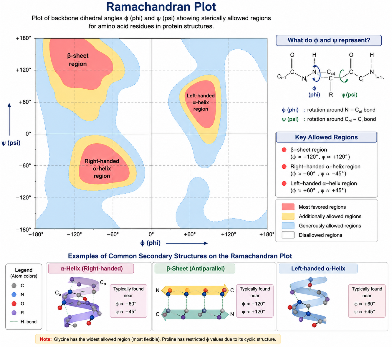
The Ramachandran plot is an important tool for understanding protein backbone conformation.
It represents the allowed combinations of the backbone dihedral angles:
- φ (phi)
- ψ (psi)
These angles describe rotations around bonds in the polypeptide backbone.
Because the peptide bond itself has restricted rotation, not all combinations of φ and ψ are sterically possible.
The Ramachandran plot therefore helps identify:
- Allowed conformations
- Favored conformations
- Sterically disallowed regions
24.1 Importance of the Ramachandran Plot
The Ramachandran plot is particularly useful for:
- Protein structure analysis
- Structural validation
- Understanding α-helices
- Understanding β-sheets
- Studying conformational restrictions
For CSIR-NET, remember:
Ramachandran plot → φ and ψ angles of the protein backbone.
25. Why Protein Structure Determines Function
A protein performs its biological function because its structure allows it to interact selectively with other molecules.
For example:
An enzyme requires an appropriate active-site architecture.
A receptor requires a ligand-binding region.
A transporter requires structural pathways through which specific molecules can move.
An antibody requires antigen-recognition regions.
A motor protein requires structural elements capable of converting chemical energy into mechanical movement.
Thus, protein function is strongly dependent on its three-dimensional structure.
A change in protein conformation can therefore change or completely abolish biological activity.
26. Protein Folding
Protein folding is the process by which a newly synthesized polypeptide acquires its biologically functional three-dimensional conformation.
The information required for folding is largely encoded in the amino-acid sequence, although cellular conditions and molecular chaperones can strongly influence the folding process.
Protein folding is not simply random movement until the protein “finds” its shape. It involves a complex energy landscape containing numerous possible conformational states.
The biologically functional state is generally referred to as the native state.
27. Energy Landscape of Protein Folding
Protein folding can be visualized using an energy landscape.
A newly synthesized protein can occupy many possible conformations. As folding progresses, the protein generally moves toward conformations of lower free energy.
The landscape is often represented conceptually as a funnel.
At the top:
Many unfolded conformations
↓
Intermediate conformations
↓
Fewer folded conformations
↓
Native functional state
This model explains why protein folding can involve many possible pathways rather than a single predetermined sequence of structural events.
28. Molecular Chaperones
Cells contain specialized proteins called molecular chaperones that assist other proteins in achieving or maintaining proper conformations.
Chaperones do not generally provide the structural information specifying the final protein sequence. Instead, they can:
- Prevent inappropriate aggregation
- Provide protected environments for folding
- Assist refolding
- Help maintain proteins under stressful conditions
Important chaperone systems include:
- Hsp70
- Hsp60/chaperonins
- Hsp90
Chaperones are particularly important during cellular stress, when proteins are more likely to misfold or aggregate.
29. Protein Denaturation
Denaturation refers to disruption of the native three-dimensional structure of a protein without necessarily breaking all peptide bonds.
Denaturation may be caused by:
- High temperature
- Extreme pH
- Organic solvents
- Detergents
- Chaotropic agents
- Heavy metals
- High concentrations of certain chemicals
During denaturation, secondary, tertiary and/or quaternary structures may be disrupted.
The primary structure generally remains intact unless conditions are severe enough to cause chemical degradation or peptide-bond hydrolysis.
30. Renaturation
In some proteins, removal of the denaturing condition can allow the protein to regain its native structure.
This process is called renaturation.
The classical experiments of Christian Anfinsen with ribonuclease provided strong evidence that amino-acid sequence contains substantial information required for the native fold of a protein.
However, not every protein can efficiently renature after denaturation. Many proteins require cellular assistance, cofactors or specific conditions for correct folding.
31. Protein Stability
Protein stability refers to the tendency of a protein to remain in its functional native state under given conditions.
Protein stability depends on a balance between multiple interactions, including:
- Hydrophobic interactions
- Hydrogen bonds
- Ionic interactions
- Van der Waals interactions
- Disulfide bonds
- Interactions with solvent
- Metal coordination where applicable
Protein stability is therefore not determined by a single bond or interaction.
A stable protein is not necessarily rigid. Proteins are dynamic molecules and often undergo conformational changes as part of their biological functions.
32. Globular Proteins
Globular proteins generally have compact, approximately spherical structures.
They often perform dynamic functions such as:
- Enzymatic catalysis
- Transport
- Regulation
- Signalling
- Immune defence
Examples include:
- Hemoglobin
- Myoglobin
- Many enzymes
- Albumin
Their hydrophobic residues are commonly enriched in the interior, while many polar and charged residues are exposed to water.
33. Fibrous Proteins
Fibrous proteins generally have elongated structures and frequently serve structural or mechanical roles.
Examples include:
- Collagen
- Keratin
- Fibroin
These proteins often contain repetitive sequence patterns and structural arrangements suited to mechanical strength.
33.1 Collagen
Collagen is a major structural protein in animals.
Its characteristic structure consists of three polypeptide chains forming a triple helix.
Glycine occurs frequently in collagen sequences, allowing tight packing of the chains.
Proline and hydroxyproline also contribute to collagen structure.
34. Protein Functions
Proteins perform an enormous range of functions in living organisms.
34.1 Enzymatic Function
Enzymes are biological catalysts.
Examples include:
- DNA polymerase
- RNA polymerase
- ATP synthase
- Proteases
- Kinases
Enzymes accelerate biochemical reactions without being consumed in the overall reaction.
35. Structural Function
Structural proteins provide mechanical strength and organization.
Examples include:
Collagen – connective tissues
Keratin – hair, nails and epithelial structures
Actin and tubulin – cytoskeletal organization
36. Transport Function
Proteins can transport molecules and ions.
Examples include:
Hemoglobin – oxygen transport
Membrane transporters – movement of ions and metabolites
Albumin – transport of several molecules in blood
37. Storage Function
Some proteins function as reservoirs for particular molecules.
Examples include:
- Ferritin – iron storage
- Myoglobin – oxygen storage in muscle
38. Hormonal and Signalling Functions
Many signalling molecules are proteins or peptides.
Examples include:
- Insulin
- Growth hormone
- Several cytokines
Protein receptors recognize signalling molecules and convert extracellular information into intracellular responses.
39. Defense Function
Proteins participate extensively in immune defence.
Examples include:
- Antibodies
- Complement proteins
- Antimicrobial proteins
- Cytokines
Antibodies recognize specific molecular structures called antigens and participate in immune responses.
40. Contractile and Motor Functions
Proteins are essential for cellular movement.
Important examples include:
- Actin
- Myosin
- Kinesin
- Dynein
These proteins participate in muscle contraction, intracellular transport, chromosome movement and other forms of cellular motion.
41. Receptor Proteins
Receptors are proteins that recognize specific ligands.
A ligand may be:
- Hormone
- Neurotransmitter
- Growth factor
- Metabolite
- Extracellular signal
Binding of a ligand can produce a conformational change in the receptor and initiate a signalling cascade.
Examples include:
- G-protein-coupled receptors
- Receptor tyrosine kinases
- Ligand-gated ion channels
42. Protein-Ligand Interactions
Proteins interact with ligands through a combination of molecular forces.
These may include:
- Hydrogen bonding
- Electrostatic interactions
- Hydrophobic interactions
- Van der Waals forces
- Metal coordination
- Occasionally covalent interactions
The specificity of a protein-ligand interaction depends on complementary chemical and structural features.
This concept is particularly important for understanding enzyme-substrate recognition, receptor-ligand binding and drug interactions.
43. Protein-Protein Interactions
Proteins rarely work in complete isolation.
Many biological processes depend on protein-protein interactions.
Examples include:
- Receptor signalling complexes
- Transcription-factor complexes
- Enzyme complexes
- Cytoskeletal assemblies
- Antigen-antibody interactions
The interaction surface depends on complementary shapes, charges and chemical groups.
44. Protein-Protein Interaction Forces
Protein-protein interactions can involve:
Hydrogen bonds
These provide directional interactions between suitable donor and acceptor groups.
Electrostatic interactions
Charged residues can attract or repel one another.
Hydrophobic interactions
Nonpolar surfaces can associate to minimize unfavorable exposure to water.
Van der Waals interactions
Close contact between atoms can provide additional stabilizing contributions.
Together, many relatively weak interactions can create a strong and specific protein complex.
45. Protein Modification
Proteins can undergo chemical modifications after translation.
These are called post-translational modifications (PTMs).
Important examples include:
- Phosphorylation
- Acetylation
- Methylation
- Glycosylation
- Ubiquitination
- Lipidation
- Proteolytic cleavage
Post-translational modifications can alter:
- Protein activity
- Stability
- Localization
- Protein-protein interactions
- Degradation
- Cellular signalling
46. Protein Phosphorylation
Phosphorylation commonly occurs on:
- Serine
- Threonine
- Tyrosine
Protein kinases catalyze phosphorylation, whereas phosphatases remove phosphate groups.
Phosphorylation is one of the most important mechanisms of reversible regulation in cell signalling.
It can activate or inhibit proteins depending on the protein and the location of the modification.
47. Protein Glycosylation
Glycosylation involves the attachment of carbohydrate groups to proteins.
It is particularly important for proteins entering the secretory pathway.
Glycosylation can influence:
- Protein folding
- Stability
- Cellular trafficking
- Cell-cell recognition
- Immune recognition
48. Protein Ubiquitination
Ubiquitination involves attachment of ubiquitin to a target protein.
Ubiquitin can act as a signal for protein degradation, particularly through the 26S proteasome, although ubiquitination can also regulate functions other than degradation.
This illustrates an important principle:
Protein modification does not necessarily mean activation or degradation; the biological consequence depends on the modification and cellular context.
49. Protein Degradation
Cells continuously synthesize and degrade proteins.
Protein degradation is necessary for:
- Removal of damaged proteins
- Regulation of protein abundance
- Cell-cycle control
- Adaptation to environmental changes
- Maintenance of protein quality
Major degradation systems include:
- Ubiquitin-proteasome system
- Lysosomal degradation
- Autophagy-related pathways
50. Misfolding and Protein Aggregation
Incorrectly folded proteins can lose their normal functions and may form aggregates.
Protein misfolding is associated with several human diseases.
Examples include:
- Alzheimer’s disease
- Parkinson’s disease
- Huntington’s disease
- Prion diseases
The biological consequences of protein aggregation depend on the specific protein, cell type and aggregation pathway.
This is why cells have extensive protein-quality-control mechanisms.
51. Prions
Prions are infectious protein conformations associated with transmissible neurodegenerative diseases.
They provide a remarkable example of how a change in protein conformation can influence the behavior of another protein molecule.
Prion biology demonstrates that protein structure can sometimes carry biologically important information independently of changes in nucleic-acid sequence.
52. Protein Isoforms
A single gene can sometimes give rise to multiple protein isoforms.
This can occur through:
- Alternative splicing
- Alternative promoter usage
- Alternative translation initiation
- Post-translational processing
Different isoforms may have different:
- Cellular locations
- Activities
- Regulatory properties
- Interaction partners
This increases the functional diversity of the proteome.
53. Proteome
The complete set of proteins expressed by a cell, tissue or organism under particular conditions is called the proteome.
Unlike the genome, which is relatively stable, the proteome can change substantially depending on:
- Cell type
- Developmental stage
- Environmental conditions
- Disease
- Nutritional status
- Signalling state
Therefore:
Genome → relatively stable genetic information
Proteome → dynamic expression of functional proteins
54. Protein Structure and Mutation
Mutations can alter the amino-acid sequence of a protein.
A mutation may:
- Have no detectable effect
- Change protein stability
- Alter enzyme activity
- Modify ligand binding
- Affect protein localization
- Cause misfolding
- Produce disease
The effect depends strongly on where the amino-acid substitution occurs and how the substituted residue differs chemically from the original residue.
For example, replacement of a small nonpolar residue with a bulky charged residue in a protein core can have a much larger structural effect than a chemically conservative substitution on an exposed surface.
55. Protein Sequence and Evolution
Protein sequences contain information about evolutionary relationships.
Proteins derived from a common ancestral protein may retain conserved amino-acid residues, especially at positions essential for:
- Catalysis
- Structural stability
- Ligand binding
- Molecular interactions
Highly conserved residues are therefore often functionally important.
Sequence comparison can be used to identify:
- Homologous proteins
- Conserved domains
- Functional motifs
- Evolutionary relationships
56. Protein Structure and Bioinformatics
Modern biological research combines experimental and computational approaches to understand protein structure.
Protein sequences can be analyzed to predict:
- Conserved domains
- Functional motifs
- Secondary structure
- Structural similarity
- Potential binding sites
- Evolutionary relationships
Three-dimensional structures can be obtained experimentally using techniques such as:
- X-ray crystallography
- Nuclear magnetic resonance spectroscopy
- Cryo-electron microscopy
Computational approaches are increasingly used alongside these experimental methods for protein structure prediction and analysis.
57. Protein Structure Hierarchy
For quick conceptual revision, the structural hierarchy can be summarized as follows:
Primary structure
→ Linear amino-acid sequence
Secondary structure
→ Local α-helices, β-sheets and turns
Tertiary structure
→ Complete three-dimensional structure of one polypeptide
Quaternary structure
→ Association of multiple polypeptide subunits
This hierarchy is one of the most frequently tested conceptual areas of protein biochemistry.
58. Important Comparison of Protein Structural Levels
| Structural Level | Major Feature | Major Stabilizing Factors |
|---|---|---|
| Primary | Amino-acid sequence | Peptide bonds |
| Secondary | α-helix, β-sheet | Backbone hydrogen bonds |
| Tertiary | 3D structure of one chain | Hydrophobic, ionic, H-bond, van der Waals, disulfide interactions |
| Quaternary | Association of multiple chains | Noncovalent interactions and sometimes disulfide bonds |
A crucial point is that this table describes the dominant structural determinants, not exclusive rules. Protein structures are stabilized by combinations of interactions.
59. Integrated Concept: From Gene to Functional Protein
The complete biological journey of a protein can be understood as a sequence of connected events:
DNA
↓
Transcription
↓
mRNA
↓
Translation
↓
Polypeptide
↓
Folding
↓
Post-translational modification
↓
Functional protein
↓
Biological activity
This sequence demonstrates why molecular biology, biochemistry and cell biology cannot be studied as completely separate subjects.
A mutation in DNA can ultimately alter protein sequence, structure and function.62. Protein Structure–Function Relationship
The central principle of protein biology is:
Structure determines function, and sequence influences structure.
The amino-acid sequence determines the chemical possibilities available to the polypeptide. These possibilities influence folding and ultimately generate a three-dimensional structure capable of performing a particular function.
However, protein structure is dynamic rather than completely rigid.
Many proteins undergo conformational changes during:
- Catalysis
- Ligand binding
- Signal transduction
- Transport
- Molecular recognition
- Mechanical movement
Therefore, a better conceptual model is:
Sequence → Folding → Dynamic Structure → Molecular Interactions → Function


