
1. Introduction
Cancer is characterized by uncontrolled cell proliferation, abnormal survival, altered metabolism, genomic instability, and the ability to invade surrounding tissues. One of the most important and dangerous properties of malignant tumors is metastasis.
Metastasis is the process in which cancer cells leave the original or primary tumor, travel through the body, and establish new tumors at distant sites. The newly formed tumor is called a secondary tumor or metastatic tumor.
For example, breast cancer may spread to the bone, liver, lung, or brain. When breast cancer cells form a tumor in the bone, the tumor is still biologically breast cancer, not bone cancer, because its cells originated from the breast tumor.
Metastasis is a complex, multistep process involving interactions between cancer cells, blood vessels, lymphatic vessels, immune cells, extracellular matrix, and distant tissues.
2. Definition of Metastasis
Metastasis is the spread of malignant cancer cells from a primary tumor to a physically separate site in the body, where they survive, proliferate, and form a secondary tumor.
Important features
- It occurs mainly in malignant tumors.
- Cancer cells must detach from the primary tumor.
- They invade surrounding tissues.
- They enter blood or lymphatic vessels.
- They survive during circulation.
- They leave the circulation at a distant site.
- They adapt to the new tissue environment.
- They establish a new tumor.
3. Primary Tumor and Secondary Tumor
| Feature | Primary tumor | Secondary/metastatic tumor |
|---|---|---|
| Origin | Original site of cancer | Distant site |
| Example | Breast carcinoma | Breast cancer in bone |
| Cancer identity | Original tissue | Retains origin of primary cancer |
| Formation | Initial malignant growth | Result of metastasis |
A secondary tumor does not become a different type of cancer simply because it grows in another organ.
4. Why Metastasis Is Important
Metastasis can:
- damage essential organs,
- interfere with normal organ function,
- cause pain,
- produce neurological complications,
- cause fractures when bone is involved,
- impair liver or lung function,
- produce fluid accumulation,
- contribute substantially to cancer-related morbidity and mortality.
Therefore, understanding metastasis is essential for understanding cancer progression.
5. Basic Mechanism of Metastasis
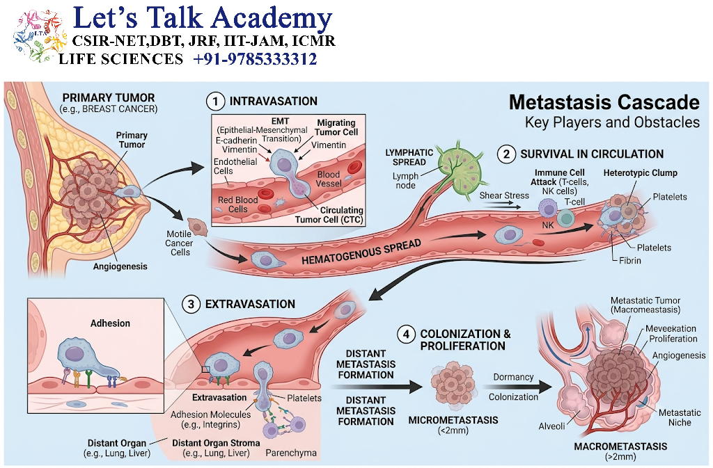
Metastasis can be represented as a sequence:
Primary tumor formation
↓
Loss of cell-cell adhesion
↓
Epithelial-to-mesenchymal transition
↓
Local invasion
↓
Extracellular matrix degradation
↓
Intravasation
↓
Survival in circulation
↓
Arrest in distant organ
↓
Extravasation
↓
Survival in new tissue
↓
Colonization
↓
Angiogenesis
↓
Secondary tumor formation
This entire process is sometimes described as the metastatic cascade.
6. Metastatic Cascade
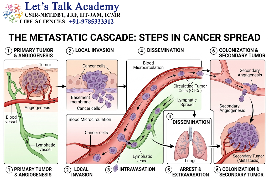
The metastatic cascade consists of multiple biological stages.
Major stages
- Local tumor growth
- Loss of cell adhesion
- Tumor-cell migration
- Extracellular matrix degradation
- Local invasion
- Intravasation
- Survival in circulation
- Arrest at distant tissue
- Extravasation
- Adaptation to the new environment
- Micrometastasis formation
- Angiogenesis
- Macrometastasis formation
Each step represents a major biological barrier.
7. Loss of Cell-Cell Adhesion
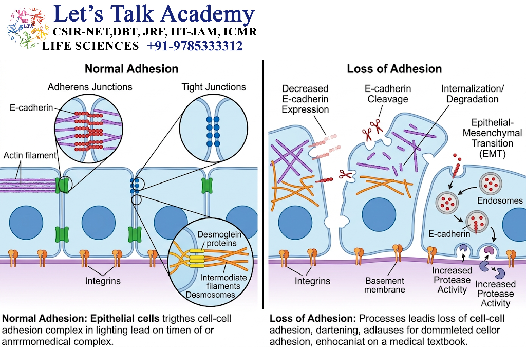
Normal epithelial cells are strongly attached to neighboring cells.
One important adhesion molecule is E-cadherin.
E-cadherin helps maintain:
- cell-cell adhesion,
- tissue organization,
- epithelial structure.
During cancer progression, E-cadherin expression or function may be reduced.
As a result:
Reduced E-cadherin
→ weaker cell-cell adhesion
→ increased cellular mobility
→ separation of tumor cells
→ increased invasion.
Loss of adhesion therefore facilitates the first stages of metastasis.
8. Epithelial-to-Mesenchymal Transition
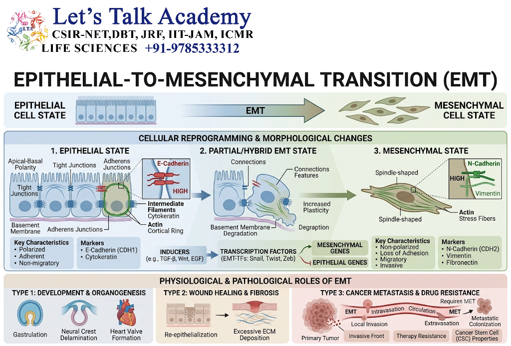
Epithelial-to-mesenchymal transition (EMT) is a biological process in which epithelial cells acquire characteristics associated with mesenchymal cells.
During EMT, cells may:
- lose epithelial polarity,
- reduce cell-cell adhesion,
- increase migration,
- increase invasiveness,
- alter cytoskeletal organization,
- become more resistant to some forms of cell death.
Important EMT-related transcription factors
- SNAIL
- SLUG
- TWIST
- ZEB1
- ZEB2
These factors can repress epithelial genes such as E-cadherin and promote a more migratory phenotype.
Simplified mechanism
Epithelial tumor cell
→ EMT signals
→ reduced E-cadherin
→ cytoskeletal remodeling
→ increased migration
→ invasive tumor cell.
EMT is not simply an irreversible switch; cancer cells can display partial or reversible epithelial and mesenchymal characteristics.
9. Role of the Extracellular Matrix
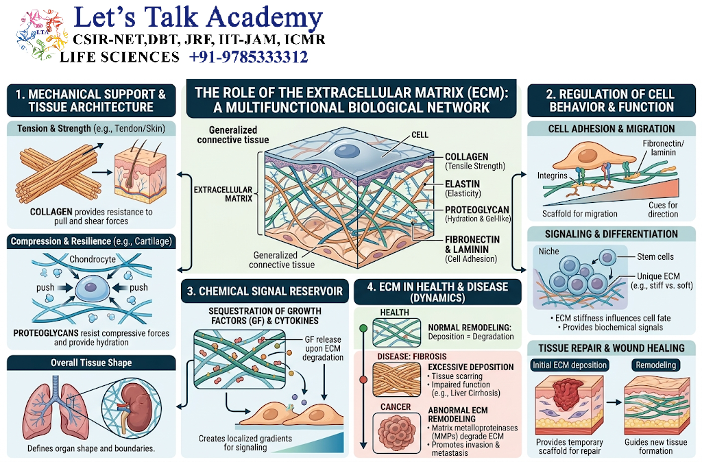
The extracellular matrix (ECM) is a network of proteins surrounding cells.
Major ECM components include:
- collagen,
- laminin,
- fibronectin,
- proteoglycans.
The basement membrane is an important physical barrier between epithelial cells and underlying tissues.
For invasion to occur, tumor cells must interact with and modify the ECM.
10. Matrix Metalloproteinases
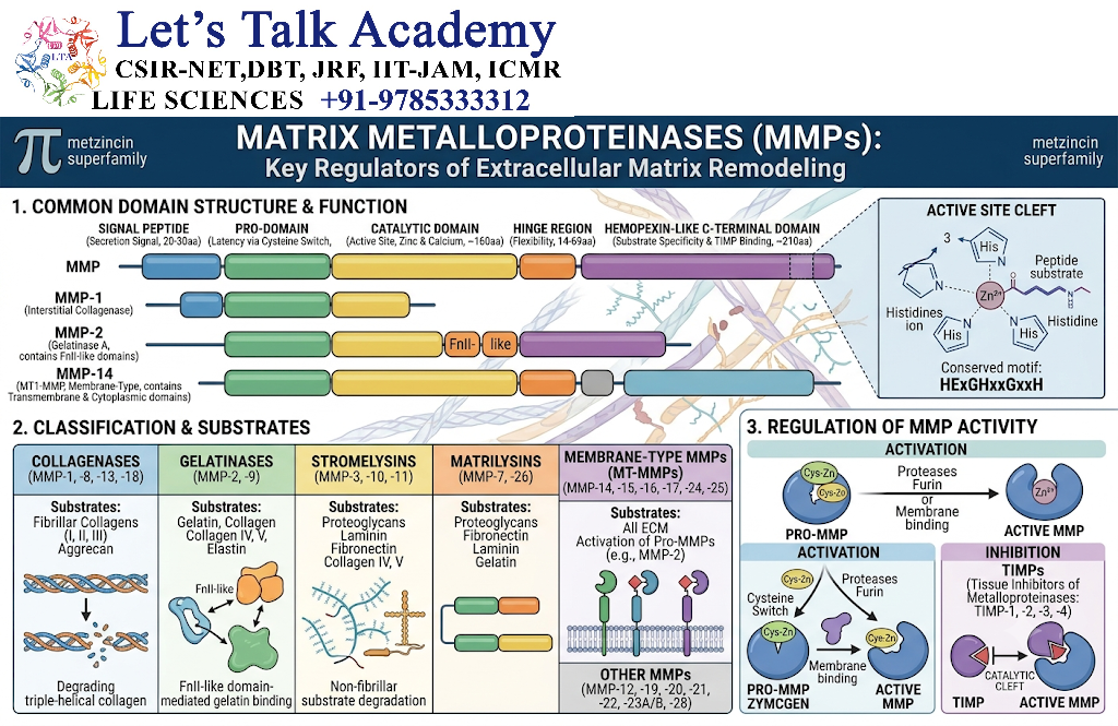
Cancer cells and associated stromal cells can produce enzymes called matrix metalloproteinases (MMPs).
Examples include:
- MMP-2
- MMP-9
- MMP-14
These enzymes can degrade components of the extracellular matrix and basement membrane.
Mechanism
Tumor cell
→ MMP production
→ ECM degradation
→ basement membrane disruption
→ tissue invasion.
MMP activity is regulated and can be influenced by tumor cells, fibroblasts, inflammatory cells, and signaling molecules.
11. Local Invasion
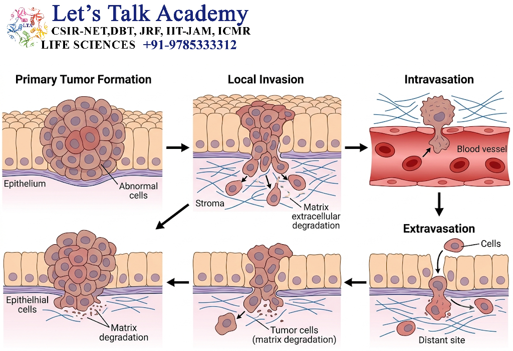
After acquiring increased motility and degrading surrounding barriers, cancer cells invade neighboring tissues.
Tumor cells can:
- migrate through extracellular matrix,
- move between stromal cells,
- interact with fibroblasts,
- modify the surrounding tissue,
- invade blood and lymphatic vessels.
Local invasion is an essential step before distant dissemination.
12. Intravasation
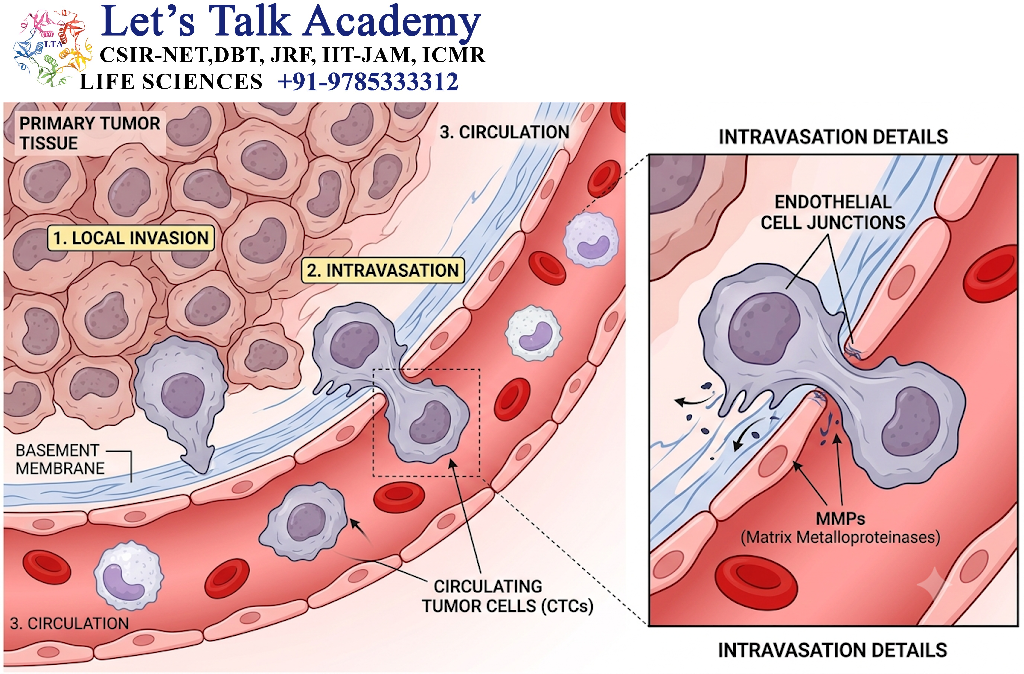
Intravasation is the entry of cancer cells into blood vessels or lymphatic vessels.
Tumor cells may enter vessels through regions where the vessel wall is altered or where interactions between tumor cells, endothelial cells, and stromal cells facilitate entry.
Mechanism
Primary tumor
→ local invasion
→ interaction with endothelial cells
→ penetration of vessel wall
→ entry into blood/lymph
→ circulating tumor cell.
13. Role of Tumor-Associated Macrophages
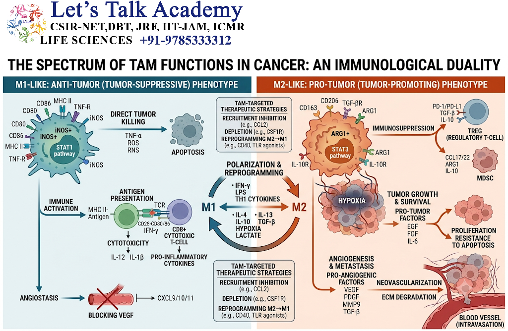
Tumor-associated macrophages can influence metastasis.
They may produce:
- growth factors,
- cytokines,
- proteases,
- angiogenic factors.
These signals can promote:
- tumor-cell migration,
- blood-vessel formation,
- ECM remodeling,
- invasion.
Thus, metastasis is not performed by cancer cells alone; the tumor microenvironment is also important.
14. Survival in the Circulation
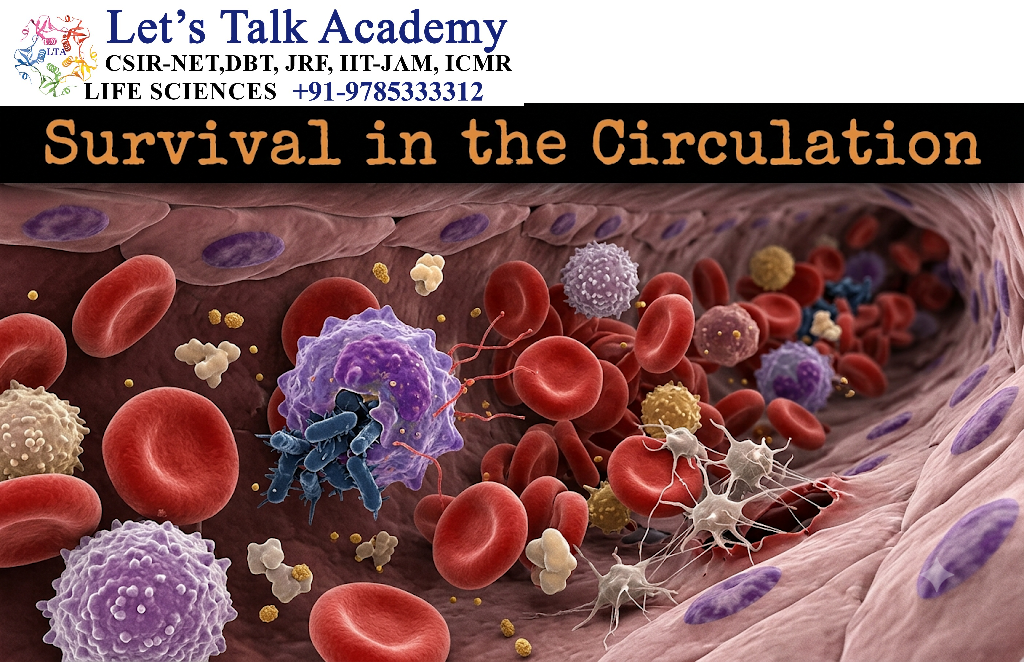
Cancer cells entering the bloodstream face several challenges.
These include:
- physical shear forces,
- lack of normal tissue support,
- immune attack,
- oxidative stress,
- anoikis.
Anoikis
Anoikis is a form of programmed cell death that normally occurs when cells lose appropriate attachment to the extracellular matrix.
Metastatic cancer cells may acquire mechanisms that allow them to resist anoikis.
This allows them to survive while detached from their original tissue.
15. Circulating Tumor Cells
Cancer cells present in blood are called circulating tumor cells (CTCs).
CTCs may exist as:
- individual cells,
- clusters of tumor cells,
- tumor cells associated with platelets or other blood components.
Tumor-cell clusters can have increased metastatic potential in some contexts.
16. Platelets and Metastasis
Platelets can interact with circulating tumor cells.
Platelet-tumor cell interactions may:
- physically shield tumor cells,
- modify immune recognition,
- facilitate adhesion to endothelial cells,
- release signaling molecules,
- promote vascular interactions.
Therefore, platelets can contribute to the survival and dissemination of tumor cells.
17. Immune Evasion During Circulation
The immune system can recognize and destroy abnormal cells.
However, metastatic tumor cells may avoid immune destruction through mechanisms such as:
- reduced antigen presentation,
- expression of inhibitory molecules,
- secretion of immunosuppressive factors,
- recruitment of regulatory immune cells,
- interaction with platelets,
- alteration of the tumor microenvironment.
This allows some circulating tumor cells to survive long enough to reach distant tissues.
18. Arrest in Distant Organs
Circulating tumor cells eventually become trapped or adhere to the small blood vessels of distant tissues.
This process depends on:
- blood flow,
- vascular anatomy,
- endothelial adhesion molecules,
- chemokines,
- tumor-cell surface receptors,
- properties of the target tissue.
The first organ reached through a particular vascular route is not always the final metastatic site.
19. Extravasation
Extravasation is the process by which circulating tumor cells leave the blood vessel and enter surrounding tissue.
Mechanism
Circulating tumor cell
→ adhesion to endothelium
→ interaction with endothelial cells
→ crossing endothelial barrier
→ movement through basement membrane
→ entry into tissue.
This resembles some aspects of leukocyte extravasation.
20. Colonization
Extravasation alone does not guarantee successful metastasis.
The cancer cell must survive and adapt to the new tissue.
This process is called colonization.
Colonization is often considered one of the most difficult steps of metastasis.
A tumor cell may reach an organ but remain dormant or die because the new environment does not support its growth.
21. Metastatic Dormancy
Some disseminated cancer cells can remain in a relatively inactive state for long periods.
This is called tumor dormancy or metastatic dormancy.
Dormant cells may later resume proliferation.
Factors involved include:
- cellular signaling,
- immune surveillance,
- angiogenic balance,
- extracellular matrix,
- tissue-specific growth factors.
This helps explain why some cancers can recur years after apparently successful treatment.
22. Seed and Soil Hypothesis
The seed and soil hypothesis, proposed by Stephen Paget, describes metastasis as an interaction between:
- the seed = metastatic cancer cell,
- the soil = target tissue environment.
According to this concept, successful metastasis depends not only on the ability of tumor cells to travel but also on whether the target organ provides a suitable environment.
Example
A tumor cell may reach many organs through circulation, but only some organs may provide the signals required for its survival and proliferation.
23. Organ Tropism
Some cancers show preferential patterns of metastatic spread.
This is called organ tropism.
Examples include:
| Primary cancer | Common metastatic sites |
|---|---|
| Breast cancer | Bone, liver, lung, brain |
| Prostate cancer | Bone, lymph nodes |
| Colorectal cancer | Liver, lung |
| Lung cancer | Brain, bone, liver, adrenal glands |
| Kidney cancer | Lung, bone, liver, brain |
| Melanoma | Skin/subcutaneous tissue, lymph nodes, lung, liver, brain |
These are general patterns; individual patients can show different metastatic distributions.
24. Pre-Metastatic Niche
Before metastatic cells arrive, primary tumors can influence distant organs.
Tumor-derived factors and extracellular vesicles can modify distant tissues.
This may create a pre-metastatic niche.
It can involve:
- recruitment of stromal cells,
- extracellular matrix remodeling,
- vascular changes,
- inflammatory signaling,
- immune-cell changes.
Simplified mechanism
Primary tumor
→ secretion of factors/extracellular vesicles
→ distant-organ changes
→ formation of favorable microenvironment
→ arrival of tumor cells
→ improved metastatic colonization.
25. Angiogenesis in Metastasis
A growing metastatic tumor requires nutrients and oxygen.
Therefore, successful colonization is often associated with angiogenesis, the formation of new blood vessels.
Important angiogenic factors include:
- VEGF,
- FGF,
- angiopoietins.
Mechanism
Tumor growth
→ hypoxia
→ HIF signaling
→ increased VEGF
→ endothelial-cell activation
→ new blood vessels
→ increased nutrient supply.
26. Hypoxia and HIF
Rapidly growing tumors may develop areas of low oxygen.
This condition is called hypoxia.
Hypoxia stabilizes hypoxia-inducible factors (HIFs).
HIF signaling can influence:
- angiogenesis,
- metabolism,
- cell survival,
- migration,
- invasion.
Therefore, hypoxia can contribute to metastatic progression.
27. Role of Cancer-Associated Fibroblasts
Cancer-associated fibroblasts (CAFs) are stromal cells found in many tumor microenvironments.
They can produce:
- extracellular matrix proteins,
- growth factors,
- cytokines,
- chemokines,
- matrix-remodeling enzymes.
CAFs can therefore influence:
- invasion,
- angiogenesis,
- tumor-cell migration,
- immune suppression,
- metastatic colonization.
28. Chemokines and Chemokine Receptors
Chemokines are signaling proteins that regulate cell migration.
Tumor cells can express chemokine receptors that respond to chemokines produced by distant organs.
An important example studied in metastasis is the CXCL12-CXCR4 axis.
Such interactions can contribute to:
- tumor-cell migration,
- tissue localization,
- survival,
- metastatic colonization.
29. Integrins and Cell Migration
Integrins are cell-surface adhesion receptors that connect cells with the extracellular matrix.
They participate in:
- adhesion,
- migration,
- survival signaling,
- invasion.
Altered integrin expression can change how tumor cells interact with different tissues.
30. Signaling Pathways Involved in Metastasis
Several signaling pathways contribute to metastatic behavior.
Important pathways include:
- RAS-MAPK
- PI3K-AKT-mTOR
- TGF-β
- WNT-β-catenin
- NF-κB
- JAK-STAT
- Hippo-YAP/TAZ
These pathways can regulate:
- proliferation,
- survival,
- migration,
- EMT,
- invasion,
- angiogenesis.
31. TGF-β and Metastasis
Transforming growth factor-beta (TGF-β) has complex effects.
Depending on cellular and tumor context, TGF-β can:
- regulate epithelial differentiation,
- influence EMT,
- modify the extracellular matrix,
- affect immune responses,
- promote invasion in advanced tumors.
Thus, its role can change during tumor progression.
32. WNT/β-Catenin Signaling
The WNT/β-catenin pathway regulates:
- cell proliferation,
- differentiation,
- stem-cell properties,
- tissue organization.
Abnormal WNT signaling can contribute to tumor progression and invasive behavior.
For example, alterations in APC, a tumor suppressor involved in WNT regulation, are important in colorectal cancer.
33. Tumor Microenvironment
The tumor microenvironment consists of cells and extracellular components surrounding a tumor.
It includes:
- cancer-associated fibroblasts,
- macrophages,
- endothelial cells,
- immune cells,
- extracellular matrix,
- blood vessels,
- signaling molecules.
The tumor microenvironment can either restrict or promote metastatic progression depending on its composition and signaling state.
34. Role of Immune Cells
Immune cells have complex roles in metastasis.
They can:
- destroy tumor cells,
- recognize abnormal antigens,
- produce inflammatory cytokines,
- modify the tumor microenvironment,
- sometimes promote tumor progression through chronic inflammation and immunosuppressive signaling.
Important cells include:
- T cells,
- NK cells,
- macrophages,
- neutrophils,
- dendritic cells,
- myeloid-derived suppressor cells.
35. Immune Escape at the Metastatic Site
Cancer cells may suppress immune responses by:
- expressing immune-checkpoint ligands,
- reducing antigen presentation,
- secreting immunosuppressive cytokines,
- recruiting regulatory immune populations,
- creating metabolically hostile environments.
For example, interaction between PD-1 on T cells and PD-L1 on tumor or other cells can inhibit T-cell activity.
This pathway is therapeutically important in cancer immunotherapy.
36. Metastatic Niches
A metastatic niche is a tissue environment that supports disseminated tumor cells.
It may provide:
- growth factors,
- nutrients,
- survival signals,
- extracellular matrix support,
- vascular support,
- immune protection.
Successful metastasis therefore depends on cooperation between tumor cells and the host tissue.
37. Metastasis and Cancer Stem-Like Properties
Some tumor cells may display stem-like properties, including:
- self-renewal,
- differentiation potential,
- resistance to stress,
- ability to initiate tumor growth.
Cancer stem-like properties have been studied in relation to:
- metastasis,
- recurrence,
- therapy resistance,
- dormancy.
The cancer stem-cell concept remains an active area of research and can vary between tumor types.
38. Genetic and Epigenetic Changes
Metastatic progression can involve genetic and epigenetic alterations.
Genetic changes
- mutations,
- chromosomal abnormalities,
- gene amplification,
- gene deletion.
Epigenetic changes
- DNA methylation,
- histone modification,
- chromatin remodeling,
- non-coding RNA regulation.
These changes can alter expression of genes controlling:
- adhesion,
- migration,
- invasion,
- survival,
- immune response.
39. Exosomes and Extracellular Vesicles
Tumor cells release extracellular vesicles, including exosome-like vesicles.
These vesicles can contain:
- proteins,
- lipids,
- DNA,
- RNA,
- microRNAs.
They can influence distant cells and contribute to changes in the metastatic microenvironment.
40. Lymphatic Spread
Cancer cells can enter lymphatic vessels and travel to regional lymph nodes.
The sequence may be:
Primary tumor
→ lymphatic invasion
→ lymphatic vessel
→ regional lymph node
→ additional lymphatic spread
→ bloodstream or distant organs.
Lymph-node involvement is important in staging many cancers.
41. Hematogenous Spread
Hematogenous spread occurs through blood vessels.
Tumor cells may enter:
- veins,
- venules,
- capillaries.
Venous circulation can transport tumor cells to distant organs.
The anatomical pattern of blood flow influences where some tumor cells initially lodge.
42. Transcoelomic Spread
Some cancers spread across body cavities.
This is called transcoelomic dissemination.
For example, cancers arising in abdominal or pelvic organs may disseminate across the peritoneal surfaces.
This can result in multiple tumor deposits within a body cavity.
43. Common Routes of Metastasis
| Route | Description |
|---|---|
| Lymphatic | Spread through lymphatic vessels |
| Hematogenous | Spread through blood |
| Transcoelomic | Spread across body cavities |
| Direct extension | Local invasion into neighboring structures |
44. Lymphatic vs Hematogenous Metastasis
| Feature | Lymphatic spread | Hematogenous spread |
|---|---|---|
| Main route | Lymphatic vessels | Blood vessels |
| Common early site | Regional lymph nodes | Distant organs |
| Importance | Regional staging | Distant dissemination |
| Example | Carcinomas frequently involve lymph nodes | Sarcomas often show prominent hematogenous spread |
This is a general pattern rather than an absolute rule.
45. Metastasis and Angiogenesis
Metastatic tumors need vascular support when they grow beyond a small size.
The angiogenic switch can involve:
Hypoxia
→ HIF activation
→ VEGF production
→ endothelial-cell proliferation
→ new blood vessels
→ metastatic tumor growth.
46. Metastasis and Inflammation
Chronic inflammation can promote several stages of metastasis.
Inflammatory mediators can influence:
- cell proliferation,
- angiogenesis,
- ECM remodeling,
- immune suppression,
- migration.
Important mediators include:
- TNF,
- IL-1,
- IL-6,
- chemokines,
- prostaglandins.
Thus, inflammation can become part of the tumor-promoting microenvironment.
47. Metastasis and Matrix Remodeling
The ECM is continuously remodeled during tumor progression.
Important components include:
- collagen,
- fibronectin,
- laminin,
- proteoglycans,
- MMPs,
- integrins.
Altered ECM can provide pathways that facilitate tumor-cell movement.
48. Metastatic Colonization as a Major Barrier
Many tumor cells may enter the circulation, but only a small fraction successfully form clinically detectable metastases.
This occurs because metastatic cells must overcome multiple barriers:
- survive detachment,
- survive circulation,
- evade immune attack,
- exit the vessel,
- survive in a foreign tissue,
- adapt metabolically,
- establish supportive interactions,
- induce or access blood supply,
- proliferate successfully.
Therefore, metastasis is highly inefficient.
49. Metastasis and Metabolic Adaptation
Metastatic cells may need to adapt to the metabolism of the new tissue.
They can alter:
- glucose metabolism,
- lipid metabolism,
- amino-acid utilization,
- mitochondrial function,
- redox balance.
The ability to adapt metabolically can influence survival in different organs.
50. Metastasis and Therapy Resistance
Metastatic tumors can show resistance to treatment because of:
- genetic heterogeneity,
- altered signaling,
- drug efflux,
- changes in cell state,
- tumor dormancy,
- microenvironmental protection,
- immune evasion.
Therefore, treatment of metastatic disease can be biologically complex.


