
Principles of Isozymes
Isozymes, also called isoenzymes, are different molecular forms of an enzyme that catalyze the same overall biochemical reaction but differ in their molecular structure, physicochemical properties, kinetic behavior, tissue distribution, regulatory characteristics, or cellular localization. The existence of isozymes allows organisms to perform the same basic chemical transformation in different tissues while adapting the reaction to the specific physiological requirements of each tissue.
The concept of isozymes is particularly important because metabolism is not identical in every cell of a multicellular organism. A cardiac muscle cell has a continuous and very high demand for ATP, whereas a skeletal muscle cell may experience sudden increases in energy demand. A liver cell is extensively involved in nutrient processing, biosynthesis, detoxification, and maintenance of blood metabolite concentrations. A brain cell requires a highly continuous supply of energy and has specialized metabolic requirements. Although these tissues may use the same biochemical reactions, the conditions under which those reactions occur are different.
Isozymes provide a molecular mechanism for this biochemical specialization. Different forms of the same enzyme can possess different values of Kₘ, k₍cat₎, catalytic efficiency, regulatory sensitivity, thermal stability, pH dependence, and substrate-response characteristics. Consequently, a particular tissue can express the enzyme form that is best suited to its metabolic environment.
The catalytic reaction itself remains fundamentally the same. For example, two isozymes may catalyze: S ⇌ P
However, their responses to substrate concentration, inhibitors, activators, cellular conditions, and regulatory signals may differ substantially.
Isozymes therefore provide an important connection between molecular structure and physiological function. They demonstrate how small differences in protein sequence or subunit composition can generate significant differences in cellular metabolism.
1. Introduction to Isozymes
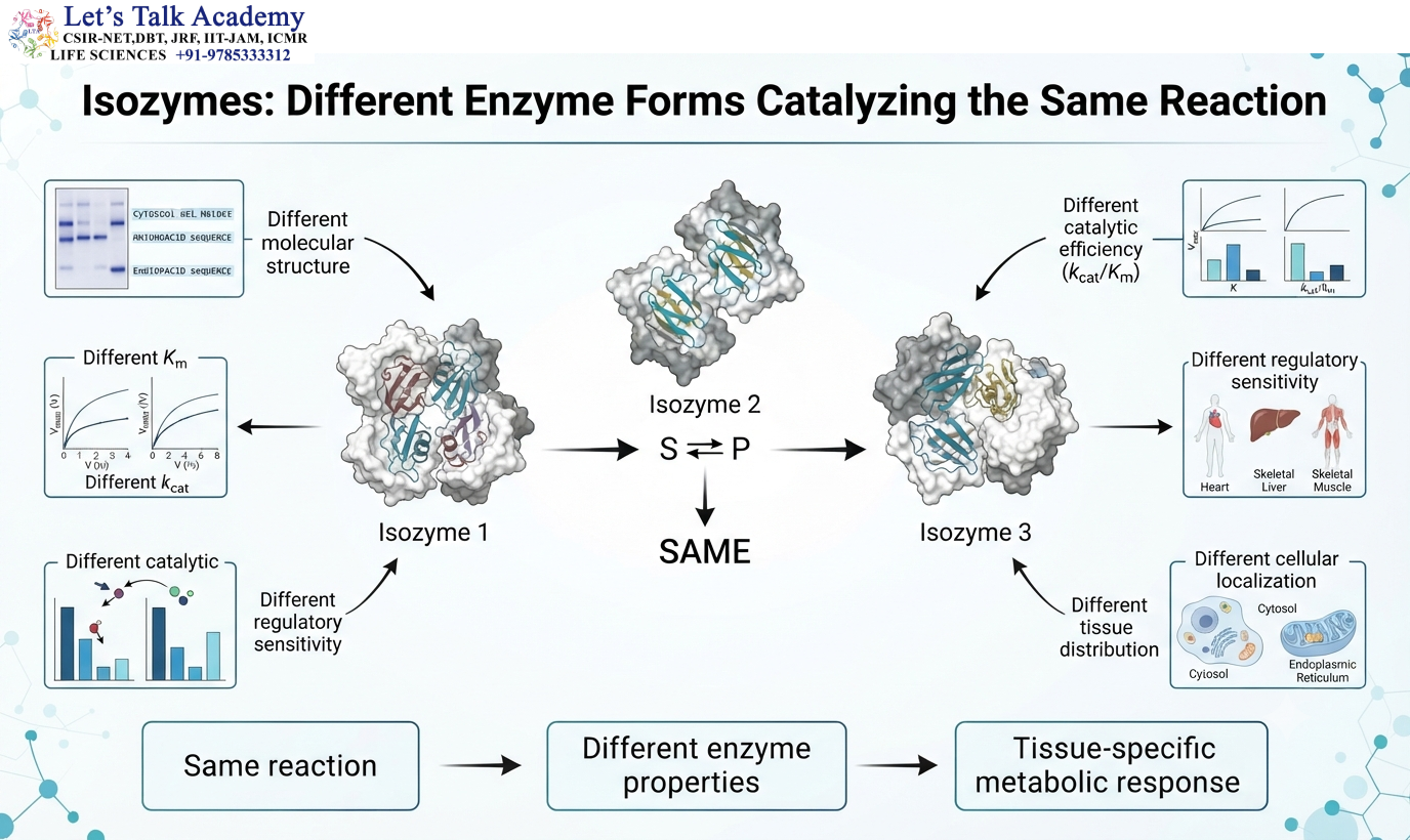
The term isozyme refers to different molecular forms of an enzyme that catalyze the same overall reaction.
The basic concept can be represented as:
Isozyme 1: S ⇌ P
Isozyme 2: S ⇌ P
Isozyme 3: S ⇌ P
All of these enzyme forms catalyze the same overall chemical transformation, but their molecular properties may differ.
For example, one isozyme may interact more effectively with a substrate when its concentration is low, whereas another may become highly active only when substrate concentration increases. One form may be strongly inhibited by a metabolic product, while another may be relatively insensitive to that product. One isozyme may be particularly stable under the conditions found in one tissue, whereas another may be better adapted to a different cellular environment.
These differences are not accidental. They arise from differences in protein sequence, subunit composition, molecular structure, cellular localization, or regulatory interactions.
The concept of isozymes therefore extends beyond simply identifying different forms of the same enzyme. It explains how organisms generate metabolic specialization while retaining a common biochemical reaction.
2. Definition and Basic Characteristics of Isozymes
Isozymes are molecularly distinct forms of an enzyme that catalyze the same reaction.
A simple catalytic representation is:
E₁ + S ⇌ E₁S → E₁ + P
E₂ + S ⇌ E₂S → E₂ + P
Both E₁ and E₂ convert S into P, but they may not behave identically.
For example:
Kₘ₁ ≠ Kₘ₂ and k₍cat₎₁ ≠ k₍cat₎₂
Their catalytic efficiencies may therefore also differ:
(k₍cat₎ ÷ Kₘ)₁ ≠ (k₍cat₎ ÷ Kₘ)₂
The two proteins may also respond differently to inhibitors, activators, pH, temperature, or changes in substrate concentration.
The conservation of catalytic function together with differences in other properties is the defining biochemical feature of isozymes.
Isozymes can be produced through different genetic and molecular mechanisms. Classical isozyme systems commonly involve different genes encoding related enzyme forms or different combinations of subunits encoded by distinct genes.
3. Biological Need for Isozymes
A multicellular organism contains many tissues that perform different physiological functions. Even when two tissues require the same biochemical reaction, their metabolic requirements may be very different.
Consider a reaction that occurs in both cardiac muscle and skeletal muscle. The chemical conversion may be identical, but the metabolic conditions surrounding the reaction can differ because the tissues have different energy demands, substrate availability, oxygen supply, and regulatory requirements.
If both tissues expressed exactly the same enzyme form, they would have fewer possibilities for independently adjusting the reaction.
Isozymes solve this problem.
A tissue can express one molecular form of an enzyme, while another tissue expresses a different form. Both enzymes perform the same basic reaction, but their kinetic and regulatory properties allow the reaction to be adapted to the local cellular environment.
This means that isozyme expression contributes to metabolic flexibility.
The general relationship can be expressed as:
Tissue requirement → Isozyme expression → Catalytic properties → Metabolic response
Thus, the same biochemical pathway can operate differently in different tissues without requiring a completely different chemical reaction.
4. Molecular Basis of Isozyme Formation
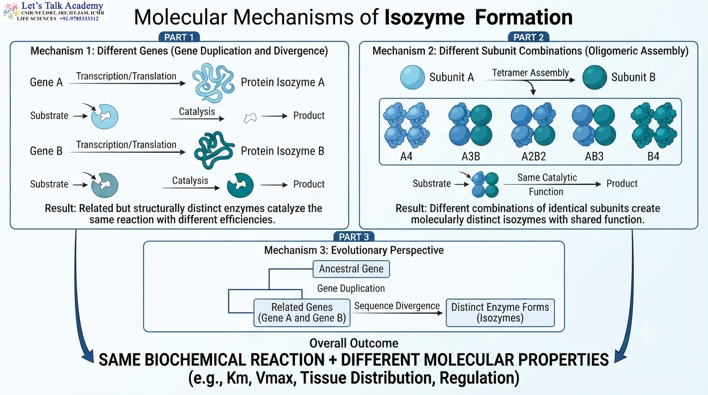
Isozyme diversity can arise through several molecular mechanisms.
One important mechanism is the presence of different genes that encode related enzymes capable of catalyzing the same reaction.
Another important mechanism involves the association of different types of protein subunits.
Suppose an enzyme contains two types of subunits, A and B. If the functional enzyme is a tetramer, different subunit combinations can produce:
A₄
A₃B
A₂B₂
AB₃
B₄
These molecular forms can have the same basic catalytic function while differing in kinetic or regulatory properties.
The exact number of possible forms depends on the number of subunit types, the structure of the enzyme, and whether all possible combinations are biologically stable and functional.
This mechanism is particularly important in classical isozyme systems such as lactate dehydrogenase.
Subunit composition therefore provides an efficient mechanism for producing molecular diversity from a relatively small number of protein components.
5. Isozymes and Multimeric Proteins
Many classical isozymes are associated with multimeric proteins.
A protein composed of identical subunits is called a homomeric or homooligomeric protein.
A protein composed of different types of subunits is called a heteromeric or heterooligomeric protein.
The subunits can influence one another through interactions at their interfaces.
As a result, changing the composition of the multimer can alter the overall conformation, stability, catalytic behavior, and regulatory properties of the enzyme.
For example, consider two subunit types: A and B
A tetrameric enzyme may exist as: A₄ or A₂B₂ or B₄
Even though these proteins may catalyze the same overall reaction, their molecular environments are not identical.
Differences in subunit composition can influence substrate binding, communication between subunits, conformational flexibility, inhibitor sensitivity, and electrophoretic mobility.
Therefore, quaternary structure can become an important source of isozyme diversity.
6. Structural Differences Between Isozymes
Isozymes are not necessarily identical proteins.
They can differ in their amino acid sequence, subunit composition, three-dimensional structure, surface charge, flexibility, stability, and regulatory regions.
The differences may be relatively small but still biologically meaningful.
For example, an amino acid substitution near the active site can change the geometry of the substrate-binding pocket. A substitution farther from the active site can influence protein flexibility or alter the communication between regulatory and catalytic regions.
Changes in surface residues can alter the net charge of the protein and therefore its migration during electrophoresis.
Changes at subunit interfaces can alter the stability of a multimeric enzyme.
Thus, a small molecular difference can produce a measurable difference in enzyme behavior.
7. Isozymes and Primary Structure
The primary structure of a protein is its amino acid sequence.
Different isozymes may have related but nonidentical amino acid sequences.
A simplified representation is:
Isozyme A → Protein sequence A
Isozyme B → Protein sequence B
Although the sequences differ, important catalytic residues may remain conserved.
The conservation of catalytic residues allows both proteins to retain the same chemical function.
Other regions of the protein can accumulate sequence changes without completely destroying catalytic activity.
These variable regions can influence substrate recognition, protein stability, subunit interactions, regulatory properties, and tissue-specific expression.
This creates an important evolutionary balance.
The catalytic core remains sufficiently conserved to preserve the biochemical reaction, while other regions can diverge and generate functional specialization.
8. Active-Site Similarity and Isozyme Function
Because isozymes catalyze the same reaction, the essential catalytic features of their active sites are generally conserved.
However, the surrounding residues may differ.
These surrounding differences can modify the shape, charge, flexibility, and chemical environment of the active site.
Consequently, two isozymes may use the same fundamental catalytic mechanism while displaying different kinetic properties.
For example:
Kₘ₁ ≠ Kₘ₂
The difference in Kₘ may reflect differences in substrate interaction and catalytic steps.
Similarly:
k₍cat₎₁ ≠ k₍cat₎₂
can indicate differences in the rate of chemical conversion or another rate-limiting step.
Therefore, identical reaction chemistry does not require identical kinetic behavior.
9. Kinetic Properties of Isozymes
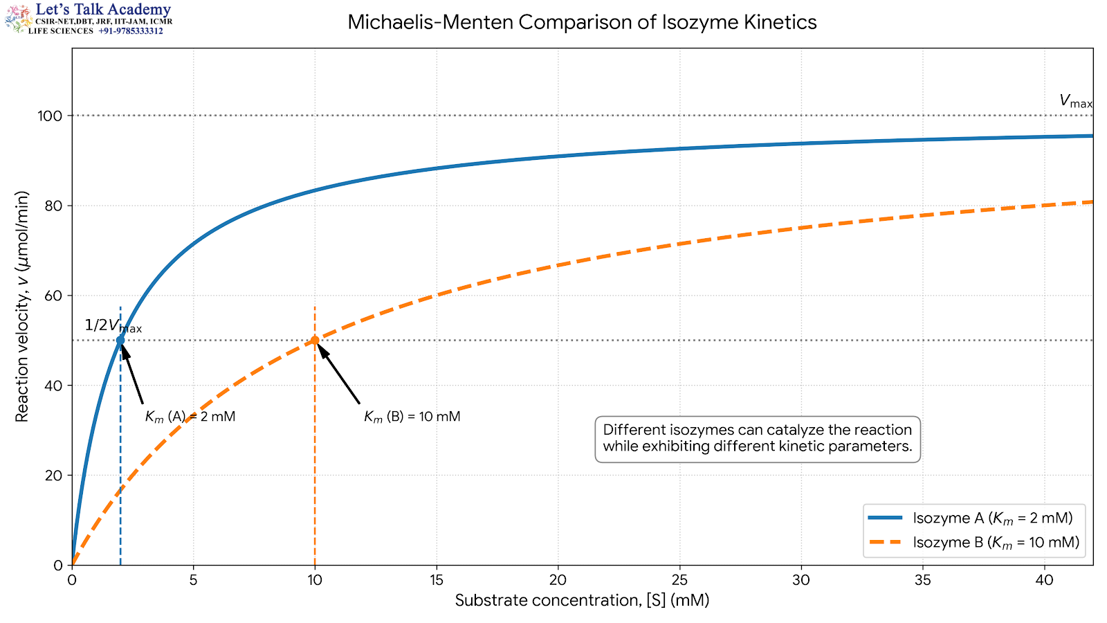
Kinetic differences are among the most useful characteristics for distinguishing isozymes.
For a simple enzyme-catalyzed reaction:
E + S ⇌ ES → E + P
the Michaelis-Menten relationship is:
v = Vₘₐₓ[S] ÷ (Kₘ + [S])
Different isozymes may possess different Kₘ and k₍cat₎ values.
Suppose:
Isozyme A: Kₘ = 2 mM
Isozyme B: Kₘ = 10 mM
At relatively low substrate concentration, Isozyme A may show a greater response to the substrate because its Kₘ is lower.
However, Kₘ must be interpreted carefully. It is not universally identical to a substrate-binding equilibrium constant. For the simple Michaelis-Menten mechanism:
Kₘ = (k₋₁ + k₍cat₎) ÷ k₁
Therefore, Kₘ reflects both the dissociation of the enzyme-substrate complex and the catalytic conversion step.
This distinction is important when comparing the biochemical properties of different isozymes.
10. Catalytic Turnover of Isozymes
Isozymes can also differ in their catalytic turnover.
The turnover number is:
k₍cat₎ = Vₘₐₓ ÷ [E]ₜ
where [E]ₜ represents total active enzyme concentration.
If:
k₍cat₎₁ > k₍cat₎₂
Isozyme 1 can potentially process substrate more rapidly than Isozyme 2 when the enzymes are operating under saturating substrate conditions.
This difference can be physiologically significant.
A tissue that requires rapid metabolic flux may benefit from an isozyme with a high catalytic turnover.
Another tissue may require stronger regulation rather than maximum catalytic speed and may therefore express a form with different kinetic characteristics.
11. Catalytic Efficiency of Isozymes
Catalytic efficiency is commonly represented as: k₍cat₎ ÷ Kₘ
This parameter combines information about catalytic turnover and substrate behavior.
Suppose two isozymes have the following relationship:
(k₍cat₎ ÷ Kₘ)₁ > (k₍cat₎ ÷ Kₘ)₂
Isozyme 1 is more efficient under conditions where the catalytic-efficiency approximation is appropriate, particularly when substrate concentration is low relative to Kₘ.
Catalytic efficiency therefore provides a useful way of comparing related enzyme forms.
However, enzyme performance in a cell cannot be predicted from k₍cat₎ and Kₘ alone. Protein concentration, substrate availability, inhibitors, activators, cofactors, compartmentation, and metabolic feedback also contribute to actual pathway flux.
12. Tissue-Specific Distribution of Isozymes
A major characteristic of isozymes is their tissue-specific distribution.
Different tissues can express different molecular forms of the same enzyme.
This distribution reflects the metabolic requirements of each tissue.
Cardiac muscle has a continuous requirement for ATP because contraction occurs continuously.
Skeletal muscle can experience sudden and intense increases in ATP demand.
The liver performs extensive metabolic processing and maintains systemic nutrient homeostasis.
The brain requires a continuous supply of energy and has specialized metabolic constraints.
Because these tissues experience different metabolic conditions, the same enzyme reaction may need to be regulated differently.
Isozyme expression provides a molecular mechanism for achieving this specialization.
13. Lactate Dehydrogenase as a Classical Example
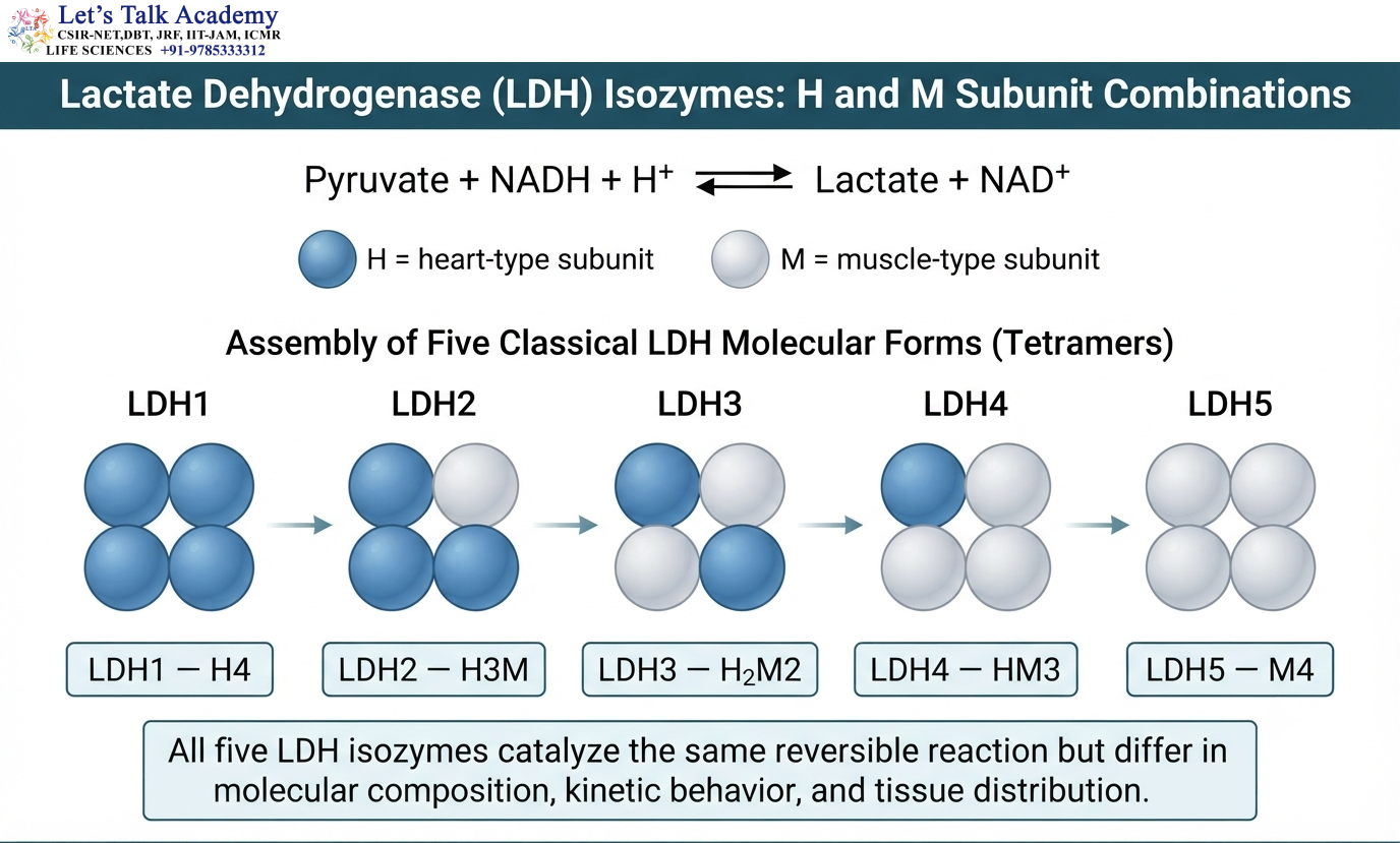
Lactate dehydrogenase, commonly abbreviated as LDH, is one of the best-known examples of an isozyme system.
LDH catalyzes the reversible conversion between pyruvate and lactate:
Pyruvate + NADH + H⁺ ⇌ Lactate + NAD⁺
The enzyme is a tetramer.
Two major types of subunits are traditionally designated:
H = heart-type subunit
M = muscle-type subunit
Different combinations of these subunits generate five classical molecular forms:
H₄
H₃M
H₂M₂
HM₃
M₄
These forms are traditionally designated:
LDH₁ = H₄
LDH₂ = H₃M
LDH₃ = H₂M₂
LDH₄ = HM₃
LDH₅ = M₄
All five forms catalyze the same overall reaction, but their kinetic properties and tissue distributions differ.
14. Tissue Distribution of LDH Isozymes
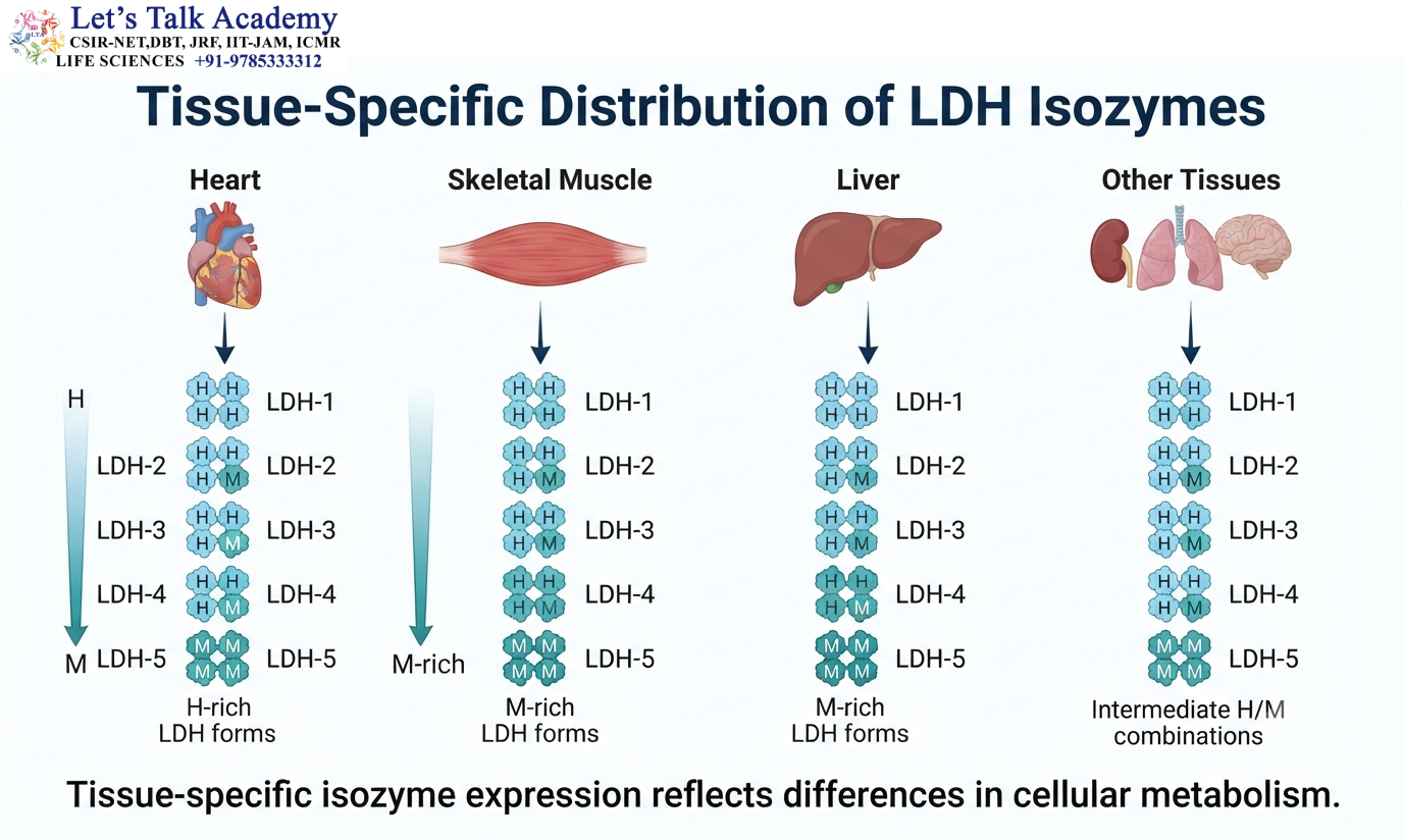
LDH isozymes occur in different proportions in different tissues.
H-rich forms are particularly abundant in tissues with strong oxidative metabolism, especially cardiac muscle.
M-rich forms are prominent in tissues such as skeletal muscle and liver.
Intermediate combinations occur in varying proportions in other tissues.
The distribution reflects differences in cellular metabolism.
A tissue with high oxidative activity may require an enzyme form suited to its pattern of lactate and pyruvate utilization.
A tissue with substantial glycolytic activity may benefit from a different molecular form.
Therefore, LDH isozyme distribution provides an example of how enzyme structure can be adapted to tissue physiology.
15. LDH Isozymes and Metabolic Direction
LDH catalyzes a reversible reaction:
Pyruvate + NADH + H⁺ ⇌ Lactate + NAD⁺
The direction of this reaction depends on the concentrations of pyruvate, lactate, NADH, and NAD⁺ as well as the thermodynamic state of the cell.
An enzyme does not independently force a reaction in one direction.
The cellular environment determines the overall direction, while the isozyme influences the kinetic response of the enzyme system.
M-rich LDH forms are commonly associated with tissues capable of substantial glycolytic metabolism and lactate production.
H-rich forms are associated with tissues in which lactate oxidation and oxidative metabolism are prominent.
The physiological significance therefore emerges from the interaction between isozyme properties and the metabolic environment.
16. Isozymes and Metabolic Adaptation
Isozymes allow biochemical reactions to adapt to changing metabolic conditions.
Consider two tissues exposed to different substrate concentrations.
One tissue may require an enzyme that remains highly active at low substrate concentration.
Another may require an enzyme that becomes strongly active only when substrate concentration rises.
Different isozymes can provide these different responses.
The same principle applies to changes in energy demand, hormonal signals, metabolic products, pH, cofactors, and regulatory molecules.
Isozyme expression therefore provides a molecular mechanism for metabolic adaptation.
17. Isozymes and Allosteric Regulation
Some enzyme systems contain regulatory sites separate from the catalytic site.
An allosteric regulator binds to such a site and alters enzyme conformation.
The interaction can be represented as:
E + R ⇌ E·R
where R represents a regulatory molecule.
Binding of R may increase or decrease enzyme activity.
Different isozymes can have different sensitivities to the same regulator because differences in protein structure can alter the regulatory site or communication between the regulatory and catalytic regions.
This means that the same metabolite can produce different effects in different tissues depending on which isozyme is expressed.
18. Isozymes and Feedback Regulation
Metabolic pathways frequently contain feedback regulation.
Consider a simplified pathway:
A → B → C → D
If D accumulates, it may inhibit an enzyme involved in an earlier step.
This prevents unnecessary production of additional D.
Different isozymes of the regulated enzyme may respond differently to D.
One tissue may express a strongly inhibited form, whereas another may express a less sensitive form.
This allows the same pathway to respond differently to metabolic demand in different tissues.
Isozyme diversity therefore increases the regulatory flexibility of metabolic pathways.
19. Isozymes and pH Dependence
Different isozymes may display different pH-activity profiles.
The reason is that amino acid substitutions can alter the local chemical environment around catalytic residues.
The protonation state of a catalytic residue can be represented as:
HA ⇌ H⁺ + A⁻
If an enzyme requires a residue to be protonated for catalysis, changes in pH can affect its activity.
A neighboring amino acid can alter the pKₐ of that residue by changing its electrostatic environment.
Consequently, two isozymes can contain corresponding catalytic residues but display different responses to pH.
This difference may contribute to tissue-specific adaptation.
20. Isozymes and Thermal Stability
Isozymes can differ in their resistance to thermal denaturation.
Protein stability depends on many interactions, including hydrophobic packing, hydrogen bonding, ionic interactions, van der Waals forces, disulfide bonds where present, and subunit interactions.
Amino acid substitutions can alter these interactions.
As a result, one isozyme may retain its folded structure under conditions that destabilize another.
Differences in thermal stability can be important when comparing enzyme forms from different tissues, developmental stages, or organisms.
21. Isozymes and Electrophoresis
Isozymes can often be distinguished experimentally using electrophoresis.
Electrophoresis separates proteins according to their movement through a medium under an electric field.
Protein migration is influenced by properties such as charge and, depending on the electrophoretic method, molecular size and shape.
Different isozymes may have different net charges because of differences in amino acid composition.
Consequently, they may migrate to different positions during electrophoresis.
After separation, enzyme-specific staining can reveal the positions of catalytically active molecular forms.
This produces an isozyme pattern characteristic of the sample.
22. Isozyme Electrophoretic Patterns
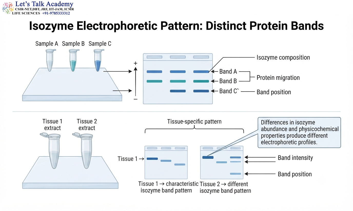
An electrophoretic sample containing several isozymes can produce several distinct bands.
A simplified representation is:
Isozyme A → Band A
Isozyme B → Band B
Isozyme C → Band C
The presence, absence, and relative intensity of these bands can provide information about enzyme composition.
Different tissues can produce different patterns because the relative abundance of the isozymes varies.
For example, a tissue rich in H-type LDH subunits produces a different LDH electrophoretic profile from a tissue rich in M-type subunits.
Isozyme electrophoresis therefore provides a direct biochemical method for studying tissue-specific enzyme composition.
23. LDH and Electrophoretic Separation
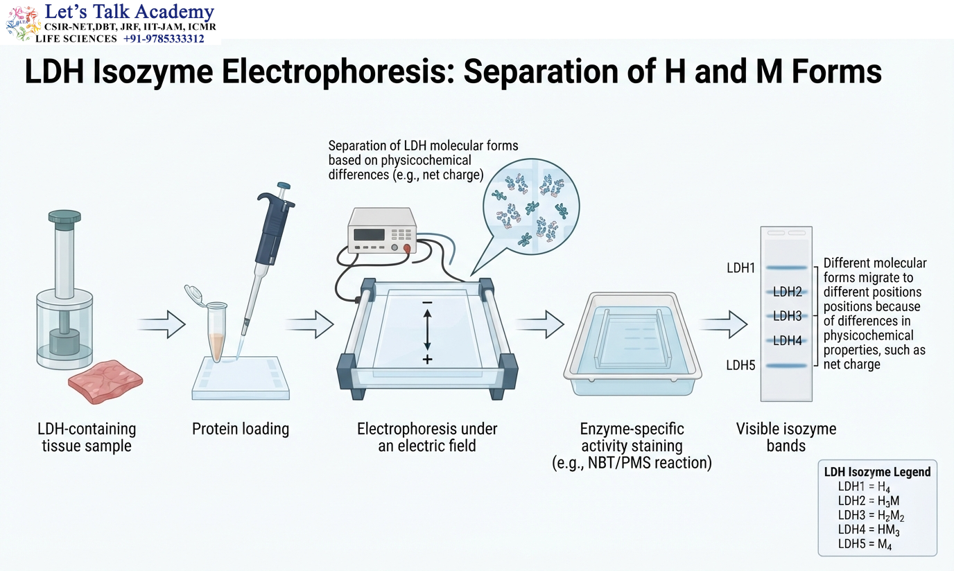
LDH provides a classical example of electrophoretic isozyme analysis.
The five LDH forms differ in their subunit composition.
Because these molecular forms differ in charge and other physicochemical properties, they migrate differently during electrophoresis.
The resulting pattern allows the different forms to be distinguished.
Historically, LDH isozyme electrophoresis became an important biochemical method for investigating tissue-specific enzyme distribution and certain patterns of tissue injury.
24. Isozymes in Clinical Biochemistry
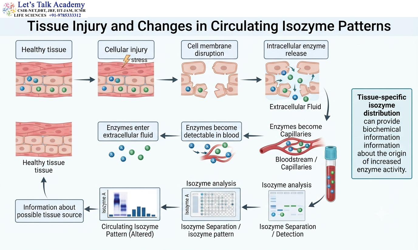
The tissue-specific distribution of isozymes can provide information about the origin of enzymes released into the bloodstream.
Normally, many enzymes are located inside cells.
When cellular membranes are damaged, intracellular enzymes can enter extracellular fluids and eventually become detectable in blood.
If different tissues contain different proportions of an enzyme’s molecular forms, analysis of the isozyme pattern can sometimes provide information about the tissue contributing to the increased enzyme activity.
The general principle is:
Tissue-specific isozyme distribution → Cellular injury → Enzyme release → Altered circulating isozyme pattern
LDH is a classical example.
Although modern clinical diagnostics often use more specific biomarkers and advanced analytical methods, the biochemical basis of tissue-specific isozyme distribution remains important.
25. Creatine Kinase Isozymes
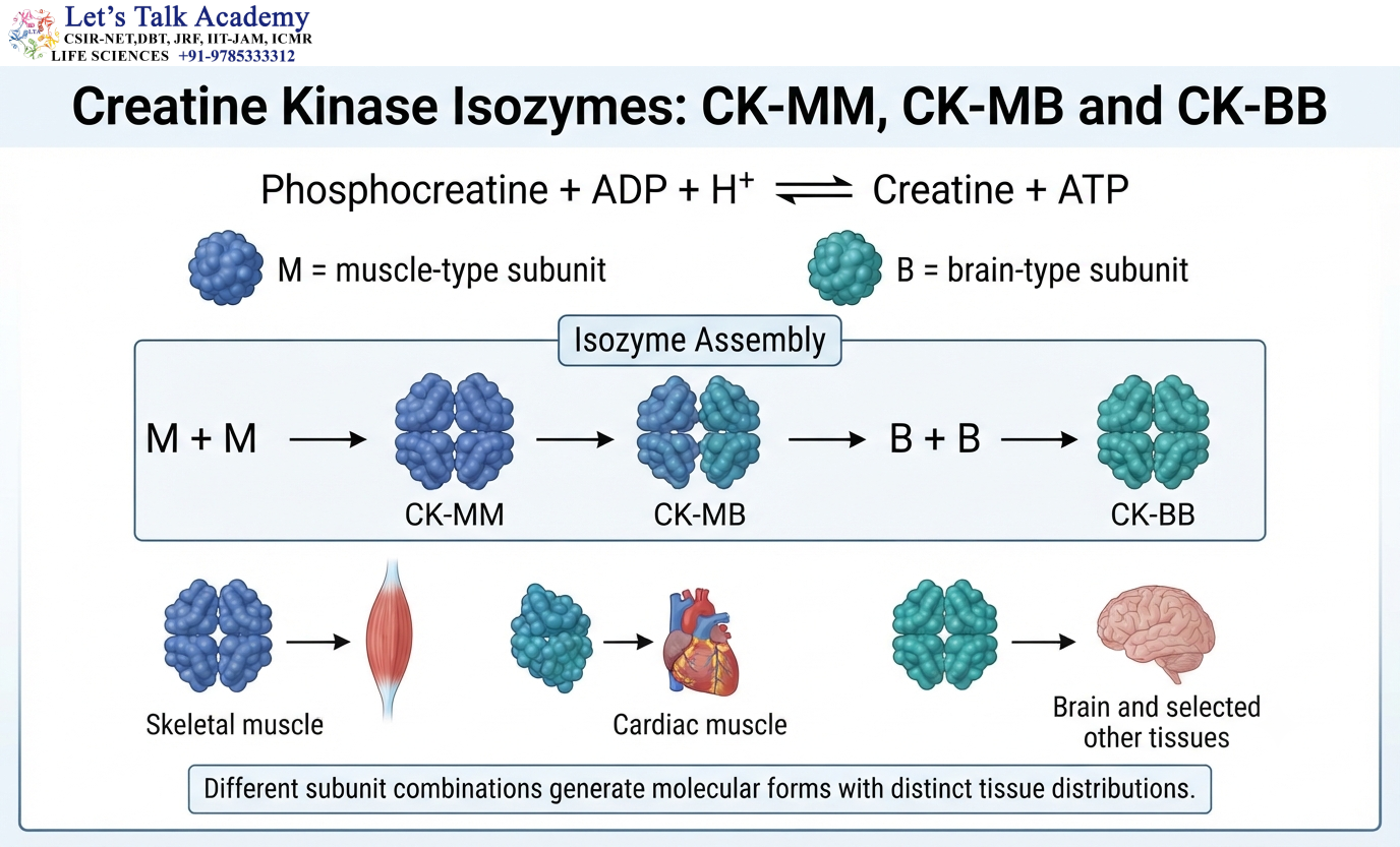
Creatine kinase, abbreviated CK or CPK, is another classical isozyme system.
Creatine kinase participates in rapid energy buffering by catalyzing the reversible transfer of a phosphate group between phosphocreatine and ADP.
The reaction can be represented as:
Phosphocreatine + ADP + H⁺ ⇌ Creatine + ATP
This reaction is particularly important in tissues where ATP demand can change rapidly.
Creatine kinase contains different types of subunits.
The major subunits are designated:
M = muscle type
B = brain type
Different combinations generate:
CK-MM
CK-MB
CK-BB
These forms have different tissue distributions.
26. Tissue Distribution of Creatine Kinase Isozymes
CK-MM is particularly abundant in skeletal muscle.
CK-MB is strongly associated with cardiac muscle, although it is not completely exclusive to the heart.
CK-BB occurs prominently in tissues such as brain and some smooth-muscle-containing tissues.
The distribution reflects the role of creatine kinase in rapid ATP buffering.
The phosphocreatine system acts as an energy reservoir that can rapidly regenerate ATP when ATP consumption increases.
Different molecular forms allow this energy-transfer system to be adapted to different tissues.
27. Alkaline Phosphatase Isozymes
Alkaline phosphatase is another enzyme system containing different molecular forms associated with different tissues.
The enzyme catalyzes the hydrolysis of phosphate monoesters under alkaline conditions.
A simplified reaction is:
R−O−PO₃²⁻ + H₂O → R−OH + Pᵢ
Different alkaline phosphatase forms are associated with tissues such as liver, bone, intestine, and placenta.
The molecular forms can differ in their physicochemical properties and tissue distribution.
Changes in alkaline phosphatase activity can occur under several physiological and pathological conditions.
Analysis of molecular forms can therefore provide information about the tissue source of enzyme activity.
28. Hexokinase Isozymes
Hexokinases provide another important example of enzyme specialization.
Hexokinase catalyzes the phosphorylation of glucose:
Glucose + ATP → Glucose-6-phosphate + ADP
Several related hexokinase forms occur in mammals.
Hexokinase I is widely distributed and has a relatively high affinity for glucose.
Hexokinase II is particularly important in skeletal muscle and adipose tissue and is responsive to metabolic and hormonal conditions.
Hexokinase IV, commonly called glucokinase, is especially important in liver and pancreatic β-cells.
These enzymes catalyze the same general type of reaction but differ in kinetic properties, regulation, and tissue distribution.
This demonstrates how related enzyme forms can be adapted to different physiological roles.
29. Hexokinase and Glucokinase
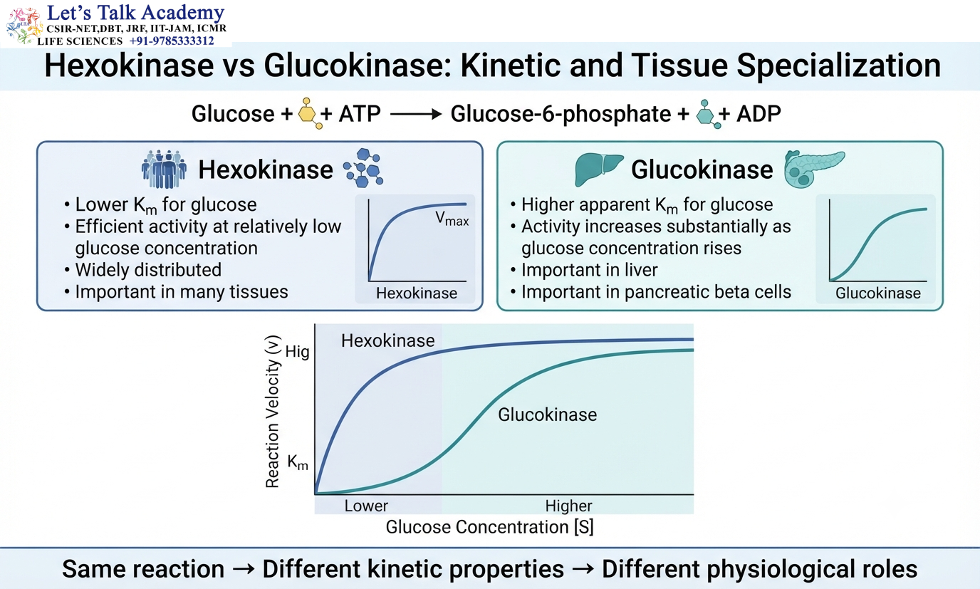
Hexokinase and glucokinase catalyze the same overall reaction:
Glucose + ATP → Glucose-6-phosphate + ADP
However, their kinetic properties are different.
Glucokinase has a higher apparent Kₘ for glucose than many other hexokinases.
As glucose concentration rises, glucokinase activity increases substantially.
This characteristic is particularly suitable for the liver, which plays a major role in handling glucose after carbohydrate intake.
Other hexokinases have lower Kₘ values and can function efficiently even when glucose concentration is relatively low.
The difference demonstrates how enzyme kinetics can be matched to tissue physiology.
30. Isozymes and Cellular Compartmentation
Isozyme specialization can also be associated with cellular localization.
Different forms of an enzyme may be preferentially localized in the cytosol, mitochondria, peroxisomes, or other cellular compartments.
Localization places the enzyme near its substrates, products, cofactors, and regulatory molecules.
For example, an enzyme operating in a mitochondrial metabolic pathway may require a different regulatory environment from a related enzyme functioning in the cytosol.
Thus, isozyme expression and cellular compartmentation can work together to organize metabolism spatially.
31. Isozymes and Development
Isozyme expression can change during development.
An enzyme form that is highly expressed during embryonic development may decrease after birth, while another form becomes predominant in the mature tissue.
This change can reflect the changing metabolic requirements of the developing organism.
During development, cells undergo differentiation, and their energy sources, signaling pathways, and metabolic activities change.
Isozyme switching can therefore accompany the transition from one physiological state to another.
32. Isozymes and Gene Expression
The tissue distribution of isozymes is closely related to gene expression.
Different tissues express different combinations of genes.
If two genes encode related enzyme forms, tissue-specific expression can determine which form is produced.
The relationship can be represented as:
Gene A expression → Isozyme A
Gene B expression → Isozyme B
Transcription factors, hormones, developmental signals, nutritional conditions, and cellular signaling pathways can all influence expression.
Thus, isozyme diversity connects gene regulation with enzyme activity and metabolism.
33. Isozymes and Hormonal Regulation
Hormones can influence enzyme expression by altering gene transcription, translation, protein stability, or degradation.
A hormonal signal may increase the expression of one enzyme form while reducing the expression of another.
This can modify the metabolic response of a tissue.
The relationship can be summarized as:
Hormonal signal → Gene regulation → Isozyme expression → Catalytic capacity → Metabolic response
This provides a mechanism through which systemic physiological signals can produce tissue-specific biochemical effects.
34. Isozymes and Post-Translational Modification
Protein diversity can also be produced after translation.
Proteins may undergo modifications such as phosphorylation, acetylation, glycosylation, methylation, lipid modification, or proteolytic processing.
These changes can influence enzyme activity, stability, localization, or molecular interactions.
However, it is important to distinguish classical isozymes from modified forms of the same protein.
Classical isozymes generally refer to genetically distinct enzyme forms or distinct gene products that catalyze the same reaction.
A post-translationally modified version of a protein may represent an enzyme state or isoform rather than a classical isozyme.
The terminology can vary between biochemical contexts, so the molecular origin of the different forms should always be considered.
35. Isozymes and Isoforms
Isozyme and isoform are related terms, but they are not completely synonymous.
An isozyme generally refers to different molecular forms of an enzyme that catalyze the same reaction and commonly arise from different genes or different gene products.
Isoform is a broader term referring to alternative molecular forms of a protein.
Protein isoforms can arise through alternative splicing, alternative promoters, different genes, or other molecular mechanisms.
Therefore, enzyme isoforms may include a wider range of molecular variants than classical isozymes.
Understanding this distinction is particularly important in modern molecular biology, where multiple mechanisms can produce related protein products.
36. Isozymes and Alternative Splicing
Alternative RNA splicing can generate multiple proteins from the same gene.
A simplified pathway is:
Pre-mRNA → mRNA₁ → Protein₁
Pre-mRNA → mRNA₂ → Protein₂
If the resulting proteins retain the same broad enzymatic function but differ in regulatory, structural, or localization properties, they may contribute to enzyme diversity.
Alternative splicing therefore provides a mechanism for generating multiple functional protein forms from a single genomic locus.
This expands the diversity of proteins available to cells without requiring a separate gene for every protein form.
37. Evolutionary Origin of Isozymes
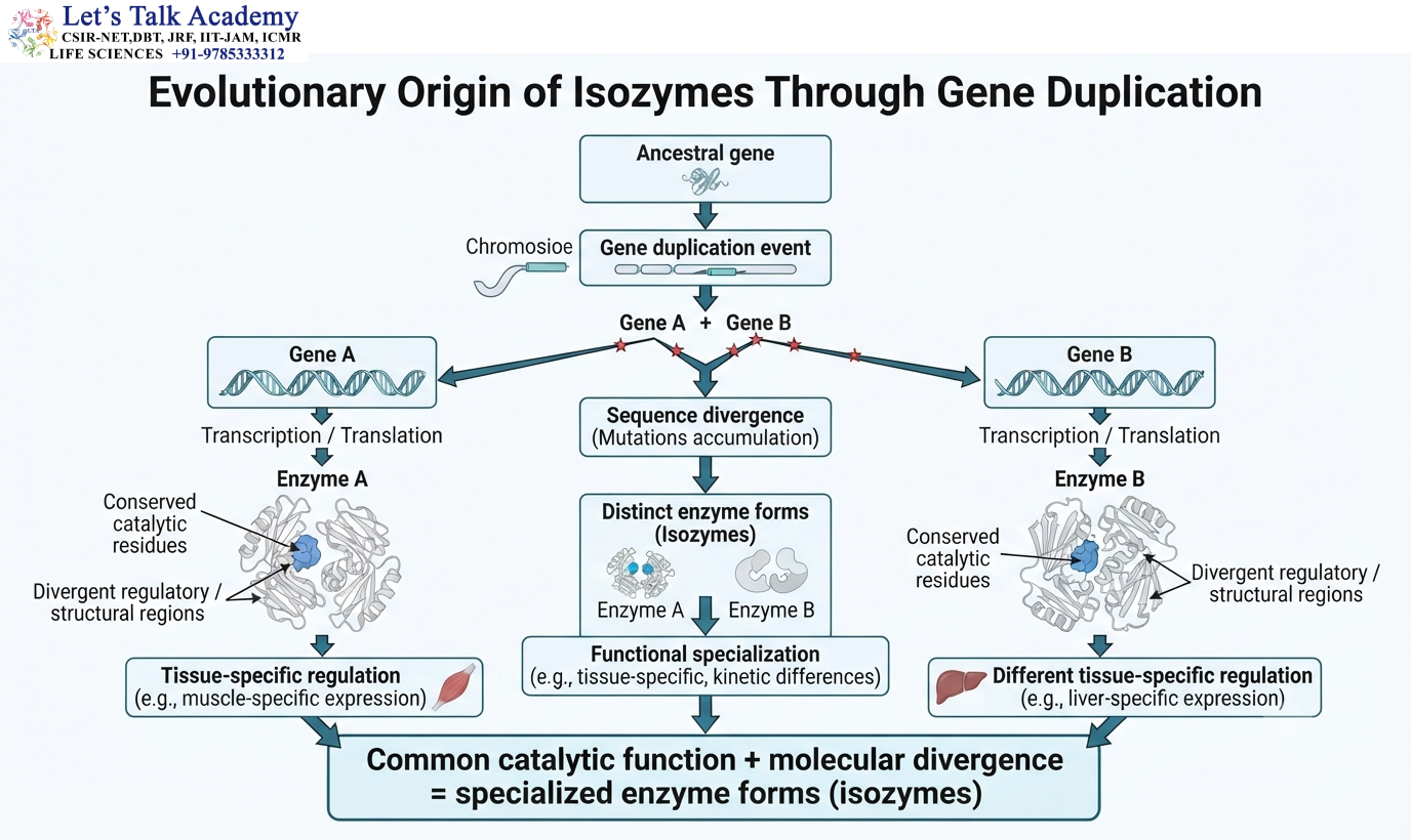
Gene duplication is an important evolutionary mechanism that can generate isozymes.
The process can be represented as:
Ancestral gene → Gene duplication → Related genes → Sequence divergence → Distinct enzyme forms
After duplication, both genes may initially encode very similar proteins.
Over evolutionary time, mutations accumulate.
If essential catalytic residues remain conserved, both proteins can retain the same overall reaction.
Other regions can diverge.
These changes may alter regulation, stability, substrate interaction, tissue expression, or cellular localization.
The result is a family of related proteins that retain a common biochemical function but have specialized properties.
38. Gene Duplication and Functional Divergence
Suppose an ancestral gene produces enzyme E.
After duplication:
Gene A → Enzyme A
Gene B → Enzyme B
Initially, Enzyme A and Enzyme B may be nearly identical.
Over time, selective pressures can favor different changes in each copy.
One protein may become specialized for expression in muscle.
Another may become specialized for expression in liver.
Both can retain the same fundamental catalytic activity.
This process is an example of functional divergence following gene duplication.
Isozymes therefore provide an important example of how evolution can generate biochemical specialization.
39. Isozymes and Metabolic Flexibility
Isozymes increase the flexibility of metabolic pathways.
Cells experience continuous changes in substrate availability, energy demand, hormonal signals, nutrient status, and metabolic products.
A single enzyme form may not be ideal under every condition.
Different isozymes allow cells to alter the behavior of a reaction according to physiological circumstances.
For example, one form may be highly active at low substrate concentration, while another may respond more strongly to increasing substrate concentration.
Another may be more strongly regulated by metabolic products.
Thus, isozyme diversity increases the range of metabolic responses available to cells.
40. Isozymes and Tissue Injury
The tissue-specific distribution of isozymes provides a biochemical basis for detecting possible tissue damage.
When cellular membranes are disrupted, intracellular enzymes can enter the extracellular environment.
If a particular isozyme is abundant in one tissue, an increase in that molecular form may provide information about the tissue source.
The principle can be represented as:
Tissue-specific isozyme → Cellular injury → Enzyme release → Circulating isozyme pattern
However, interpretation requires caution because many enzyme forms are present in multiple tissues.
Modern clinical analysis therefore combines enzyme measurements with other biochemical and molecular markers.
41. Isozyme Patterns in Biochemical Diagnosis
Isozyme patterns can provide information beyond total enzyme activity.
Suppose the total concentration of an enzyme increases in blood.
This increase may have several possible sources.
If the molecular forms are analyzed, the relative abundance of different isozymes may help determine which tissue contributed to the increase.
Historically, this approach was particularly useful for enzymes such as LDH and creatine kinase.
Modern diagnostics frequently use more specific biomarkers, but the principle remains an important example of how molecular heterogeneity can provide biological information.
42. Isozymes in Biochemical Research
Isozymes are useful research tools because their differences can be experimentally detected.
Researchers can study isozyme patterns to investigate tissue differentiation, developmental changes, metabolic specialization, genetic variation, and evolutionary relationships.
Classical methods include electrophoresis and activity staining.
Modern approaches include chromatography, immunological analysis, DNA sequencing, RNA expression analysis, mass spectrometry, and proteomics.
The combination of these methods allows researchers to connect enzyme activity with its genetic and molecular basis.
43. Isozymes and Electrophoretic Mobility
The electrophoretic mobility of a protein depends partly on its net charge.
Changes in amino acid composition can alter the net charge of an isozyme.
For example, replacing a positively charged residue with a neutral residue can reduce the positive charge of the protein.
Replacing a neutral residue with an acidic residue can increase the negative charge.
These differences can alter migration through an electrophoretic medium.
Thus, electrophoresis can convert molecular differences in protein sequence into visible biochemical patterns.
44. Isozymes and Genetic Variation
Genetic variation can produce enzyme variants within a population.
A mutation that changes one amino acid may alter the charge, size, conformation, or activity of an enzyme.
If the resulting enzyme remains functional, the variant may be detectable through biochemical analysis.
For example, a charge-changing amino acid substitution can produce a different electrophoretic mobility.
Such enzyme variants were historically important as genetic markers.
45. Isozymes in Population Biology
Before DNA-based molecular markers became widespread, isozyme electrophoresis was widely used to study genetic variation within and between populations.
Different populations can contain different frequencies of enzyme variants.
The frequency of a particular molecular form can therefore provide information about population structure.
Comparisons among populations can be used to investigate processes such as genetic drift, migration, natural selection, and population differentiation.
Although DNA-based approaches now provide much greater resolution, isozyme markers remain historically important in population genetics.
46. Isozymes and Evolutionary Relationships
Isozyme patterns can provide information about evolutionary relationships because related organisms may share similar enzyme forms inherited from common ancestors.
Differences in isozyme composition can indicate evolutionary divergence.
However, isozyme data represent only a limited subset of genetic variation.
Modern phylogenetic analysis generally relies on DNA or protein sequence data because sequence-based methods provide much greater information.
Nevertheless, isozyme analysis played an important role in the development of biochemical approaches to evolutionary biology.
47. Isozymes and Protein Charge
Differences in amino acid composition can change the overall charge of an enzyme.
Acidic residues such as aspartate and glutamate contribute negative charge under many physiological conditions.
Basic residues such as lysine and arginine contribute positive charge.
Histidine can contribute depending on its protonation state.
Therefore, changes in these residues can alter the net charge of an isozyme.
This difference can be detected by electrophoresis and can also influence protein interactions and cellular localization.
48. Isozymes and Protein Stability
The stability of an enzyme depends on the balance of many molecular interactions.
Hydrophobic residues contribute to the protein core.
Hydrogen bonds stabilize secondary and tertiary structures.
Ionic interactions contribute to structural organization.
Van der Waals interactions provide close molecular packing.
Disulfide bonds can contribute to stability in proteins where they occur.
Subunit interactions stabilize multimeric enzymes.
Differences between isozymes can alter these interactions and consequently change stability.
Therefore, two isozymes with similar catalytic activity may respond differently to temperature, pH, salt concentration, or chemical denaturants.
49. Isozymes and Substrate Interaction
Isozymes can differ in their interaction with the same substrate.
Differences in active-site geometry can alter substrate binding.
The resulting kinetic differences can be represented as:
Kₘ₁ ≠ Kₘ₂
A lower Kₘ often corresponds to stronger apparent substrate interaction in the simple Michaelis-Menten framework, but this interpretation must be made carefully because Kₘ also depends on the catalytic step.
Differences in substrate interaction can be physiologically useful because different tissues experience different substrate concentrations.
50. Isozymes and Reaction Flux
Metabolic flux refers to the rate at which metabolites move through a biochemical pathway.
Isozyme properties can influence flux by determining how rapidly a reaction responds to changes in substrate and regulatory molecules.
The relationship can be represented as:
Isozyme expression → Kinetic properties → Enzyme activity → Reaction rate → Metabolic flux
However, pathway flux is controlled by many factors.
The concentration of substrate, product, cofactors, enzyme, inhibitors, activators, and other pathway components can all influence the final metabolic rate.
Isozymes therefore represent one important layer of metabolic regulation rather than the only determinant of pathway flux.
51. Isozymes and Cellular Energy Requirements
Energy-demanding tissues often require highly specialized enzyme systems.
Skeletal muscle may experience sudden increases in ATP consumption during contraction.
Cardiac muscle requires continuous ATP production to sustain rhythmic contraction.
Brain cells require a continuous energy supply to maintain membrane potential and neuronal function.
Isozymes involved in energy metabolism can therefore be distributed according to these tissue-specific requirements.
Creatine kinase and lactate dehydrogenase illustrate this principle.
The molecular form of the enzyme influences how the reaction behaves under the metabolic conditions of the tissue.
52. Isozymes and Metabolic Pathway Integration
Isozymes operate within interconnected metabolic pathways.
An enzyme reaction does not occur in isolation.
The rate of one reaction depends on the availability of substrates, products, cofactors, and regulatory molecules.
For example:
Glucose → Pyruvate → Lactate
The activity of enzymes in this pathway is influenced by the metabolic state of the cell.
An isozyme can modify the behavior of one reaction while the surrounding pathway determines the overall physiological consequence.
Therefore, isozyme function must be interpreted within the context of the entire metabolic network.
53. Isozyme Nomenclature
Isozyme names often indicate either the enzyme involved or the subunit composition.
For lactate dehydrogenase:
LDH₁ = H₄
LDH₂ = H₃M
LDH₃ = H₂M₂
LDH₄ = HM₃
LDH₅ = M₄
For creatine kinase:
CK-MM
CK-MB
CK-BB
These designations provide information about the molecular composition of the enzyme.
Understanding this nomenclature is particularly useful when interpreting biochemical experiments and scientific literature.
54. Isozymes and Quaternary Structure
Quaternary structure refers to the organization of multiple polypeptide subunits within a functional protein complex.
Isozymes formed through different combinations of subunits demonstrate that quaternary structure can directly influence biochemical function.
Changing the subunit composition can affect:
Catalytic activity
Substrate interaction
Regulation
Stability
Protein charge
Electrophoretic mobility
Therefore, quaternary structure is not simply an architectural feature. It can be an important determinant of enzyme properties.
55. Homotetramers and Heterotetramers
A tetramer composed of four identical subunits is a homotetramer.
For example: A₄
A tetramer containing different types of subunits is a heterotetramer.
For example: A₂B₂
Different tetrameric combinations can produce distinct molecular forms.
This principle explains the multiple forms of lactate dehydrogenase.
The ability of subunits to combine in different ways allows an organism to generate several enzyme forms from a limited number of subunit types.
56. Isozymes and Metabolic Homeostasis
Metabolic homeostasis requires cells to maintain concentrations of metabolites and energy carriers within appropriate ranges.
Isozymes contribute to this process by allowing the same biochemical reaction to be controlled differently in different tissues.
One tissue may require rapid substrate utilization.
Another may require conservation of substrate.
A third may require strong feedback control.
The expression of different enzyme forms provides a molecular mechanism for these distinct requirements.
Isozymes therefore contribute to the integration of metabolism across tissues.
57. Isozymes and Cellular Differentiation
Differentiation involves the acquisition of specialized cellular functions.
As cells differentiate, their patterns of gene expression change.
This can alter the set of enzymes expressed by the cell.
The isozyme pattern can therefore change during differentiation.
The general sequence is:
Cell differentiation → Gene-expression changes → Isozyme-expression changes → Metabolic specialization
Isozyme composition can therefore serve as a molecular indicator of cellular state.
58. Isozymes and Developmental Changes
Developmental changes can alter the relative abundance of enzyme forms.
An isozyme that is abundant during embryonic development may decrease later.
Another isozyme may become dominant after tissue maturation.
These changes can reflect changes in energy sources, hormonal signals, substrate availability, and cellular function.
Isozyme switching is therefore one mechanism through which biochemical systems adapt during development.
59. Isozymes and Disease-Associated Changes
Isozyme expression can change under pathological conditions.
Changes may occur because of tissue injury, abnormal gene expression, altered cellular differentiation, metabolic stress, hormonal disturbances, or disease-associated changes in cellular metabolism.
The interpretation of these changes depends on the enzyme and tissue involved.
An altered isozyme pattern is therefore not automatically diagnostic of a particular disease.
Instead, it provides biochemical information that must be interpreted together with other molecular and physiological data.
60. Experimental Identification of Isozymes
Isozymes can be studied using a combination of biochemical and molecular techniques.
Electrophoresis can separate molecular forms according to differences in charge and other physical properties.
Activity staining can identify which separated bands retain enzyme activity.
Chromatography can separate proteins according to charge, size, affinity, or other characteristics.
Immunological techniques can distinguish molecular forms based on specific antigenic differences.
Mass spectrometry can identify differences in molecular mass and peptide composition.
DNA sequencing can reveal genetic differences.
RNA analysis can determine tissue-specific expression patterns.
Together, these techniques allow researchers to connect the molecular identity of an isozyme with its biochemical function.
61. Isozymes and Chromatographic Separation
Chromatographic methods can separate isozymes because the molecular forms may differ in their physical or chemical properties.
Ion-exchange chromatography separates proteins according to charge.
Size-exclusion chromatography separates molecules according to effective molecular size.
Affinity chromatography separates proteins according to specific molecular interactions.
If two isozymes differ in charge or molecular interaction with the stationary phase, they can elute at different positions.
This provides a useful approach for isolating and characterizing different molecular forms.
62. Isozymes and Immunological Methods
Antibodies can distinguish enzyme forms when the isozymes contain different antigenic regions.
An antibody recognizes a particular molecular structure called an epitope.
If an epitope is present in one isozyme but absent or structurally different in another, selective detection becomes possible.
The basic principle is:
Isozyme-specific structure → Antibody recognition → Selective detection
Immunological approaches can therefore complement electrophoresis, chromatography, and molecular analysis.
63. Isozymes and Molecular Biology
Modern molecular biology has greatly expanded the study of isozymes.
DNA sequencing can identify differences in coding sequences.
RNA analysis can determine where and when different enzyme genes are expressed.
Gene-expression studies can identify tissue-specific regulation.
Protein analysis can determine whether the predicted enzyme forms are actually produced.
Mass spectrometry can identify molecular differences at the protein level.
Thus, isozyme biology now connects classical enzymology with genomics, transcriptomics, and proteomics.
64. Isozymes and Enzyme Families
Isozymes often belong to broader enzyme families.
Members of an enzyme family may share conserved sequence motifs and structural features.
The catalytic residues are often strongly conserved because they are essential for the chemical reaction.
Other regions may evolve more rapidly and contribute to differences in regulation, stability, localization, or substrate interaction.
The relationship can be represented as:
Common ancestral protein → Gene duplication → Sequence divergence → Related enzyme forms
Some members retain nearly identical substrate specificity.
Others may acquire narrower or broader substrate preferences.
Thus, enzyme families provide an evolutionary framework for understanding isozyme diversity.
65. Evolutionary Conservation of Catalytic Residues
Catalytic residues are generally subject to strong evolutionary constraints.
A mutation that eliminates an essential catalytic group can severely reduce or eliminate enzyme activity.
Consequently, important catalytic residues are often conserved among related enzyme forms.
In contrast, residues involved in regulation, surface interactions, or tissue-specific properties may tolerate more variation.
This creates a characteristic pattern:
Catalytic regions → Strong conservation
Peripheral or regulatory regions → Greater variation
This balance allows isozymes to preserve the same chemical reaction while developing specialized biochemical properties.
66. Functional Specialization of Isozymes
Functional specialization is the central biological significance of isozymes.
Two enzyme forms may catalyze the same reaction but perform different physiological roles.
One may be expressed primarily in a tissue with high energy demand.
Another may be expressed in a tissue involved in storage or biosynthesis.
The chemical reaction remains the same, but the biological context differs.
Therefore:
Same catalytic reaction does not necessarily mean identical physiological function.
The properties of the enzyme determine how the reaction responds to the environment.
67. Isozymes as a Link Between Structure and Function
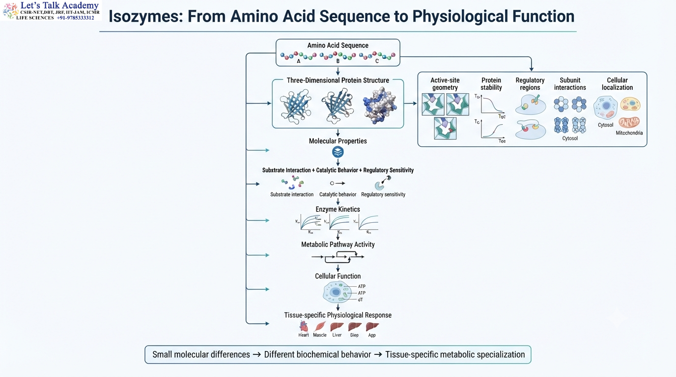
Isozymes provide a clear example of the relationship between molecular structure and biological function.
The relationship can be represented as:
Amino acid sequence → Three-dimensional structure → Molecular properties → Catalytic behavior → Cellular function
Changes in amino acid sequence can alter the structure.
Structural differences can modify substrate interaction, stability, regulatory properties, and kinetics.
These biochemical properties influence metabolic pathways.
The metabolic requirements of the tissue then influence which isozyme is preferentially expressed.
This creates a continuous connection between genes, proteins, biochemical reactions, and physiology.
68. Isozymes and Mutational Analysis
The contribution of particular amino acid residues can be investigated experimentally using site-directed mutagenesis.
A researcher can replace a specific residue and measure the resulting change in enzyme activity.
For example, replacement of a catalytic serine: Ser → Ala
removes the hydroxyl group that may be required for nucleophilic catalysis.
If enzyme activity decreases dramatically, the residue is likely to have an important catalytic role.
Similarly, mutations near the active site can alter Kₘ or k₍cat₎ without completely eliminating activity.
Such experiments help distinguish residues involved in catalysis from those involved primarily in substrate binding, protein stability, or regulation.
69. Isozymes and Transition-State Recognition
The catalytic properties of isozymes can also be studied through transition-state analogues.
If two isozymes bind the same transition-state analogue with different affinities, this suggests differences in the chemical environment of their active sites.
The interaction can be represented as:
E + I ⇌ EI
where I represents the transition-state analogue.
Differences in binding can provide information about how structural changes influence transition-state stabilization.
This connects isozyme structure with the fundamental mechanism of enzyme catalysis.
70. Isozymes and Physiological Adaptation
Isozyme expression allows organisms to adapt enzyme activity to physiological conditions.
Nutritional state, hormonal signals, developmental stage, energy demand, and tissue function can all influence the relative abundance of enzyme forms.
The same catalytic reaction can therefore be tuned differently in different physiological states.
This is particularly important in metabolic pathways that must respond rapidly to changes in nutrient availability and energy demand.
Isozymes provide one layer of this complex regulatory network.
71. Integrated Understanding of Isozymes
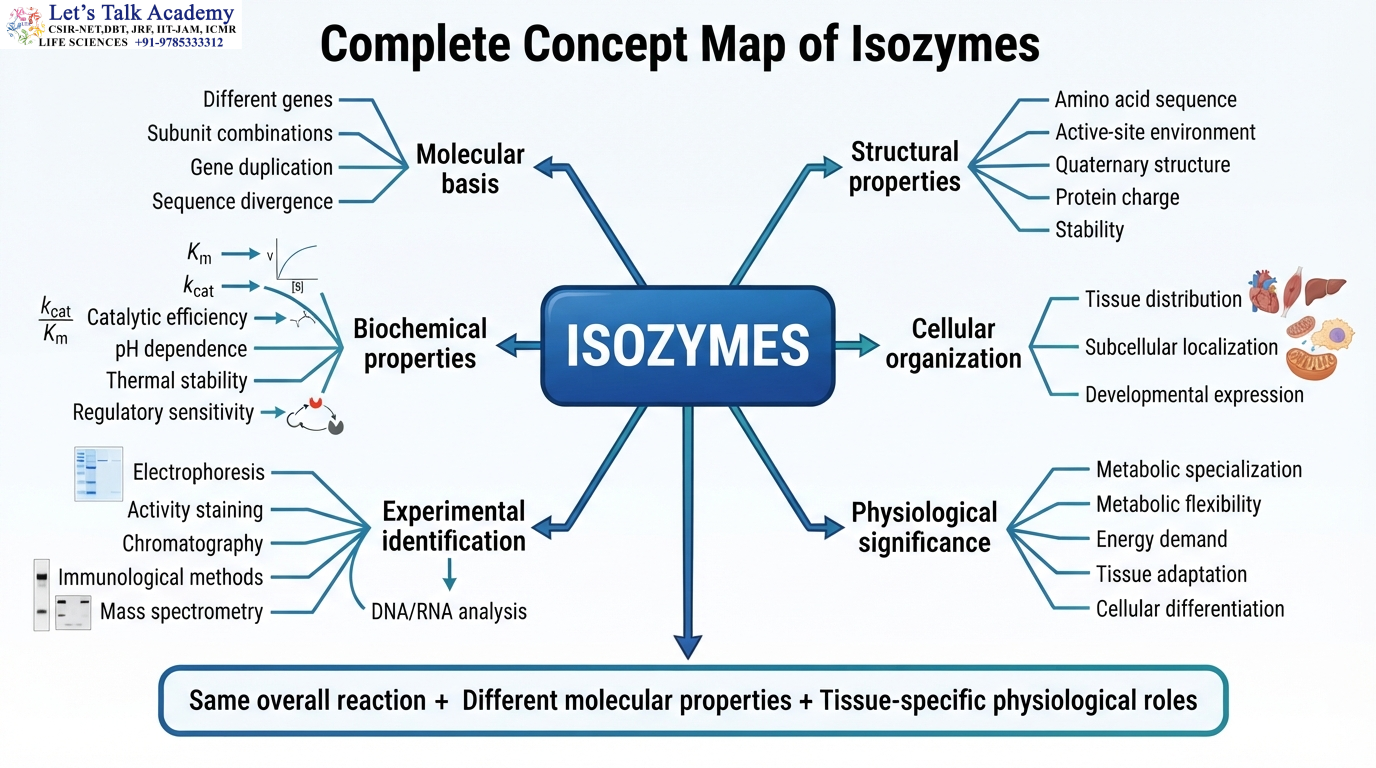
Isozymes are molecularly distinct enzyme forms that catalyze the same overall biochemical reaction.
Their molecular differences may arise from different genes, different combinations of subunits, or related mechanisms of protein diversification.
Although their catalytic function is conserved, their Kₘ, k₍cat₎, catalytic efficiency, stability, regulatory sensitivity, tissue distribution, and cellular localization can differ.
These differences allow tissues to adapt the same biochemical reaction to different physiological environments.
Lactate dehydrogenase illustrates this through different combinations of H and M subunits.
Creatine kinase illustrates tissue-associated forms through CK-MM, CK-MB, and CK-BB.
Alkaline phosphatase demonstrates tissue-associated enzyme forms.
Hexokinase and glucokinase demonstrate how related enzymes can catalyze the same reaction but operate with markedly different kinetic properties.
Together, these examples show that isozyme diversity is an important mechanism for metabolic specialization.
72. Relationship Between Isozymes and Enzyme Kinetics
For a simple enzyme-catalyzed reaction:
E + S ⇌ ES → E + P
the Michaelis-Menten relationship is:
v = Vₘₐₓ[S] ÷ (Kₘ + [S])
The maximum velocity is:
Vₘₐₓ = k₍cat₎[E]ₜ
The turnover number is:
k₍cat₎ = Vₘₐₓ ÷ [E]ₜ
The catalytic efficiency is:
k₍cat₎ ÷ Kₘ
Different isozymes may have different values for all of these parameters.
Therefore, even when:
Reaction A = Reaction B
the kinetic behavior can still differ:
Kₘₐ ≠ Kₘᵦ
and:
k₍cat₎ₐ ≠ k₍cat₎ᵦ
These differences provide the biochemical basis for tissue-specific metabolic responses.
73. Major Isozyme Examples
Several enzyme systems illustrate the principles of isozyme diversity.
Lactate dehydrogenase demonstrates different molecular forms generated by H and M subunit combinations.
Creatine kinase demonstrates different forms generated from M and B subunits.
Alkaline phosphatase demonstrates tissue-associated molecular forms.
Hexokinase and glucokinase illustrate related enzyme forms with different kinetic and regulatory properties involved in glucose phosphorylation.
These examples differ in their molecular basis, but they share the central principle that related enzyme forms allow biochemical reactions to be adapted to different cellular environments.
74. Isozymes and Metabolic Regulation
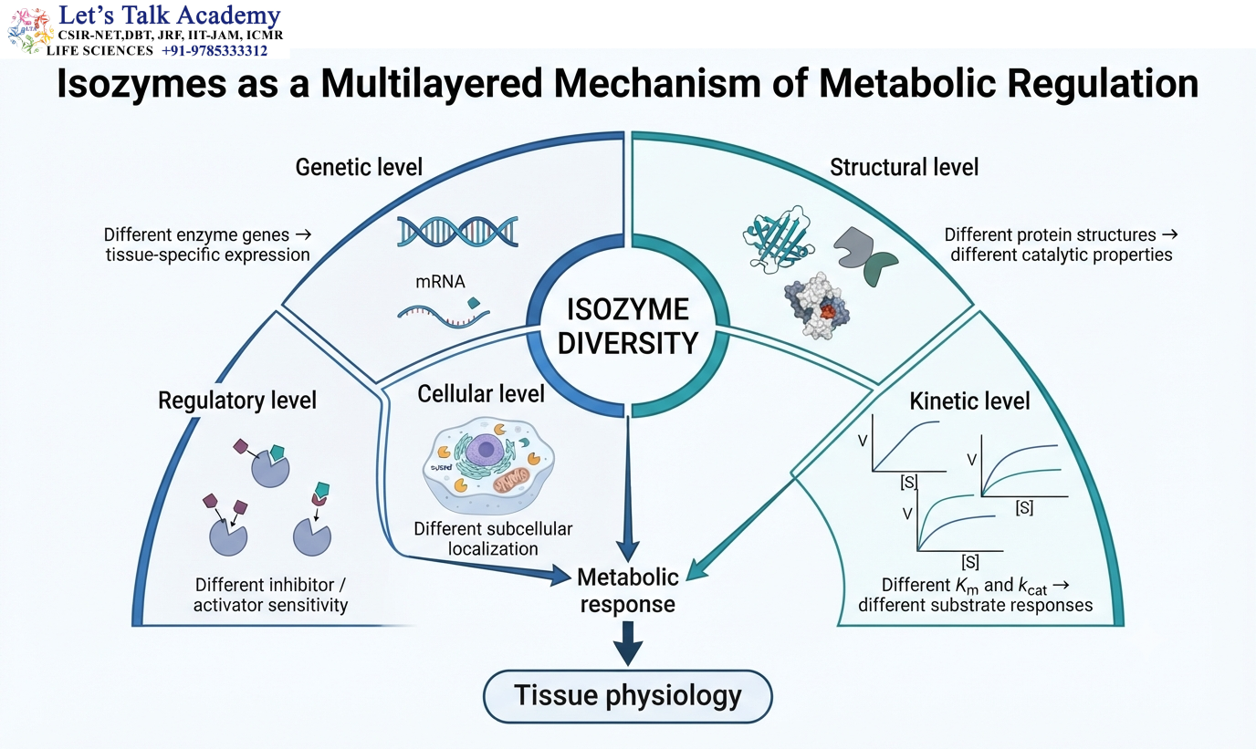
Isozymes contribute to metabolic regulation at several levels.
At the genetic level, different enzyme genes can be expressed in different tissues.
At the structural level, different proteins can have different catalytic and regulatory properties.
At the kinetic level, differences in Kₘ and k₍cat₎ influence responses to substrate concentration.
At the regulatory level, isozymes can differ in their response to inhibitors and activators.
At the cellular level, different forms can be localized to different compartments.
At the physiological level, tissue-specific isozyme expression allows metabolic pathways to respond differently to systemic signals.
Isozyme diversity is therefore a multilayered mechanism of metabolic organization.
75. Core Biochemical Relationships
The basic catalytic mechanism can be represented as:
E + S ⇌ ES → E + P
The Michaelis-Menten relationship is:
v = Vₘₐₓ[S] ÷ (Kₘ + [S])
The maximum velocity is:
Vₘₐₓ = k₍cat₎[E]ₜ
The turnover number is:
k₍cat₎ = Vₘₐₓ ÷ [E]ₜ
Catalytic efficiency is:
k₍cat₎ ÷ Kₘ
For lactate dehydrogenase:
Pyruvate + NADH + H⁺ ⇌ Lactate + NAD⁺
For creatine kinase:
Phosphocreatine + ADP + H⁺ ⇌ Creatine + ATP
For glucose phosphorylation:
Glucose + ATP → Glucose-6-phosphate + ADP
For alkaline phosphatase:
R−O−PO₃²⁻ + H₂O → R−OH + Pᵢ
These equations illustrate that different enzyme forms can participate in the same fundamental chemical transformations while displaying different molecular and physiological properties.


