
1. Introduction
Cancer is not simply a disease of abnormal cells multiplying uncontrollably. A tumor develops within a complex tissue environment in which malignant cells continuously communicate with surrounding normal and altered cells. This communication is dynamic and bidirectional: cancer cells modify the behavior of neighboring cells, while neighboring cells and the extracellular environment can, in turn, influence cancer-cell survival, proliferation, invasion, and response to therapy.
The collection of cells, extracellular matrix, signaling molecules, blood vessels, lymphatic vessels, and other components surrounding a tumor is commonly referred to as the tumor microenvironment (TME). Normal cells present within this environment include fibroblasts, endothelial cells, immune cells, pericytes, adipocytes, and other tissue-specific stromal cells. The tumor microenvironment is therefore not merely a passive scaffold around cancer cells; it is an active biological system that can influence tumor development and progression.
Cancer cells can release growth factors, cytokines, chemokines, extracellular vesicles, metabolites, and other signaling molecules. These factors can alter neighboring normal cells and sometimes convert them into a tumor-supportive state. In return, altered stromal cells can release signals that promote cancer-cell growth, survival, migration, invasion, angiogenesis, immune evasion, and resistance to treatment.
This reciprocal communication is one of the central concepts of modern tumor biology.
A useful way to understand this relationship is:
Cancer cells → modify the surrounding normal cells → modified cells support cancer cells → cancer cells become more aggressive → further modification of the microenvironment.
Thus, tumor progression can involve the development of a self-reinforcing communication network.
2. Normal Cells and Cancer Cells: A Fundamental Difference
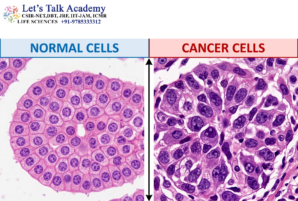
2.1 Characteristics of Normal Cells
Normal cells generally maintain a controlled relationship with neighboring cells and the surrounding tissue. Their proliferation, differentiation, survival, migration, and death are regulated by signals originating from the tissue environment.
Normal cells commonly respond to:
- Growth-promoting signals
- Growth-inhibitory signals
- Cell–cell contact
- Cell–extracellular matrix interactions
- Hormonal signals
- Nutrient availability
- Oxygen concentration
- DNA damage
- Signals promoting programmed cell death
Normal tissue therefore behaves as an organized biological system rather than as a collection of independently growing cells.
For example, many normal epithelial cells require appropriate extracellular matrix attachment and tissue-specific signals for continued survival. If these signals are lost or if serious cellular damage occurs, cells may undergo apoptosis or other forms of regulated cell death.
2.2 Characteristics of Cancer Cells
Cancer cells acquire genetic and epigenetic alterations that allow them to behave differently from normal cells.
Important characteristics include:
- Sustained proliferative signaling
- Reduced sensitivity to growth-inhibitory signals
- Resistance to cell death
- Altered metabolism
- Increased migratory capacity
- Tissue invasion
- Ability to stimulate blood-vessel formation
- Ability to interact with and manipulate immune cells
- Genomic instability
- Ability to establish supportive interactions with stromal cells
The National Cancer Institute notes that cancer cells can grow without normal growth signals, ignore signals that promote cell death, invade surrounding tissues, stimulate blood-vessel growth, and evade or manipulate immune responses.
2.3 Why Cancer Cells Need Their Surrounding Environment
Although cancer cells possess intrinsic abnormalities, many tumors cannot be understood by studying malignant cells alone.
Cancer cells require:
- Nutrients
- Oxygen
- Blood supply
- Physical support
- Growth factors
- Survival signals
- Metabolic substrates
- Routes for invasion
- Mechanisms for immune evasion
The surrounding tissue can provide many of these requirements.
Therefore, tumor progression results from an interaction between the properties of cancer cells and the properties of their surrounding microenvironment.
3. The Tumor Microenvironment
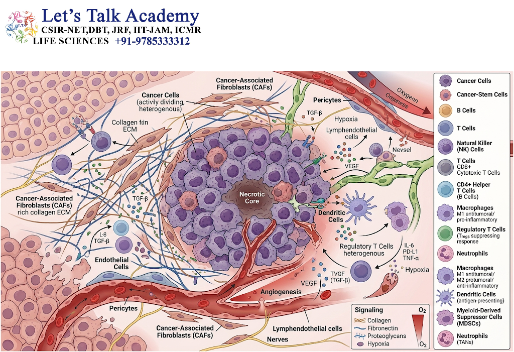
3.1 Definition of the Tumor Microenvironment
The tumor microenvironment is the local biological environment surrounding cancer cells. It includes both cellular and non-cellular components.
Major cellular components include:
- Cancer cells
- Fibroblasts
- Cancer-associated fibroblasts
- Endothelial cells
- Pericytes
- Macrophages
- T lymphocytes
- B lymphocytes
- Natural killer cells
- Dendritic cells
- Neutrophils
- Myeloid-derived suppressor cells
- Adipocytes
- Mesenchymal stromal cells
Important non-cellular components include:
- Collagen
- Fibronectin
- Laminins
- Proteoglycans
- Glycosaminoglycans
- Cytokines
- Chemokines
- Growth factors
- Extracellular vesicles
- Metabolites
- Oxygen
- Extracellular ions
- Mechanical forces
The NCI defines the tumor microenvironment as the normal cells, molecules, and blood vessels surrounding and supporting tumor cells, while emphasizing that tumors can modify this environment and that the altered environment can subsequently affect tumor growth and spread.
3.2 The Tumor Microenvironment as a Dynamic System
The tumor microenvironment changes continuously.
During early tumor development, the surrounding tissue may initially restrict abnormal cell growth. As tumor cells acquire additional abnormalities, they may alter the surrounding cells and extracellular matrix.
Over time, the microenvironment can become characterized by:
- Chronic inflammation
- Fibrosis
- Increased extracellular matrix deposition
- Abnormal vascularization
- Hypoxia
- Altered metabolism
- Immunosuppression
- Increased tissue stiffness
- Abnormal interstitial fluid pressure
Consequently, the microenvironment changes from a relatively normal tissue environment into a tumor-supportive ecosystem.
4. Direct Interaction Between Cancer and Normal Cells
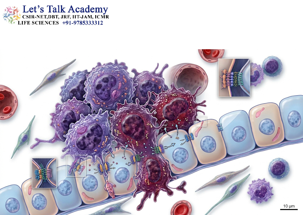
Cancer cells can interact with normal cells through both direct physical contact and indirect chemical communication.
4.1 Direct Cell–Cell Contact
Direct contact occurs when membranes of neighboring cells interact through cell-adhesion molecules, receptors, and other membrane proteins.
Important molecules include:
- Cadherins
- Integrins
- Selectins
- Immunoglobulin-superfamily adhesion molecules
- Eph receptors and ephrin ligands
- Notch receptors and ligands
These interactions can regulate:
- Cell proliferation
- Differentiation
- Migration
- Survival
- Polarity
- Tissue organization
Alteration of these interactions can contribute to cancer progression.
4.2 Cell–Matrix Interaction
Cells also communicate with the extracellular matrix through receptors such as integrins.
Integrins connect extracellular matrix proteins to the intracellular cytoskeleton and signaling machinery.
Therefore, the extracellular matrix is not simply a structural material. It acts as a signaling platform.
Changes in matrix composition or stiffness can influence:
- Cell shape
- Cytoskeletal organization
- Migration
- Proliferation
- Survival
- Differentiation
- Invasion
Recent research continues to emphasize the physical properties of the tumor microenvironment, including tissue stiffness, mechanical stress, fluid pressure, and altered microarchitecture.
5. Interaction Through Signaling Molecules
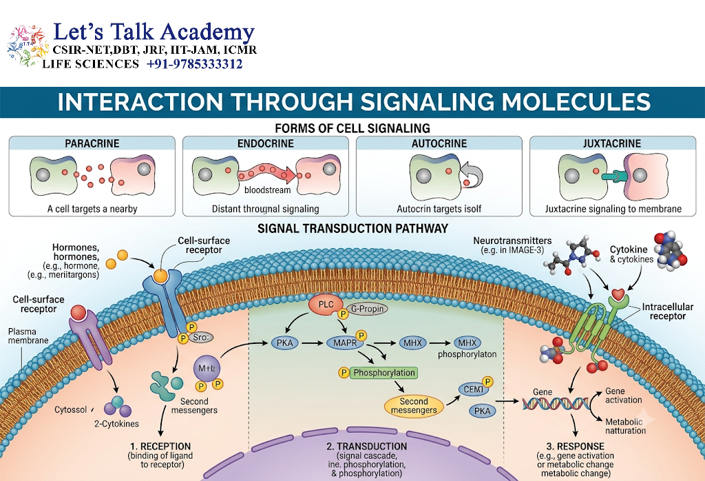
5.1 Growth Factors
Cancer cells and surrounding stromal cells can produce growth factors that act through paracrine signaling.
Important examples include:
- Epidermal growth factor
- Transforming growth factor beta
- Vascular endothelial growth factor
- Fibroblast growth factors
- Hepatocyte growth factor
- Platelet-derived growth factors
These molecules can stimulate neighboring cells and establish feedback loops.
For example:
Cancer cell → growth factor → fibroblast activation → fibroblast-derived growth factor → cancer-cell proliferation
Such reciprocal signaling can amplify tumor growth.
5.2 Cytokines
Cytokines are important mediators of communication between cancer cells and immune or stromal cells.
Examples include:
- Interleukins
- Tumor necrosis factor
- Transforming growth factor beta
- Interferons
Depending on the context, cytokines can promote inflammation, immune activation, immune suppression, proliferation, migration, or tissue remodeling.
5.3 Chemokines
Chemokines are particularly important for controlling cell migration.
Cancer cells and stromal cells can produce chemokines that attract:
- Macrophages
- Neutrophils
- T cells
- Myeloid-derived suppressor cells
- Other stromal populations
Chemokine signaling can therefore influence the cellular composition of the tumor microenvironment.
6. Cancer Cells and Cancer-Associated Fibroblasts
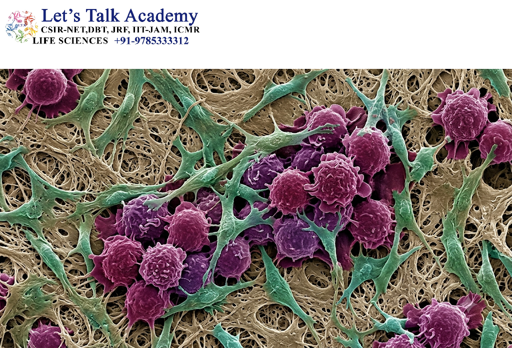
6.1 Normal Fibroblasts
Fibroblasts are connective-tissue cells that contribute to:
- Extracellular matrix production
- Tissue organization
- Wound repair
- Collagen synthesis
- Tissue homeostasis
Under normal conditions, fibroblasts contribute to maintaining tissue structure.
6.2 Conversion of Fibroblasts into Cancer-Associated Fibroblasts
Cancer cells can release signals that alter nearby fibroblasts.
The resulting cells are commonly called cancer-associated fibroblasts (CAFs).
CAFs are heterogeneous rather than being a single uniform cell type. They may originate from different cellular populations and display different molecular and functional properties.
Signals involved in fibroblast activation can include:
- TGF-β
- IL-1-related inflammatory signaling
- Growth factors
- Chemokines
- Reactive oxygen species
- Hypoxia-related signals
- Extracellular vesicles
Reviews of CAF biology describe reciprocal interactions between cancer cells and fibroblasts that can support tumor proliferation, angiogenesis, metastasis, extracellular matrix remodeling, and treatment resistance.
6.3 Functions of Cancer-Associated Fibroblasts
CAFs can:
- Produce extracellular matrix proteins
- Remodel collagen
- Alter tissue stiffness
- Secrete growth factors
- Produce cytokines and chemokines
- Promote angiogenesis
- Influence immune-cell recruitment
- Support cancer-cell survival
- Facilitate invasion
- Contribute to therapy resistance
Thus, the fibroblast is transformed from a normal tissue-supporting cell into a component of a tumor-supportive ecosystem.
6.4 Reciprocal Interaction Between Cancer Cells and CAFs
The relationship is not one-directional.
Cancer cells activate fibroblasts.
Activated fibroblasts then produce factors that influence cancer cells.
This creates a positive feedback loop:
Cancer cells → fibroblast activation → CAF formation → growth-factor and matrix production → increased cancer-cell survival and invasion → further tumor signaling.
This reciprocal relationship is an important example of how cancer progression involves both malignant and non-malignant cells.
7. Cancer Cells and Immune Cells
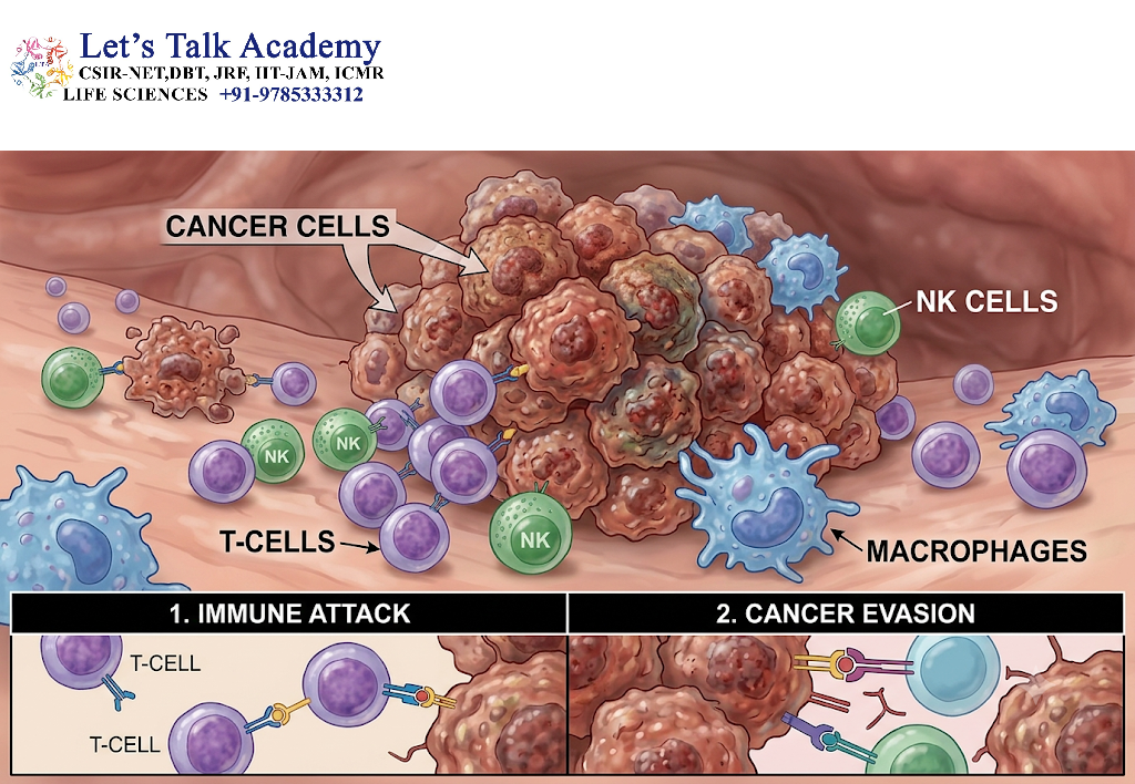
7.1 Immune Surveillance
The immune system can recognize and eliminate abnormal cells.
Important antitumor immune cells include:
- Cytotoxic T lymphocytes
- Natural killer cells
- Dendritic cells
- Macrophages
- Other innate and adaptive immune populations
However, established tumors can develop mechanisms that reduce effective immune attack.
7.2 Immune Evasion
Cancer cells may:
- Reduce immune recognition
- Alter antigen presentation
- Express immune checkpoint ligands
- Release immunosuppressive molecules
- Recruit immunosuppressive cells
- Alter cytokine signaling
- Create metabolically hostile conditions for immune cells
Therefore, the tumor microenvironment can become immunosuppressive.
7.3 Tumor-Associated Macrophages
Macrophages are highly plastic immune cells.
Within tumors, macrophages can acquire phenotypes and functional states influenced by local signals.
Tumor-associated macrophages can participate in:
- Tumor growth
- Angiogenesis
- Extracellular matrix remodeling
- Immune regulation
- Cancer-cell migration
- Metastasis
The precise behavior of macrophages depends on the tumor type, tissue context, cytokine environment, metabolic conditions, and other signals. Research emphasizes their role as important components of the tumor microenvironment rather than treating all macrophages as biologically identical.
7.4 T Cells
T lymphocytes can either contribute to antitumor immunity or become functionally suppressed within tumors.
Cytotoxic T cells can kill susceptible cancer cells, whereas regulatory T cells can suppress immune responses.
The balance among different T-cell populations can therefore influence tumor progression.
7.5 Natural Killer Cells
Natural killer cells recognize stressed or abnormal cells without requiring the same antigen-specific recognition mechanism used by conventional T cells.
Tumors can nevertheless develop mechanisms that reduce NK-cell activity.
Consequently, cancer–NK-cell interaction represents another important component of tumor immune surveillance.
8. Cancer Cells and Endothelial Cells
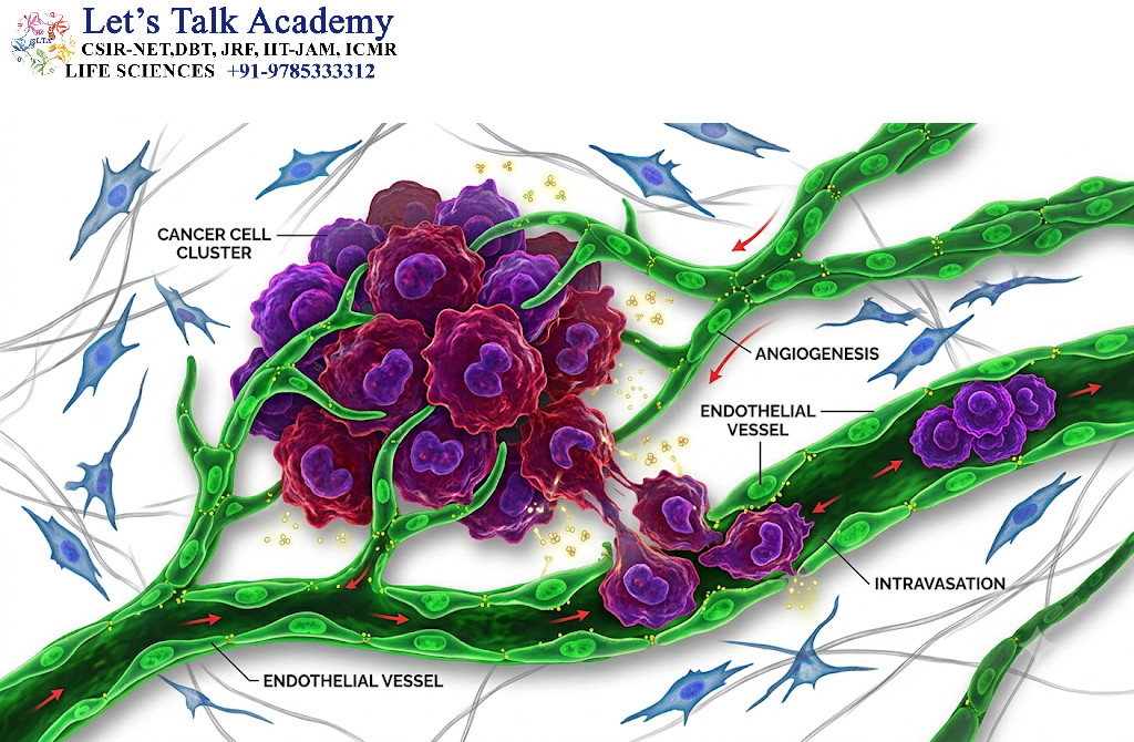
8.1 Importance of Blood Vessels
A growing tumor requires access to oxygen and nutrients.
As tumor mass increases, diffusion alone becomes insufficient to meet metabolic demands.
Cancer cells therefore release signals that stimulate blood-vessel formation.
This process is called angiogenesis.
8.2 Vascular Endothelial Growth Factor
One of the major angiogenic signaling molecules is vascular endothelial growth factor (VEGF).
Hypoxic tumor regions can increase expression of angiogenic factors, stimulating endothelial cells.
The simplified sequence is:
Tumor growth → oxygen limitation → hypoxia signaling → angiogenic factor production → endothelial-cell activation → new vessel formation.
8.3 Abnormal Tumor Vasculature
Tumor-associated blood vessels are frequently abnormal.
They may show:
- Irregular architecture
- Abnormal permeability
- Disorganized branching
- Incomplete maturation
- Abnormal endothelial junctions
- Altered blood flow
These vascular abnormalities can contribute to hypoxia and abnormal drug delivery.
The relationship between cancer cells and blood vessels is therefore bidirectional: tumors stimulate vascular changes, while the resulting vascular environment influences tumor growth and therapeutic response.
9. Cancer Cells and Extracellular Matrix
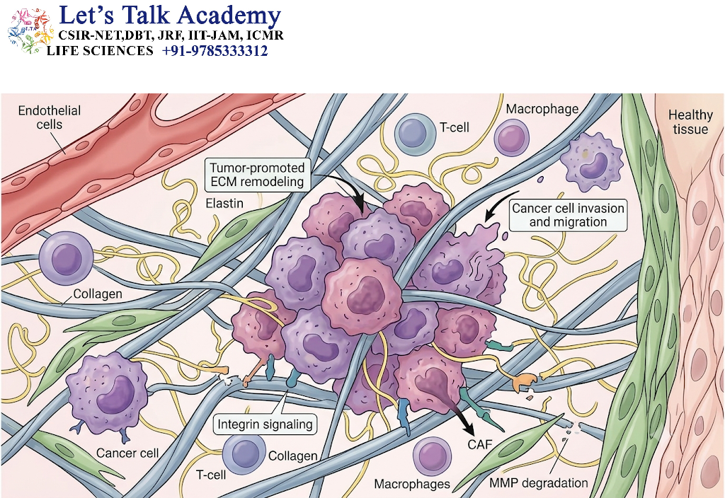
9.1 Structure of the Extracellular Matrix
The extracellular matrix (ECM) is a complex network of proteins and carbohydrates surrounding cells.
Important components include:
- Collagen
- Fibronectin
- Laminin
- Proteoglycans
- Glycosaminoglycans
The ECM provides structural support but also functions as a signaling environment.
9.2 ECM Remodeling
Cancer cells and stromal cells can alter ECM composition.
Matrix remodeling may involve:
- Increased collagen deposition
- Collagen cross-linking
- Proteolytic degradation
- Fibronectin remodeling
- Altered matrix stiffness
Matrix remodeling can generate conditions favorable for tumor-cell migration and invasion.
9.3 Matrix Metalloproteinases
Matrix metalloproteinases (MMPs) are proteolytic enzymes capable of degrading components of the extracellular matrix.
In cancer, increased protease activity can facilitate:
- Basement-membrane disruption
- ECM remodeling
- Tumor-cell invasion
- Release of matrix-bound signaling molecules
However, MMPs have diverse functions, and their biological effects depend on their specific substrate and tissue context.
9.4 Mechanical Properties of the Tumor
Tumors can become mechanically different from normal tissues.
Increased matrix deposition and cross-linking can increase tissue stiffness.
Mechanical signals can then influence cell behavior through mechanotransduction pathways.
Thus:
Biochemical signaling + mechanical signaling = integrated tumor-cell regulation.
10. Cancer Cells and Adipocytes
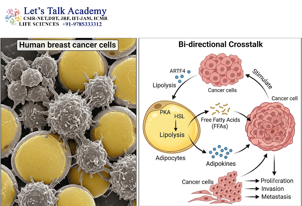
Adipocytes are increasingly recognized as important components of the tumor microenvironment in several cancers.
They can provide:
- Fatty acids
- Metabolic substrates
- Cytokines
- Adipokines
- Growth-supporting signals
Cancer cells can alter nearby adipocytes, while adipocyte-derived factors can influence cancer-cell metabolism and proliferation.
This interaction is particularly relevant in tissues rich in adipose cells.
The relationship illustrates another important principle: cancer cells do not depend exclusively on glucose or on their own intracellular metabolic pathways. They can exploit nutrients and signals supplied by neighboring cells.
11. Cancer Cells and Other Stromal Cells
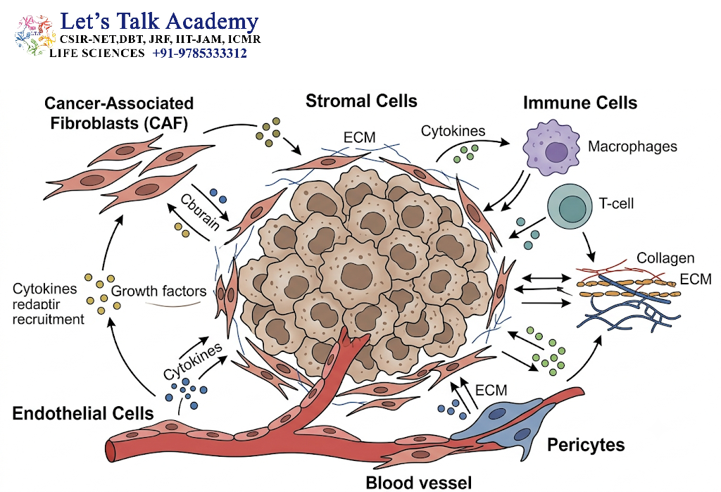
11.1 Pericytes
Pericytes associate with blood vessels and contribute to vascular stability.
Alterations in pericyte–endothelial interactions can affect tumor vascular structure and permeability.
11.2 Mesenchymal Stromal Cells
Mesenchymal stromal cells can interact with tumor cells through soluble factors, extracellular vesicles, and cell contact.
Their effects can vary according to tissue and experimental context.
11.3 Dendritic Cells
Dendritic cells are important antigen-presenting cells.
Tumors can interfere with dendritic-cell maturation and function, thereby weakening effective antitumor immune responses.
11.4 Neutrophils
Neutrophils can be recruited to tumor tissues by chemokines and inflammatory signals.
Depending on the context, tumor-associated neutrophils can participate in inflammation, matrix remodeling, angiogenesis, and immune regulation.
12. Hypoxia and Metabolic Interaction
12.1 Hypoxia in Tumors
Rapid tumor growth can outpace the development of an adequate blood supply.
This produces regions of reduced oxygen availability known as hypoxic regions.
Hypoxia is not merely a consequence of tumor growth; it can actively change cellular behavior.
12.2 Hypoxia-Inducible Factors
Hypoxia-inducible factors, particularly HIF signaling, help cells adapt to reduced oxygen availability.
Hypoxia can promote:
- Angiogenesis
- Metabolic adaptation
- Cell survival
- Migration
- Invasion
- Altered immune behavior
12.3 Metabolic Reprogramming
Cancer cells frequently display altered metabolic patterns.
One well-known phenomenon is increased reliance on glycolysis even when oxygen is available, commonly associated with the Warburg effect.
However, cancer metabolism is more complex than simply “glycolysis versus oxidative phosphorylation.”
Cancer cells may use:
- Glucose
- Glutamine
- Fatty acids
- Lactate
- Other nutrients
The surrounding normal cells can contribute to this metabolic network.
12.4 Metabolic Cooperation
Cancer cells can exchange metabolites with stromal cells.
For example, stromal cells may alter their metabolism in response to tumor-derived signals and subsequently provide metabolites that cancer cells can use.
This creates metabolic coupling between tumor and normal cells.
13. Inflammation and Cancer Progression
13.1 Chronic Inflammation
Inflammation is a major component of many tumor microenvironments.
Acute inflammation can be protective and help eliminate damaged cells or pathogens.
However, persistent inflammation can produce an environment containing:
- Cytokines
- Chemokines
- Reactive oxygen species
- Growth factors
- Proteases
These factors can influence tumor development.
13.2 Inflammatory Signaling
Important inflammatory pathways include:
- NF-κB
- STAT3
- MAPK-related pathways
- Cytokine signaling networks
Persistent activation of inflammatory pathways can support cancer-cell survival and proliferation.
13.3 Inflammation as a Communication Network
Inflammation should not be viewed as a single pathway.
It is a network involving cancer cells, immune cells, fibroblasts, endothelial cells, and extracellular mediators.
Therefore, tumor-associated inflammation is an excellent example of multicellular communication.
14. Cell Adhesion and Cancer Cell Invasion
14.1 Cell Adhesion
Normal tissues depend on controlled cell adhesion.
Cell adhesion molecules help maintain:
- Tissue architecture
- Cell polarity
- Cell–cell communication
- Basement-membrane attachment
Cancer progression may involve changes in these adhesion systems.
14.2 Loss of Epithelial Characteristics
Many epithelial cancers show reduced cell–cell adhesion and increased migratory properties during progression.
Alterations in proteins such as E-cadherin can weaken epithelial cell–cell adhesion.
This can facilitate changes in cellular organization.
14.3 Integrin-Mediated Signaling
Integrins connect cells to the extracellular matrix.
When integrins bind ECM proteins, they can activate intracellular signaling pathways controlling:
- Survival
- Proliferation
- Migration
- Cytoskeletal organization
Therefore, cancer cells can use altered matrix interactions to gain a survival and migratory advantage.
15. Epithelial–Mesenchymal Transition
15.1 Concept of EMT
Epithelial–mesenchymal transition (EMT) describes a biological program in which epithelial cells acquire some mesenchymal-like characteristics.
During EMT-associated changes, cells may show:
- Reduced epithelial adhesion
- Increased motility
- Cytoskeletal remodeling
- Altered polarity
- Increased interaction with extracellular matrix
EMT is not necessarily a complete binary switch. In many tumors, cells can occupy intermediate or hybrid states.
15.2 Role of TGF-β
Transforming growth factor beta is one of the major signals associated with EMT-related programs.
TGF-β can influence:
- Fibroblast activation
- ECM deposition
- Immune regulation
- Cell differentiation
- Migration
- Invasion
This demonstrates why a single signaling molecule can influence several different components of the tumor microenvironment.
16. Interaction During Metastasis
16.1 Definition of Metastasis
Metastasis is the process by which cancer cells leave a primary tumor, travel to another location, establish themselves in a distant tissue, and form a secondary tumor.
The process involves multiple steps:
- Local invasion
- Intravasation
- Survival in circulation
- Extravasation
- Colonization
- Growth in the new tissue
16.2 Local Invasion
Cancer cells interact with:
- Basement membranes
- Fibroblasts
- ECM proteins
- Immune cells
- Endothelial cells
Matrix remodeling and altered adhesion facilitate movement through surrounding tissue.
16.3 Intravasation
During intravasation, cancer cells enter blood or lymphatic vessels.
Tumor-associated endothelial cells, macrophages, fibroblasts, and ECM remodeling can influence this process.
16.4 Survival in Circulation
Circulating tumor cells face several challenges:
- Shear stress
- Immune attack
- Loss of normal tissue attachment
- Anoikis
- Competition for survival signals
Some tumor cells interact with platelets and other blood components, which may contribute to survival during circulation.
16.5 Extravasation
Cancer cells eventually interact with endothelial cells at distant sites.
They may attach to the vascular endothelium and cross the vessel wall.
Tumor-associated vascular abnormalities can influence this process.
16.6 Colonization of a Distant Organ
Reaching a distant organ does not automatically result in successful metastasis.
Cancer cells must adapt to the new tissue environment.
The target organ provides a distinct:
- Cellular environment
- ECM composition
- Immune environment
- Metabolic environment
- Vascular environment
- Growth-factor environment
This explains why the interaction between cancer cells and normal cells is important even after the cancer cell has left the primary tumor.
17. Role of Extracellular Vesicles and Exosomes
17.1 Extracellular Vesicles
Cells communicate not only through soluble molecules but also through extracellular vesicles.
These membrane-bound particles can contain:
- Proteins
- Lipids
- Messenger RNAs
- MicroRNAs
- Other non-coding RNAs
- DNA-associated material
17.2 Cancer-Derived Vesicles
Cancer cells can release extracellular vesicles that influence neighboring stromal cells.
They may contribute to:
- Fibroblast activation
- Immune modulation
- Angiogenesis
- Metabolic changes
- ECM remodeling
17.3 Pre-Metastatic Niche
One important concept is the pre-metastatic niche.
Before tumor cells arrive at certain distant organs, tumor-derived signals can alter those tissues.
These changes may include:
- Recruitment of bone-marrow-derived cells
- ECM remodeling
- Altered vascular behavior
- Immune changes
The resulting environment may become more favorable for future tumor-cell colonization.
18. Reciprocal Communication Between Cancer and Normal Cells
Cancer–normal cell interaction should be understood as a two-way communication system.
A simplified model is:
Step 1: Cancer cells release signals.
Step 2: Normal stromal cells detect those signals.
Step 3: Stromal cells undergo functional changes.
Step 4: Modified stromal cells release new signals.
Step 5: Cancer cells respond to those signals.
Step 6: Cancer-cell behavior becomes altered.
Step 7: The modified cancer cells further remodel their environment.
This creates a feedback loop.
The National Cancer Institute describes tumor biology research as focusing specifically on these bidirectional interactions, including cell–cell, cell–matrix, cytokine, growth-factor, and extracellular-vesicle-mediated communication.
19. Consequences of Cancer–Normal Cell Interaction
The interaction between cancer cells and normal cells can influence several major biological processes.
19.1 Increased Proliferation
Growth factors and cytokines from stromal cells can stimulate cancer-cell proliferation.
19.2 Enhanced Survival
Normal cells can provide survival signals that protect cancer cells from apoptosis or other forms of cell death.
19.3 Angiogenesis
Tumor and stromal cells can stimulate endothelial cells and promote formation of abnormal tumor-associated blood vessels.
19.4 Immune Evasion
Tumors can alter immune cells and create an environment in which effective immune responses are reduced.
19.5 Invasion
ECM remodeling, altered adhesion, protease activity, and stromal-cell interactions can facilitate invasion.
19.6 Metastasis
Interactions with endothelial cells, fibroblasts, immune cells, platelets, and distant tissue cells contribute to the metastatic process.
19.7 Metabolic Adaptation
Tumor cells can exploit nutrients and metabolic products supplied by neighboring cells.
19.8 Therapy Resistance
The tumor microenvironment can influence drug penetration, cell survival, immune responses, and cellular states associated with resistance.
Studies of the TME emphasize its influence on tumor progression and therapeutic response, including the contribution of stromal cells and abnormal physical conditions.
20. Biological and Therapeutic Significance
20.1 Why Target the Tumor Microenvironment?
Traditional cancer biology has often emphasized abnormalities within malignant cells.
However, cancer cells are surrounded by genetically and functionally diverse non-malignant cells.
Targeting the microenvironment may therefore provide additional therapeutic opportunities.
Potential targets include:
- Angiogenic signaling
- Fibroblast activity
- Immune checkpoints
- Cytokine signaling
- Chemokine pathways
- ECM remodeling
- Stromal metabolism
- Abnormal vascular function
20.2 Tumor Microenvironment Normalization
An important therapeutic concept is not necessarily to eliminate every stromal cell.
Instead, researchers may attempt to normalize abnormal tumor-associated functions.
For example, modifying abnormal vasculature may improve tissue perfusion and potentially improve delivery of therapeutic agents.
Similarly, changing immunosuppressive signals may improve antitumor immune responses.
20.3 Challenges of Stromal Targeting
The tumor microenvironment is highly heterogeneous.
A cell type that promotes tumor progression in one context may perform a different or even protective function in another.
For example, CAF populations are functionally diverse, making indiscriminate elimination potentially problematic.
This is one reason current research increasingly emphasizes functional and molecular heterogeneity rather than treating all stromal cells as a single population.
21. Integrated Model of Cancer–Normal Cell Interaction
The complete process can be understood as a sequence of interconnected events.
21.1 Initiation
A normal cell acquires molecular alterations that provide a growth or survival advantage.
21.2 Early Tumor Development
The abnormal cells proliferate and begin interacting with neighboring cells.
21.3 Stromal Recruitment
Fibroblasts, immune cells, endothelial cells, and other stromal populations are recruited or functionally altered.
21.4 Microenvironment Remodeling
Cancer cells and stromal cells alter:
- ECM
- Cytokines
- Chemokines
- Growth factors
- Metabolism
- Blood vessels
- Immune responses
21.5 Tumor Progression
The remodeled environment increasingly supports:
- Proliferation
- Survival
- Angiogenesis
- Invasion
- Immune evasion
21.6 Metastatic Dissemination
Cancer cells invade surrounding tissues, enter circulation, survive transport, and reach distant organs.
21.7 Formation of a New Tumor Microenvironment
At the distant site, cancer cells interact with resident normal cells and establish a new tumor-supportive niche.
Thus, tumor development can be viewed as an evolving ecosystem rather than an isolated population of malignant cells.


