
1. Introduction
Inflammation is one of the most important protective mechanisms of the immune system. Whenever tissue is damaged by microorganisms, physical injury, chemicals, toxins, foreign substances or other harmful stimuli, the body initiates a coordinated response called inflammation.
Inflammation is not simply the accumulation of immune cells at a site of injury. It is a highly organized process involving:
- Blood vessels
- Endothelial cells
- Plasma proteins
- Neutrophils
- Macrophages
- Lymphocytes
- Mast cells
- Platelets
- Complement proteins
- Cytokines
- Chemokines
- Lipid mediators
- Growth factors
The primary purpose of inflammation is to remove the harmful stimulus and restore tissue homeostasis.
For example, when bacteria enter a wound, the immune system detects microbial molecules and activates inflammatory pathways. Blood vessels near the wound dilate, their permeability increases, and leukocytes leave the circulation and migrate toward the bacteria. These cells then recognize, engulf and destroy the microorganisms.
Therefore, inflammation represents an important connection between innate immunity, adaptive immunity and tissue repair.
2. Definition of Inflammation
Inflammation may be defined as:
A protective response of vascularized living tissue to infection, tissue injury or harmful stimuli that involves the coordinated action of blood vessels, immune cells, plasma proteins and chemical mediators.
The response attempts to:
- Detect the harmful stimulus.
- Contain the damage.
- Eliminate microorganisms or foreign material.
- Remove dead and damaged cells.
- Activate appropriate immune responses.
- Initiate tissue repair.
- Restore normal tissue function.
3. Why Is Inflammation Necessary?
A damaged tissue cannot simply return to normal without removing the cause of damage.
Suppose bacteria enter damaged skin.
The body must perform several tasks:
Bacteria enter tissue
↓
Recognition of bacteria
↓
Release of inflammatory mediators
↓
Blood vessel dilation
↓
Recruitment of neutrophils
↓
Phagocytosis and killing of bacteria
↓
Removal of dead cells and debris
↓
Tissue repair
Therefore, inflammation provides the biological environment necessary for effective defense and repair.
4. Causes of Inflammation
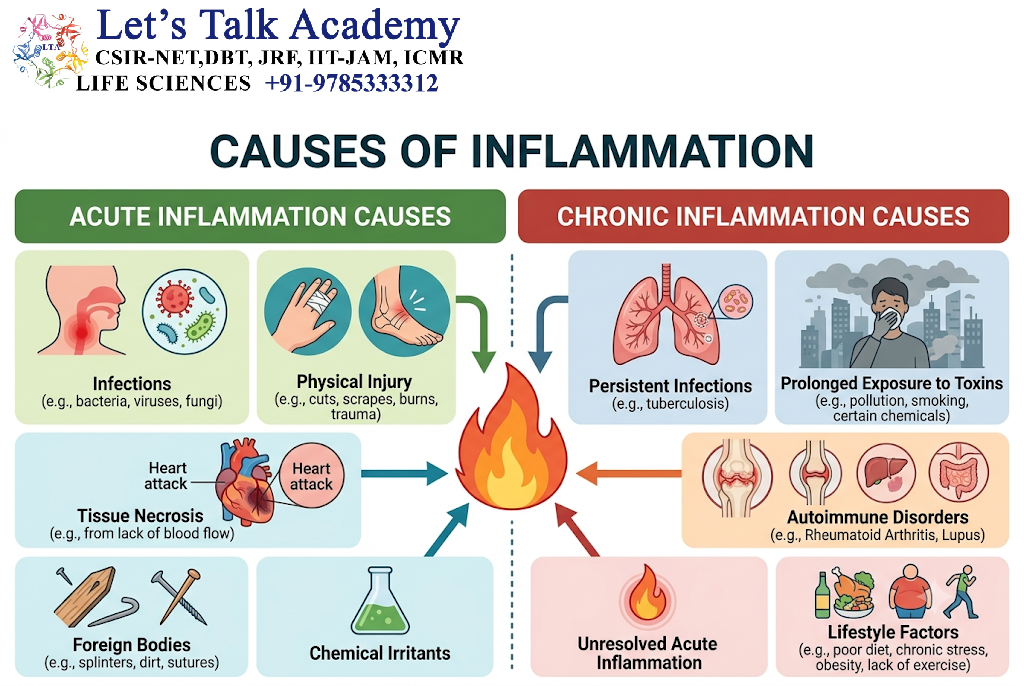
Inflammation can be caused by both external and internal stimuli.
4.1 Microbial Infections
Microorganisms contain molecular structures that are recognized by immune receptors.
Examples include:
- Bacteria
- Viruses
- Fungi
- Protozoa
- Helminths
Microbial molecules are known as pathogen-associated molecular patterns (PAMPs).
Examples include:
- Bacterial lipopolysaccharide
- Flagellin
- Viral nucleic acids
- Fungal cell-wall components
These structures are detected by pattern-recognition receptors (PRRs).
5. Tissue Injury
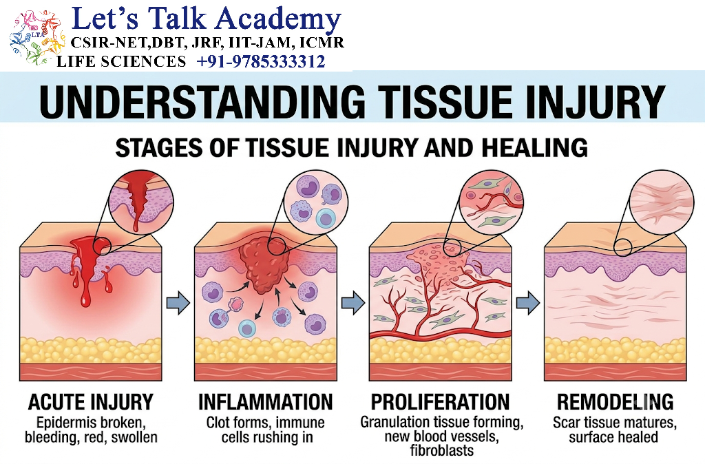
Inflammation may occur even in the absence of infection.
Physical injury can result from:
- Cuts
- Burns
- Trauma
- Frostbite
- Radiation
- Mechanical stress
Damaged cells release intracellular molecules that function as danger signals.
These are called damage-associated molecular patterns (DAMPs).
Examples include:
- ATP
- HMGB1
- DNA fragments
- Uric acid crystals
DAMPs activate inflammatory pathways.
6. Foreign Materials
Foreign substances can trigger inflammation.
Examples include:
- Splinters
- Dust particles
- Crystals
- Surgical materials
- Certain implanted materials
The immune system attempts to eliminate or isolate these substances.
7. Immune-Mediated Inflammation
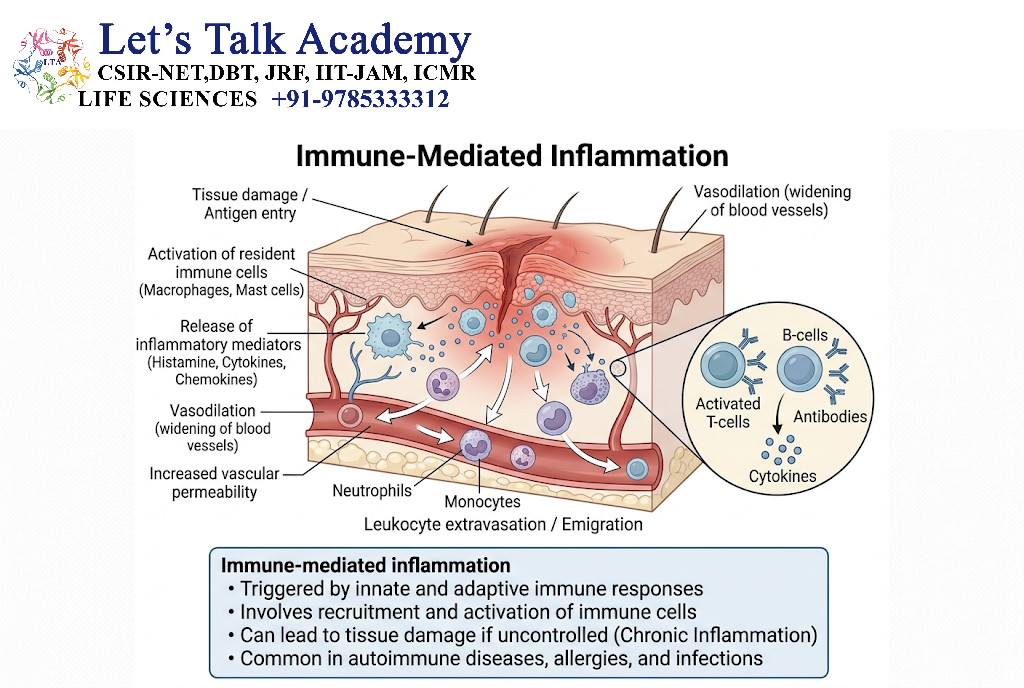
Sometimes inflammation occurs because the immune system itself becomes activated against an inappropriate target.
Examples include:
- Allergic inflammation
- Autoimmune reactions
- Immune-complex reactions
In such situations, inflammation may continue even after the original stimulus is absent.
8. Recognition of the Inflammatory Stimulus
The first important event is recognition.
Cells such as macrophages, dendritic cells, mast cells and epithelial cells contain receptors capable of detecting danger signals.
These receptors include:
- Toll-like receptors
- NOD-like receptors
- RIG-I-like receptors
- C-type lectin receptors
Collectively, these receptors are called pattern-recognition receptors.
Recognition activates intracellular signaling pathways that result in production of inflammatory mediators.
9. Role of Pattern-Recognition Receptors
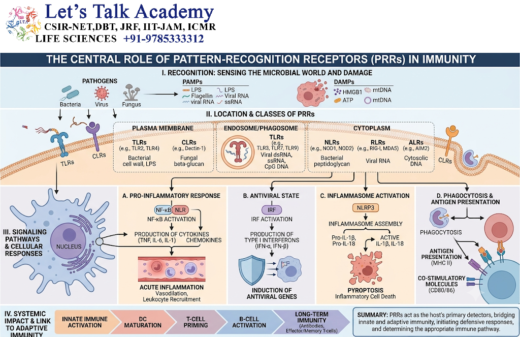
Pattern-recognition receptors recognize:
PAMPs + DAMPs
This activates signaling pathways such as:
- NF-κB pathway
- MAP kinase pathways
- Inflammasome pathways
- Interferon regulatory pathways
These pathways increase the production of:
- TNF
- IL-1
- IL-6
- Chemokines
- Interferons
- Other inflammatory mediators
10. Classification of Inflammation
Inflammation can be classified according to its duration and characteristics.
The two major forms are:
Acute inflammation
Rapid and usually short-lived.
Chronic inflammation
Persistent and prolonged.
The two forms can also overlap. Persistent acute inflammation can eventually develop into chronic inflammation.
11. Acute Inflammation
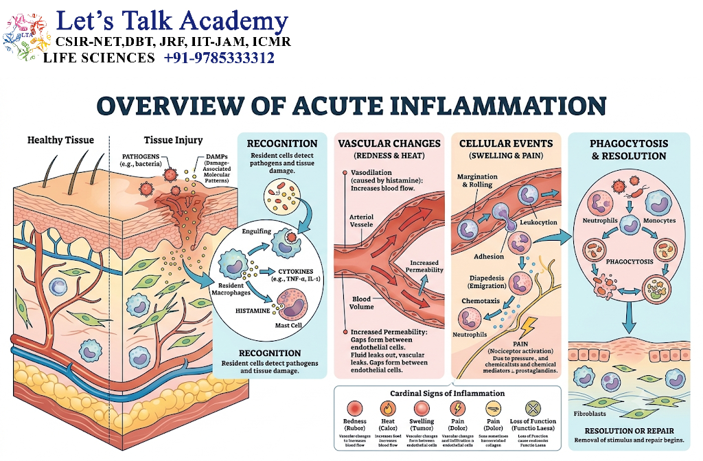
Acute inflammation is the immediate response of vascularized tissue to injury or infection.
It generally develops within minutes to hours and may last for hours or several days depending on the cause.
Its two major components are:
Vascular changes
and
Cellular changes
12. Vascular Changes in Acute Inflammation
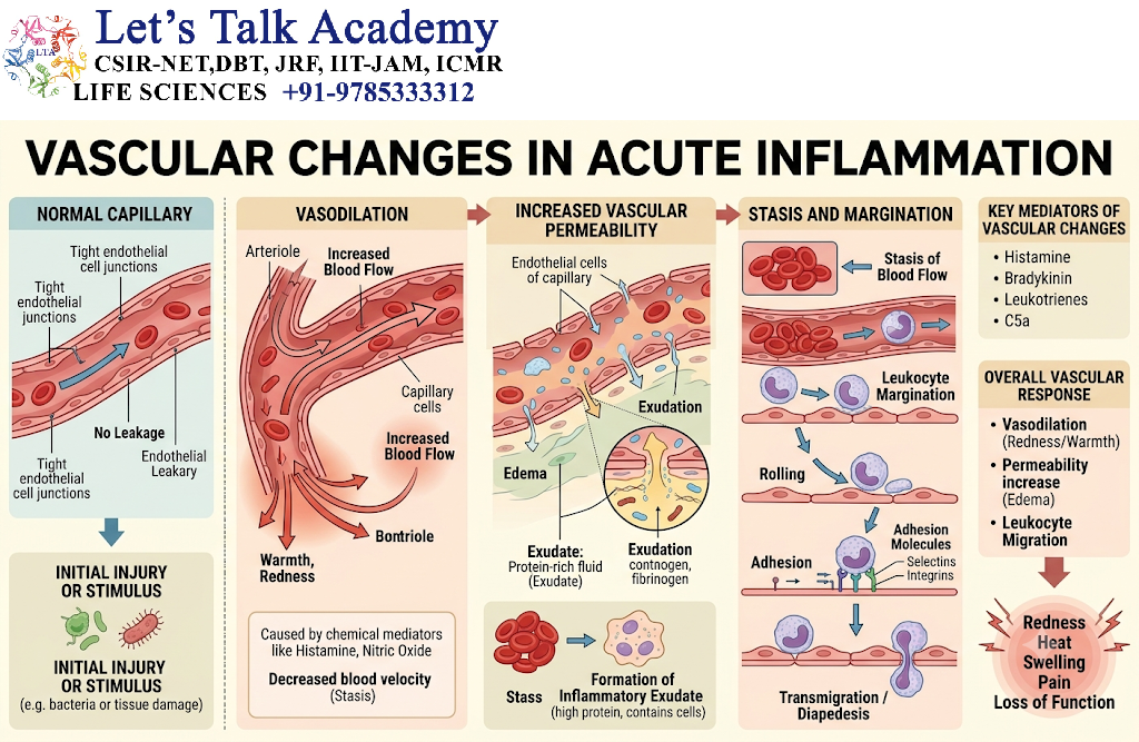
Vascular changes are essential because circulating immune cells and plasma proteins must reach the damaged tissue.
The major changes include:
- Vasodilation
- Increased blood flow
- Increased vascular permeability
- Plasma protein leakage
- Slowing of blood flow
13. Vasodilation
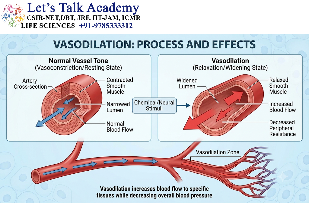
Vasodilation means an increase in the diameter of blood vessels.
Initially, small arterioles dilate.
This causes increased blood flow to the affected tissue.
This increased blood flow produces:
- Redness
- Increased local temperature
Important mediators include:
- Histamine
- Nitric oxide
- Prostaglandins
14. Increased Vascular Permeability
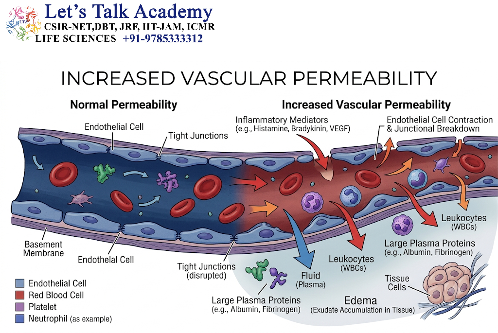
During inflammation, endothelial cells become more permeable.
Normally, blood vessels restrict movement of plasma proteins.
During inflammation, endothelial permeability increases, allowing:
- Water
- Electrolytes
- Albumin
- Complement proteins
- Immunoglobulins
- Fibrinogen
to move into the tissue.
This produces inflammatory exudate.
15. Formation of Edema
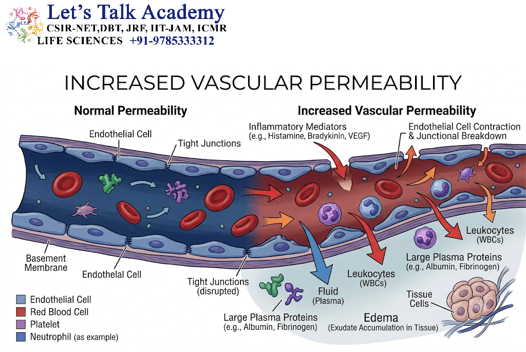
The accumulation of fluid in tissue is called edema.
The sequence is:
Inflammatory mediator release
↓
Vascular permeability increases
↓
Plasma proteins leave blood vessels
↓
Osmotic pressure in tissue increases
↓
Water moves into tissue
↓
Edema develops
Edema contributes to swelling and can increase pressure on sensory nerve endings, producing pain.
16. Exudate
Exudate is protein-rich fluid that accumulates in tissue because of increased vascular permeability.
It may contain:
- Water
- Albumin
- Antibodies
- Complement proteins
- Fibrinogen
- Leukocytes
The presence of antibodies and complement in exudate helps immune cells eliminate microorganisms.
17. Transudate
Transudate is generally a protein-poor fluid produced mainly because of changes in hydrostatic or osmotic pressure rather than inflammation.
Thus:
Exudate → inflammatory vascular permeability
Transudate → pressure-related fluid movement
18. Leukocyte Recruitment
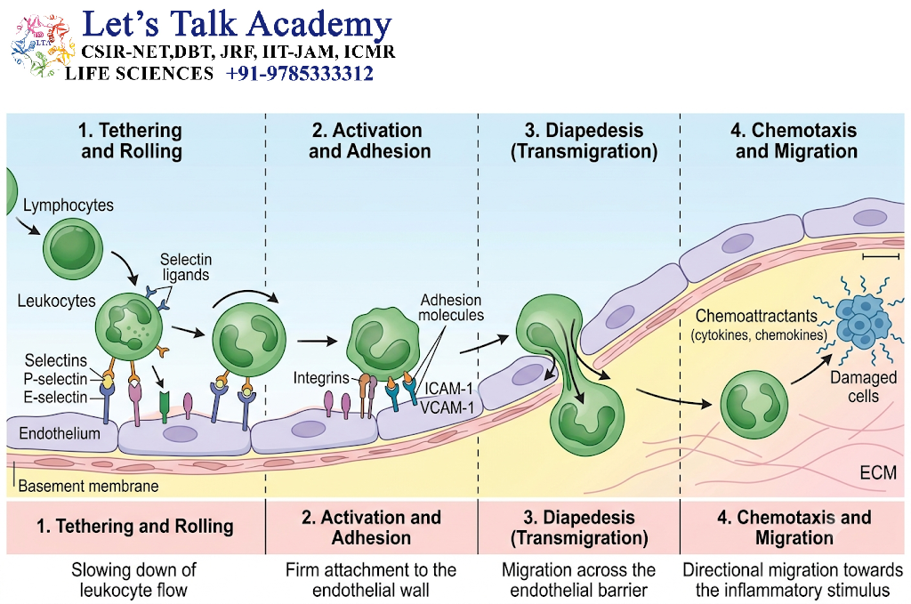
The most important cellular event in inflammation is recruitment of leukocytes from blood to tissue.
The major sequence is:
Margination
↓
Rolling
↓
Adhesion
↓
Transmigration
↓
Chemotaxis
↓
Activation
↓
Phagocytosis / Effector functions
19. Margination
During normal blood flow, erythrocytes occupy the central portion of the blood vessel.
When vascular permeability increases, plasma leaves the vessel.
Blood flow becomes slower.
Leukocytes move toward the endothelial surface.
This movement is called margination.
20. Rolling
Leukocytes temporarily attach to endothelial cells.
The attachments are weak, so leukocytes repeatedly attach and detach while moving along the vessel wall.
This produces a characteristic rolling movement.
The major molecules responsible are selectins.
Important Selectins
| Selectin | Main location |
|---|---|
| E-selectin | Endothelial cells |
| P-selectin | Endothelial cells and platelets |
| L-selectin | Leukocytes |
21. Adhesion
Rolling is followed by firm adhesion.
Inflammatory cytokines such as:
- TNF
- IL-1
stimulate endothelial cells to increase adhesion molecules.
Important adhesion molecules include:
- ICAM-1
- VCAM-1
Leukocyte integrins bind strongly to these molecules.
As a result, the leukocyte becomes firmly attached to the endothelium.
22. Transmigration
After adhesion, leukocytes must leave the blood vessel.
They move between endothelial cells through a process called transmigration or diapedesis.
This occurs mainly in post-capillary venules.
PECAM-1 (CD31) contributes to this process.
The leukocyte then enters the surrounding tissue.
23. Chemotaxis
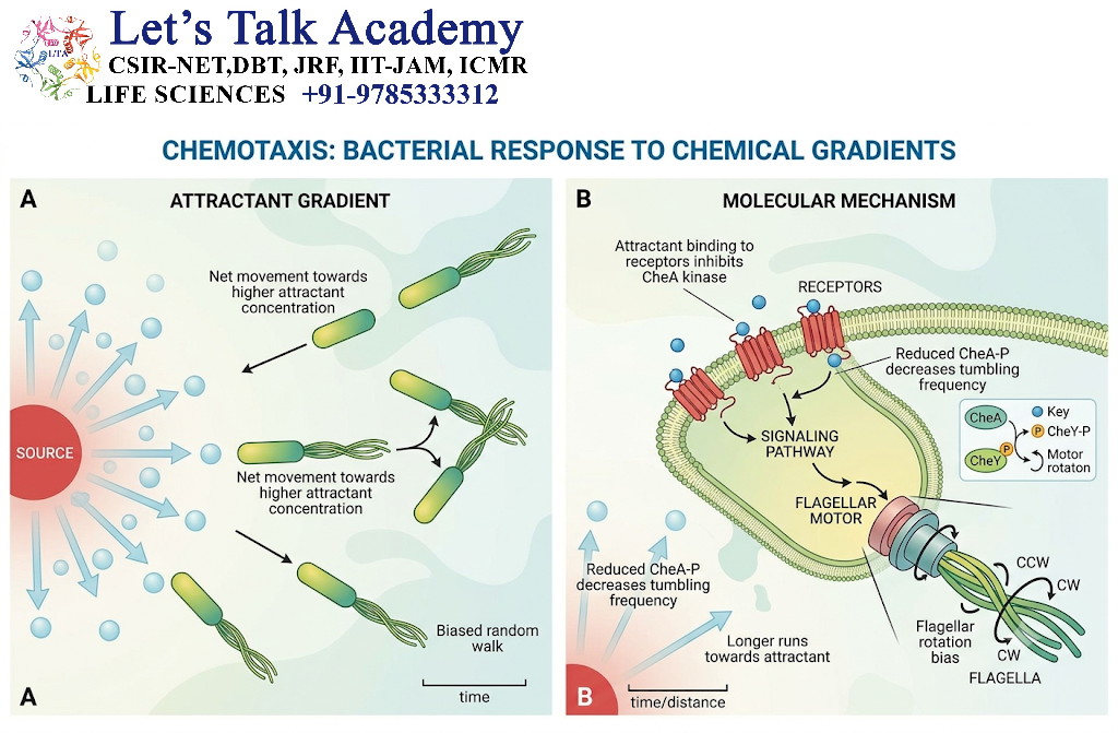
Once leukocytes enter tissue, they must locate the site of injury.
They follow a chemical concentration gradient.
This process is called chemotaxis.
Important chemoattractants include:
- C5a
- LTB4
- CXCL8/IL-8
- Bacterial peptides
The leukocyte moves from a region with lower concentration toward a region with higher concentration of the chemotactic substance.
24. Leukocyte Activation
After reaching the inflammatory site, leukocytes become activated.
Activation causes:
- Increased phagocytosis
- Increased production of reactive oxygen species
- Degranulation
- Cytokine production
- Increased antimicrobial activity
The type of response depends on the microorganism and inflammatory environment.
25. Phagocytosis
Phagocytosis is the process through which specialized cells engulf and destroy foreign particles.
Major professional phagocytes are:
- Neutrophils
- Macrophages
The three major stages are:
Recognition and attachment
The phagocyte recognizes the target.
Engulfment
The target is surrounded by pseudopods.
Killing and degradation
The microorganism is destroyed inside the phagolysosome.
26. Opsonization
Opsonization means coating of a microorganism with molecules that make it easier for phagocytes to recognize and ingest it.
Important opsonins include:
- IgG
- C3b
- Certain collectins
For example:
Bacterium + IgG
↓
IgG-coated bacterium
↓
Fc receptor on phagocyte recognizes IgG
↓
Phagocytosis becomes more efficient
27. Phagosome Formation
After engulfment, the microorganism becomes enclosed in a membrane-bound vesicle called the phagosome.
The phagosome then fuses with lysosomes.
This produces the:
Phagolysosome
Inside the phagolysosome, microorganisms are exposed to:
- Acidic conditions
- Reactive oxygen species
- Reactive nitrogen species
- Lysosomal enzymes
- Antimicrobial proteins
28. Respiratory Burst
Activated phagocytes increase oxygen consumption and generate reactive oxygen species.
This is called the respiratory burst.
A major enzyme involved is:
NADPH oxidase
The pathway begins with production of superoxide.
Superoxide can subsequently generate other reactive oxygen species.
These molecules contribute to microbial killing.
29. Myeloperoxidase System
Neutrophils contain an enzyme called myeloperoxidase (MPO).
MPO uses hydrogen peroxide and chloride ions to generate hypochlorous acid (HOCl).
HOCl is highly effective against many microorganisms.
Thus:
NADPH oxidase
↓
Reactive oxygen species
↓
H₂O₂
↓
MPO
↓
HOCl
↓
Microbial killing
30. Nitric Oxide in Inflammation
Macrophages can produce nitric oxide through inducible nitric oxide synthase.
Nitric oxide and related reactive nitrogen species can contribute to:
- Microbial killing
- Regulation of vascular tone
- Inflammatory signaling
31. Major Inflammatory Cells
Different immune cells dominate different inflammatory situations.
| Cell | Major function |
|---|---|
| Neutrophils | Early antimicrobial response |
| Macrophages | Phagocytosis, cytokines, repair |
| Mast cells | Early mediator release |
| Eosinophils | Parasites and allergic inflammation |
| Lymphocytes | Adaptive immune responses |
| NK cells | Killing infected/abnormal cells |
| Dendritic cells | Antigen presentation |
32. Neutrophils
Neutrophils are usually the first major leukocytes recruited during acute inflammation.
Their functions include:
- Chemotaxis
- Phagocytosis
- Respiratory burst
- Degranulation
- Production of inflammatory mediators
- NET formation
Neutrophils have a short lifespan and are eventually removed from the inflammatory site.
33. Neutrophil Extracellular Traps
Neutrophils can release networks composed of DNA and antimicrobial proteins.
These structures are called NETs (neutrophil extracellular traps).
NETs help trap microorganisms and expose them to antimicrobial molecules.
However, excessive NET formation can also contribute to tissue injury and pathological inflammation.
34. Macrophages
Macrophages are important both in inflammation and tissue repair.
They can:
- Phagocytose microorganisms
- Remove dead cells
- Produce cytokines
- Present antigens
- Release growth factors
- Promote tissue remodeling
Macrophages may therefore have both inflammatory and repair-associated functions.
35. Mast Cells
Mast cells are strategically located near blood vessels and epithelial surfaces.
When activated, they rapidly release stored mediators.
The most important early mediator is:
Histamine
Histamine causes:
- Vasodilation
- Increased vascular permeability
- Endothelial activation
Mast cells also synthesize lipid mediators and cytokines after activation.
36. Eosinophils
Eosinophils are particularly important in:
- Helminth infections
- Allergic inflammation
They contain granules containing toxic proteins such as:
- Major basic protein
- Eosinophil peroxidase
- Eosinophil cationic protein
37. Lymphocytes
Lymphocytes become particularly important in chronic inflammation and adaptive immune responses.
They include:
- T lymphocytes
- B lymphocytes
- Plasma cells
Different T-cell subsets produce different cytokines and regulate different inflammatory processes.
38. Chemical Mediators
Inflammatory mediators can be classified into:
Cell-derived mediators
Produced by:
- Mast cells
- Macrophages
- Neutrophils
- Platelets
- Endothelial cells
Examples:
- Histamine
- Prostaglandins
- Leukotrienes
- Cytokines
- Chemokines
- Nitric oxide
Plasma-derived mediators
Examples:
- Complement proteins
- Bradykinin
- Coagulation-related proteins
39. Histamine
Histamine is stored mainly in mast-cell granules.
It is released rapidly after mast-cell activation.
Major effects:
Histamine
↓
Vasodilation
Increased vascular permeability
↓
Increased blood flow + edema
Histamine is particularly important during the early phase of inflammation.
40. Arachidonic Acid Metabolism
Cell membranes contain phospholipids.
When cells are activated, phospholipase A₂ releases arachidonic acid.
Arachidonic acid is then metabolized through two major pathways:
Cyclooxygenase pathway
and
Lipoxygenase pathway
41. Cyclooxygenase Pathway
The cyclooxygenase pathway produces prostanoids, including:
- Prostaglandins
- Prostacyclin
- Thromboxanes
Prostaglandins are involved in:
- Pain
- Fever
- Vasodilation
42. Lipoxygenase Pathway
The lipoxygenase pathway produces leukotrienes.
Important leukotrienes include:
- LTB4
- LTC4
- LTD4
- LTE4
LTB4 promotes leukocyte recruitment.
Some cysteinyl leukotrienes contribute to bronchoconstriction and increased vascular permeability.
43. Cytokines in Inflammation
Cytokines are small signaling proteins that coordinate immune responses.
Important inflammatory cytokines include:
TNF
Produced by activated macrophages and other cells.
Functions include:
- Endothelial activation
- Leukocyte recruitment
- Fever
- Systemic inflammatory effects
IL-1
Important functions include:
- Fever
- Endothelial activation
- Leukocyte recruitment
IL-6
Important functions include:
- Acute-phase protein production
- Systemic inflammatory responses
- Regulation of immune-cell activity
44. Chemokines
Chemokines direct leukocyte migration.
For example:
CXCL8 (IL-8)
is an important chemokine for neutrophil recruitment.
Chemokines establish gradients that guide leukocytes toward sites of infection or tissue damage.
45. Complement-Mediated Inflammation
Complement contributes to inflammation through several mechanisms.
C3a and C5a
can promote inflammatory responses.
C5a
is particularly important for:
- Chemotaxis
- Leukocyte activation
C3b
promotes opsonization and phagocytosis.
46. Bradykinin
Bradykinin is generated through plasma proteolytic pathways.
Its major effects include:
- Pain
- Vasodilation
- Increased vascular permeability
- Smooth-muscle contraction in certain tissues
It is therefore an important contributor to inflammatory pain and vascular changes.
47. Acute-Phase Response
Inflammation at a local site can produce systemic changes.
Cytokines such as IL-6, IL-1 and TNF act on the liver and other organs.
The liver increases production of acute-phase proteins.
Important acute-phase proteins include:
- C-reactive protein
- Fibrinogen
- Serum amyloid A
- Complement proteins
48. Fever
Fever is a regulated increase in body temperature during infection or inflammation.
Inflammatory cytokines stimulate pathways leading to increased production of prostaglandin E₂ in the hypothalamic region.
This raises the body’s temperature set point.
Thus:
Inflammatory stimulus
↓
Cytokine production
↓
Prostaglandin pathway
↓
Hypothalamic set-point elevation
↓
Fever
49. Chronic Inflammation
Chronic inflammation occurs when the inflammatory response continues for a prolonged period.
It may result from:
- Persistent infection
- Autoimmune reactions
- Long-term exposure to harmful substances
- Persistent tissue injury
The characteristic feature is simultaneous:
Inflammation + Tissue destruction + Repair
This distinguishes chronic inflammation from a simple short-lived inflammatory response.
50. Cells of Chronic Inflammation
The major cells include:
Macrophages
Central regulators of chronic inflammation.
Lymphocytes
Produce cytokines and regulate immune responses.
Plasma cells
Produce antibodies.
Eosinophils
Important in certain parasitic and allergic conditions.
51. Tissue Destruction in Chronic Inflammation
Persistent inflammatory cells continuously release:
- Proteases
- Reactive oxygen species
- Cytokines
- Growth factors
- Other mediators
If these substances remain elevated, they can damage normal tissue.
Thus, chronic inflammation can become a self-maintaining cycle:
Persistent stimulus
↓
Immune-cell activation
↓
Mediator production
↓
Tissue damage
↓
Release of additional danger signals
↓
Further inflammation
52. Granulomatous Inflammation
Granulomatous inflammation is a specialized type of chronic inflammation.
A granuloma is an organized collection of activated macrophages, often accompanied by lymphocytes.
It develops when the immune system attempts to contain a substance that is difficult to eliminate.
Activated macrophages may develop an epithelial-cell-like appearance and are called epithelioid cells.
Multinucleated giant cells may also develop through fusion of macrophages.
53. Resolution of Inflammation
Inflammation must eventually be terminated.
Resolution is not simply the passive disappearance of inflammation. It is an active, regulated process.
Important events include:
- Removal of the inflammatory stimulus.
- Reduction of inflammatory mediator production.
- Cessation of leukocyte recruitment.
- Apoptosis of inflammatory cells.
- Clearance of dead cells.
- Removal of inflammatory fluid.
- Restoration of vascular integrity.
- Tissue regeneration or repair.
54. Specialized Pro-Resolving Mediators
Several lipid mediators actively promote the resolution phase.
These include:
- Lipoxins
- Resolvins
- Protectins
- Maresins
They help reduce excessive leukocyte recruitment and promote clearance of inflammatory material.
55. Outcomes of Inflammation
Inflammation can have several possible outcomes.
Complete resolution
The tissue returns toward its original state.
Fibrosis
Connective tissue replaces damaged tissue when regeneration is insufficient.
Abscess
A localized collection of pus may develop.
Chronic inflammation
Persistent stimuli may cause prolonged inflammation.
56. Inflammation and Tissue Repair
Inflammation and tissue repair are closely connected.
Once the harmful stimulus is controlled, repair mechanisms become increasingly important.
Repair involves:
- Cell proliferation
- Angiogenesis
- Fibroblast activation
- Extracellular matrix production
- Collagen deposition
- Tissue remodeling
Macrophages play an important role in coordinating these processes.
57. Acute and Chronic Inflammation
| Characteristic | Acute | Chronic |
|---|---|---|
| Onset | Rapid | Slow/persistent |
| Duration | Short | Long |
| Main cells | Neutrophils | Macrophages, lymphocytes |
| Fluid exudation | Prominent | Usually less prominent |
| Tissue damage | Usually limited | Often progressive |
| Repair | After inflammation | Occurs alongside inflammation |
| Fibrosis | Usually limited | Common |


