
1. Introduction
The immune system is designed to protect the body from infectious microorganisms, abnormal cells, and potentially harmful foreign substances. Under normal conditions, immune responses are carefully regulated so that they are strong enough to eliminate harmful agents but controlled enough to prevent unnecessary injury to healthy tissues.
However, immune responses can become harmful in two major ways. In hypersensitivity, an immune response that is normally protective causes excessive or inappropriate tissue injury. In autoimmunity, the immune system loses tolerance to self-components and mounts an immune response against the body’s own cells, tissues, or molecules.
Hypersensitivity and autoimmunity are related concepts, but they are not identical. Hypersensitivity describes an excessive or inappropriate immune-mediated response that causes tissue damage, whereas autoimmunity specifically involves immune recognition of self-antigens. An autoimmune response may or may not produce clinically significant disease, while autoimmune disease occurs when such responses cause persistent tissue dysfunction or injury.
Understanding these conditions requires knowledge of immune tolerance, antigen recognition, antibodies, T lymphocytes, complement, cytokines, inflammatory mediators, and mechanisms of tissue damage.
2. Hypersensitivity
2.1 Definition of Hypersensitivity
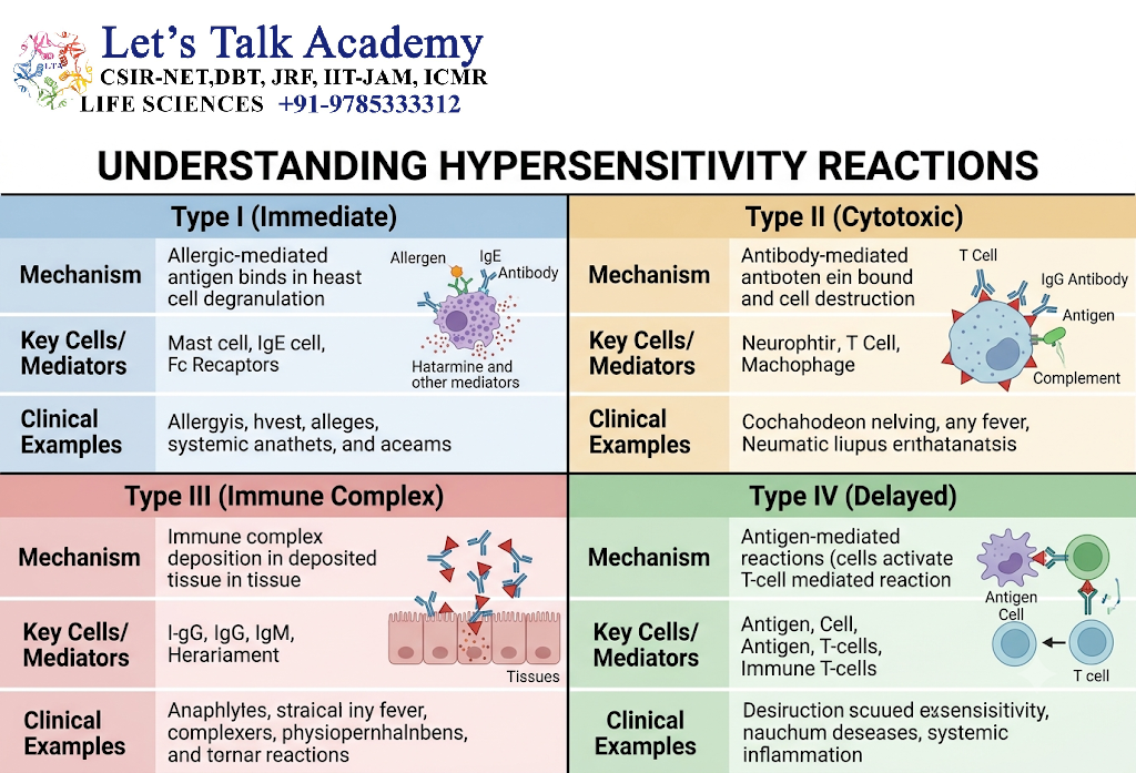
Hypersensitivity refers to an exaggerated, inappropriate, or damaging immune response that occurs after exposure to an antigen in a previously sensitized individual.
The antigen responsible for hypersensitivity may be:
- An environmental allergen
- A microbial antigen
- A drug or chemical
- A self-antigen
- An antigen associated with another biological substance
Hypersensitivity reactions can occur rapidly within minutes or may develop over several hours or days depending on the underlying immune mechanism.
The classical classification divides hypersensitivity into four major types:
- Type I – Immediate hypersensitivity
- Type II – Antibody-mediated hypersensitivity
- Type III – Immune-complex-mediated hypersensitivity
- Type IV – T-cell-mediated or delayed-type hypersensitivity
The first three types are predominantly antibody-associated, whereas Type IV hypersensitivity is primarily mediated by T cells.
3. Type I Hypersensitivity
3.1 Overview
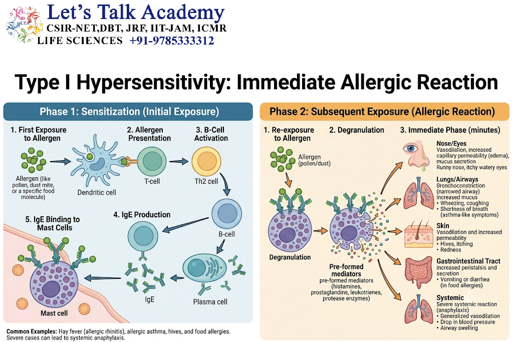
Type I hypersensitivity, also called immediate hypersensitivity, is primarily mediated by IgE antibodies, mast cells, basophils, and associated inflammatory mediators.
It is commonly associated with allergic reactions.
Examples include:
- Allergic rhinitis
- Bronchial asthma involving allergic mechanisms
- Urticaria
- Food allergy
- Atopic dermatitis
- Anaphylaxis
- Some insect-sting reactions
The reaction may develop very rapidly after exposure to the relevant allergen.
3.2 Sensitization Phase
The first exposure to an allergen usually produces a sensitization response rather than an immediate severe reaction.
The general sequence is:
Allergen exposure → antigen presentation → T-cell differentiation → cytokine production → B-cell activation → IgE production → mast-cell sensitization
A key population of T cells involved in classical allergic responses is the Th2 subset.
Th2-associated cytokines, particularly IL-4 and IL-13, promote B-cell class switching toward IgE production.
The IgE antibodies then bind to high-affinity FcεRI receptors on mast cells and basophils.
The individual is now considered sensitized to that allergen.
3.3 Re-exposure and Mast-Cell Activation
During subsequent exposure, the allergen binds to IgE molecules already attached to mast cells.
When the allergen cross-links adjacent IgE–FcεRI complexes, intracellular signaling is initiated.
This results in mast-cell activation and degranulation.
Important mediators include:
- Histamine
- Leukotrienes
- Prostaglandins
- Cytokines
- Chemokines
- Proteases
These mediators produce the clinical manifestations of immediate hypersensitivity.
3.4 Early-Phase Reaction
The early phase generally occurs rapidly after allergen exposure.
Histamine and other preformed mediators can cause:
- Vasodilation
- Increased vascular permeability
- Smooth-muscle contraction
- Increased mucus secretion
- Local edema
- Itching
For example, increased vascular permeability can contribute to swelling and tissue edema.
3.5 Late-Phase Reaction
The late phase develops after the initial reaction and involves recruitment of inflammatory cells.
Important cells include:
- Eosinophils
- Basophils
- Th2 cells
- Monocytes
- Other inflammatory leukocytes
Cytokines and chemokines released during the response maintain inflammation and can cause prolonged tissue injury.
3.6 Anaphylaxis
Anaphylaxis is a severe systemic hypersensitivity reaction that can develop rapidly after exposure to an allergen.
Mast-cell and basophil mediator release can produce widespread effects involving:
- Blood vessels
- Respiratory tract
- Skin
- Gastrointestinal tract
- Cardiovascular system
Severe reactions can result in airway compromise and circulatory collapse and require immediate medical treatment.
4. Type II Hypersensitivity
4.1 Overview
Type II hypersensitivity is primarily mediated by IgG or IgM antibodies directed against antigens associated with cell surfaces or extracellular matrix components.
Unlike Type I hypersensitivity, IgE is not the principal antibody class involved.
The target antigen may be:
- A molecule naturally present on a cell
- A receptor
- A basement-membrane component
- A foreign molecule attached to a host cell
4.2 Mechanisms of Tissue Injury
Type II hypersensitivity can cause tissue damage through several mechanisms.
4.2.1 Complement Activation
Antibody binding to a target can activate the classical complement pathway.
Complement activation can result in:
- Opsonization
- Inflammation
- Recruitment of leukocytes
- Membrane attack complex formation
4.2.2 Opsonization and Phagocytosis
Antibodies and complement fragments can coat target cells.
Phagocytes recognize these molecules through Fc and complement receptors.
This promotes engulfment and destruction of the antibody-coated cell.
4.2.3 Antibody-Dependent Cellular Cytotoxicity
Fc-receptor-bearing cells such as NK cells can recognize antibody-coated targets.
This may result in cellular cytotoxicity through mechanisms such as ADCC.
4.2.4 Functional Alteration of Receptors
Not all Type II reactions destroy cells.
An antibody may instead bind to a receptor and alter its function.
The antibody can either:
- Stimulate the receptor, or
- Block the receptor
This mechanism is particularly important in certain autoimmune disorders.
4.3 Examples of Type II Hypersensitivity
Examples include:
- Autoimmune hemolytic anemia
- Goodpasture disease
- Graves disease
- Myasthenia gravis
- Hemolytic disease of the newborn
These conditions demonstrate that antibody-mediated responses can produce tissue injury either by destroying cells or by changing the function of cellular receptors.
5. Type III Hypersensitivity
5.1 Overview
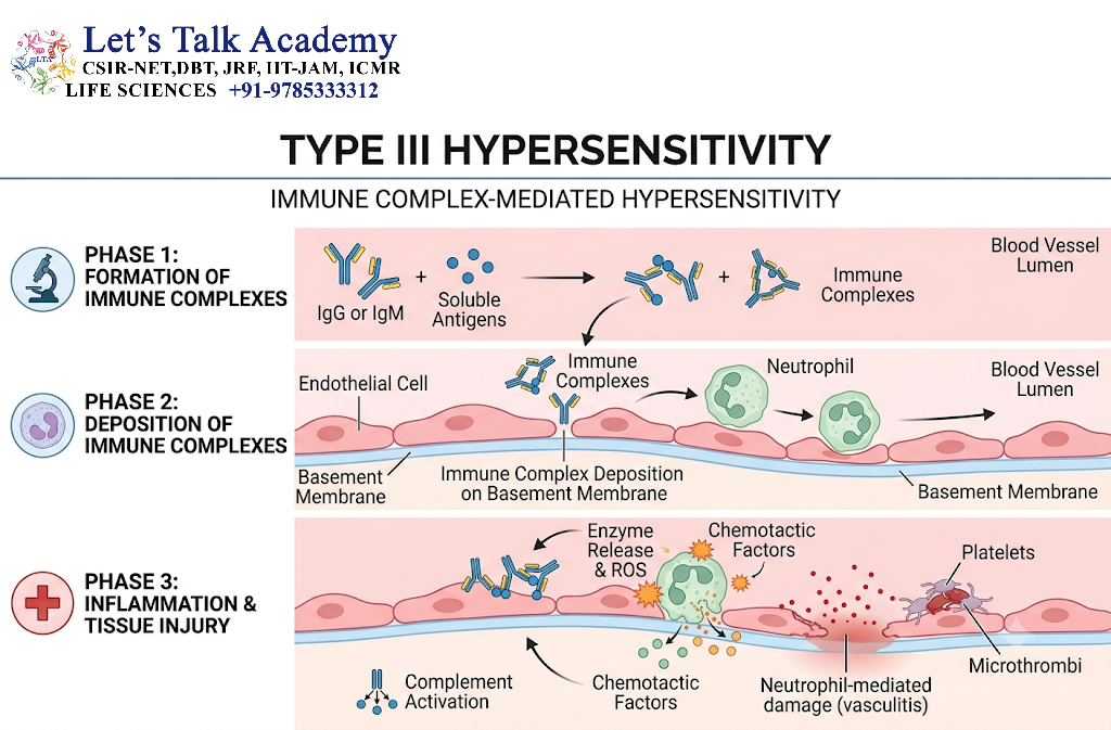
Type III hypersensitivity results from the formation and deposition of antigen–antibody immune complexes.
These immune complexes may circulate in the blood or form locally within tissues.
When they become deposited in tissues, they can activate complement and recruit inflammatory cells.
5.2 Formation of Immune Complexes
The basic process can be summarized as:
Antigen + antibody → immune complex → deposition → complement activation → inflammatory-cell recruitment → tissue injury
Immune complexes are normally removed efficiently by phagocytic cells.
Problems arise when complexes are produced in large amounts, are inadequately cleared, or become deposited in susceptible tissues.
5.3 Complement Activation
Immune-complex deposition can activate the classical complement pathway.
Complement fragments such as C3a and C5a contribute to inflammation.
C5a is particularly important as a chemotactic and activating mediator for leukocytes.
5.4 Leukocyte Recruitment
Neutrophils are recruited to the site of immune-complex deposition.
They attempt to remove the deposited complexes but may release:
- Proteolytic enzymes
- Reactive oxygen species
- Other inflammatory mediators
These substances can damage surrounding tissues.
5.5 Examples of Type III Hypersensitivity
Examples include:
- Systemic lupus erythematosus
- Serum sickness
- Arthus reaction
- Some forms of immune-complex glomerulonephritis
- Certain vasculitic disorders
6. Type IV Hypersensitivity
6.1 Overview
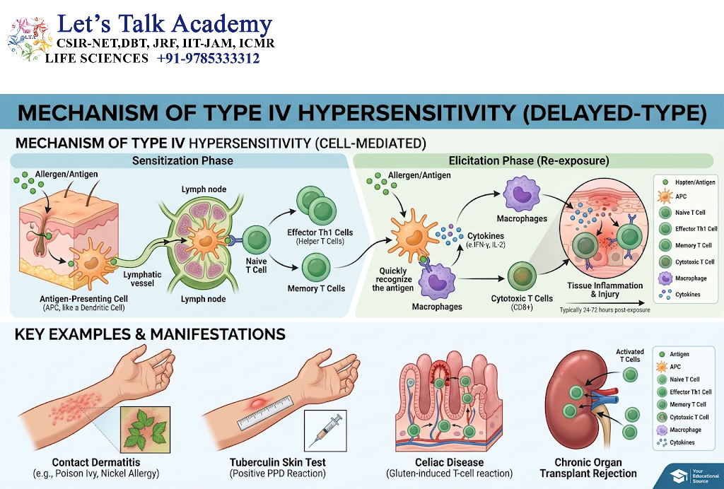
Type IV hypersensitivity differs from Types I, II, and III because it is primarily T-cell mediated rather than antibody mediated.
It is therefore also called:
- Delayed-type hypersensitivity
- T-cell-mediated hypersensitivity
The reaction usually develops more slowly than immediate hypersensitivity.
6.2 Major T-Cell Mechanisms
Different T-cell populations can contribute to Type IV hypersensitivity.
Important mechanisms include:
- Th1-mediated macrophage activation
- Th17-mediated recruitment of inflammatory cells
- CD8⁺ T-cell-mediated cytotoxicity
6.3 Th1-Mediated Responses
Th1 cells produce cytokines such as IFN-γ, which can activate macrophages.
Activated macrophages increase their antimicrobial activity but can also contribute to tissue injury when activation becomes excessive or persistent.
6.4 Th17-Mediated Responses
Th17 cells produce cytokines including IL-17.
IL-17 promotes recruitment and activation of inflammatory cells, particularly neutrophils.
Persistent Th17-mediated inflammation can contribute to chronic tissue injury.
6.5 CD8⁺ T-Cell-Mediated Injury
CD8⁺ cytotoxic T cells can directly kill target cells.
They can use:
- Perforin–granzyme mechanisms
- Fas–FasL signaling
This mechanism can produce tissue injury when the target cells are healthy host cells.
6.6 Examples of Type IV Hypersensitivity
Examples include:
- Contact dermatitis
- Tuberculin skin reaction
- Some drug-related hypersensitivity reactions
- Certain autoimmune tissue injuries
7. Comparison of the Four Types of Hypersensitivity
| Type | Major immune component | Major mechanism | Typical timing |
|---|---|---|---|
| Type I | IgE, mast cells | Mast-cell mediator release | Rapid |
| Type II | IgG/IgM | Antibody-mediated cellular or tissue injury | Variable |
| Type III | Immune complexes | Complement and inflammation | Variable |
| Type IV | T cells | Cellular inflammation or cytotoxicity | Delayed |
The classification is useful for understanding the dominant mechanism, although real diseases may involve more than one immune pathway.
8. Autoimmunity
8.1 Definition
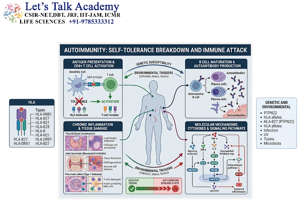
Autoimmunity is an immune response directed against the body’s own molecules, cells, or tissues.
The immune system normally develops mechanisms of self-tolerance that prevent strong immune responses against healthy self-components.
Autoimmunity develops when these mechanisms fail or become insufficient.
The presence of autoreactive lymphocytes alone does not necessarily mean that an individual has autoimmune disease. Disease develops when autoreactivity contributes to clinically significant tissue dysfunction or injury.
9. Self-Tolerance
9.1 Definition of Immune Tolerance
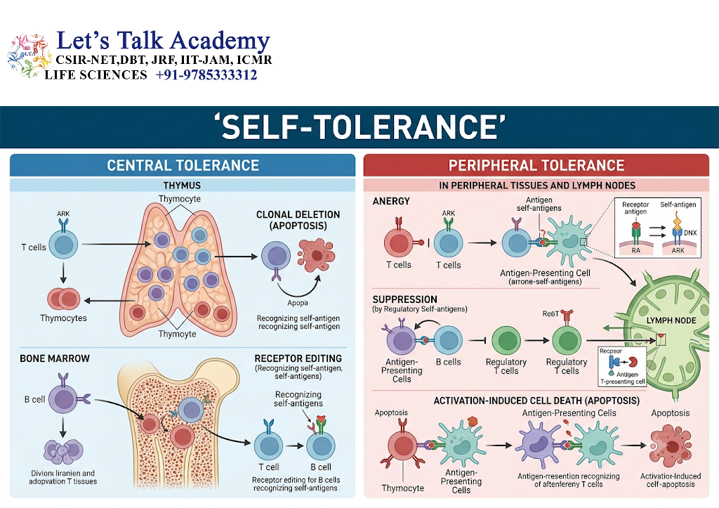
Immune tolerance is a state in which the immune system does not mount an inappropriate destructive response against a particular antigen.
Tolerance to self is essential for preventing autoimmune disease.
Tolerance is maintained through several mechanisms.
These include:
- Central tolerance
- Peripheral tolerance
- Regulatory T cells
- Anergy
- Deletion
- Inhibitory receptor signaling
- Limited accessibility of certain antigens
10. Central Tolerance
10.1 T-Cell Central Tolerance
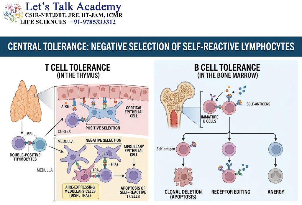
T cells develop in the thymus.
During development, immature T cells are exposed to self-antigens.
T cells with strongly self-reactive receptors can undergo negative selection, resulting in their deletion.
Some developing T cells with self-reactivity may also contribute to the regulatory T-cell population.
10.2 B-Cell Central Tolerance
B cells develop primarily in the bone marrow.
Strongly self-reactive B cells may undergo:
- Clonal deletion
- Receptor editing
- Functional inactivation
Receptor editing is particularly important because an immature B cell can rearrange its immunoglobulin light-chain genes to alter antigen specificity.
11. Peripheral Tolerance
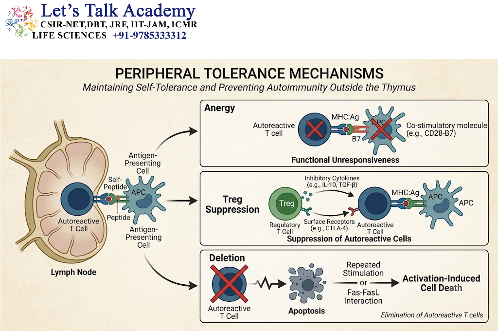
Central tolerance is not complete. Some self-reactive lymphocytes can escape into peripheral tissues.
Therefore, additional mechanisms operate outside primary lymphoid organs.
11.1 Anergy
A lymphocyte may become functionally unresponsive when it recognizes antigen without receiving the appropriate co-stimulatory signals.
This state is called anergy.
11.2 Regulatory T Cells
Regulatory T cells (Tregs) suppress potentially harmful immune responses.
They contribute to immune tolerance through mechanisms involving:
- CTLA-4
- IL-10
- TGF-β
- Suppression of effector T-cell responses
- Modulation of antigen-presenting cells
11.3 Inhibitory Receptors
Immune cells express inhibitory receptors that help prevent excessive activation.
Important inhibitory pathways include:
- CTLA-4
- PD-1
These pathways contribute to peripheral tolerance and immune regulation.
12. Mechanisms Responsible for Autoimmunity
Autoimmune disease is usually multifactorial rather than being caused by a single defect.
Several mechanisms can contribute.
12.1 Breakdown of Self-Tolerance
Failure of central or peripheral tolerance can permit autoreactive lymphocytes to become activated.
12.2 Genetic Susceptibility
Certain genetic variants can increase susceptibility to autoimmune disease.
Genes associated with antigen presentation and immune regulation are particularly important.
Human leukocyte antigen (HLA) variants are associated with susceptibility to several autoimmune disorders.
However, genetic susceptibility alone is generally insufficient to explain disease development.
12.3 Environmental Triggers
Environmental factors can contribute to autoimmune disease in genetically susceptible individuals.
Potential triggers include:
- Infections
- Certain medications
- Tissue injury
- Altered exposure to self-antigens
- Environmental factors affecting immune regulation
The specific contribution varies among diseases.
12.4 Molecular Mimicry
Molecular mimicry occurs when microbial antigens resemble host molecules sufficiently that an immune response against the microorganism may cross-react with self-components.
The simplified mechanism is:
Microbial antigen → immune response → cross-reactive lymphocytes or antibodies → recognition of similar self-antigen → tissue injury
Molecular mimicry is one proposed mechanism in some autoimmune conditions, although its contribution differs between diseases.
12.5 Release of Sequestered Antigens
Some self-antigens are normally hidden from immune surveillance.
Tissue injury may expose previously inaccessible antigens and potentially initiate an immune response.
12.6 Epitope Spreading
During chronic tissue damage, new self-epitopes may become available to the immune system.
The immune response can then expand from an initial epitope to additional epitopes.
This process is called epitope spreading.
12.7 Abnormal Co-Stimulation
Inflammatory conditions can increase expression of co-stimulatory molecules on antigen-presenting cells.
This may increase the likelihood that autoreactive T cells receive sufficient signals for activation.
13. Autoantibodies
13.1 Definition
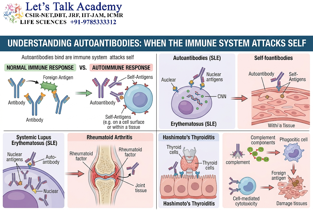
Autoantibodies are antibodies directed against self-antigens.
They can be detected in many autoimmune diseases.
However, the presence of an autoantibody does not automatically establish that an individual has an autoimmune disease.
Autoantibodies may:
- Directly damage cells
- Activate complement
- Alter receptor function
- Form immune complexes
- Serve as diagnostic biomarkers
13.2 Examples of Functionally Important Autoantibodies
Some autoantibodies have direct pathogenic effects.
For example, antibodies against the acetylcholine receptor can interfere with neuromuscular transmission in myasthenia gravis.
In Graves disease, antibodies against the thyroid-stimulating hormone receptor can stimulate receptor activity.
These examples demonstrate that antibodies can cause disease not only by destroying cells but also by altering normal physiological signaling.
14. Major Categories of Autoimmune Disease
Autoimmune diseases can broadly be classified according to the extent of tissue involvement.
14.1 Organ-Specific Autoimmunity
In organ-specific autoimmune diseases, the immune response is directed primarily toward antigens associated with a particular organ.
Examples include:
- Type 1 diabetes mellitus
- Graves disease
- Hashimoto thyroiditis
- Myasthenia gravis
- Multiple sclerosis
14.2 Systemic Autoimmunity
Systemic autoimmune diseases affect multiple organs or tissues.
Examples include:
- Systemic lupus erythematosus
- Rheumatoid arthritis
- Systemic sclerosis
- Sjögren syndrome
These diseases can involve combinations of antibody-mediated, immune-complex-mediated, and T-cell-mediated mechanisms.
15. Systemic Lupus Erythematosus
15.1 Overview
Systemic lupus erythematosus (SLE) is a systemic autoimmune disease characterized by immune responses against multiple self-antigens.
Autoantibodies can form immune complexes that contribute to inflammation and tissue injury.
15.2 Immunological Mechanisms
Important mechanisms include:
- Loss of tolerance
- Production of multiple autoantibodies
- Formation of immune complexes
- Complement activation
- Inflammatory-cell recruitment
- Tissue injury
Affected organs may include:
- Skin
- Kidneys
- Joints
- Blood cells
- Nervous system
- Cardiovascular system
The clinical presentation varies substantially between individuals.
16. Rheumatoid Arthritis
16.1 Overview
Rheumatoid arthritis is a chronic autoimmune inflammatory disease that primarily affects joints but can also involve extra-articular tissues.
Both innate and adaptive immune mechanisms contribute to disease development.
16.2 Immune Mechanisms
Important components include:
- Autoreactive T cells
- B cells
- Autoantibodies
- Macrophages
- Cytokines
- Synovial fibroblasts
- Inflammatory mediators
Cytokines such as TNF, IL-6, and other inflammatory mediators contribute to persistent synovial inflammation and joint damage.
17. Type 1 Diabetes Mellitus
Type 1 diabetes mellitus is characterized by immune-mediated destruction of insulin-producing pancreatic β cells.
T cells play an important role in the destruction of β cells.
The loss of functional β cells results in reduced insulin production and impaired glucose regulation.
Autoantibodies against pancreatic antigens can serve as useful markers of autoimmune activity, although T-cell-mediated mechanisms are central to β-cell destruction.
18. Graves Disease
Graves disease illustrates an autoimmune mechanism in which antibodies alter receptor function rather than simply destroying target cells.
Autoantibodies directed against the thyroid-stimulating hormone receptor can stimulate thyroid-cell activity.
This results in increased thyroid hormone production.
Thus, Graves disease is an important example of antibody-mediated receptor stimulation.
19. Myasthenia Gravis
Myasthenia gravis demonstrates the opposite type of receptor dysfunction.
Autoantibodies commonly target components of the neuromuscular junction, particularly the acetylcholine receptor.
These antibodies can interfere with normal neuromuscular transmission.
The resulting physiological effect is muscle weakness that typically worsens with repeated activity.
This condition illustrates that autoimmune antibodies can produce disease through functional blockade or receptor disruption.
20. Hashimoto Thyroiditis
Hashimoto thyroiditis is an autoimmune disorder involving immune-mediated damage to the thyroid gland.
Autoantibodies against thyroid-associated antigens can be detected, while T-cell-mediated mechanisms also contribute to tissue injury.
Progressive destruction of thyroid tissue can result in reduced thyroid hormone production.
21. Relationship Between Hypersensitivity and Autoimmunity
Hypersensitivity and autoimmunity are closely connected because autoimmune diseases can use one or more hypersensitivity mechanisms to produce tissue injury.
For example:
- Autoantibodies against cell-surface antigens can produce Type II hypersensitivity.
- Immune complexes containing autoantibodies can produce Type III hypersensitivity.
- Autoreactive T cells can produce Type IV hypersensitivity.
Therefore, autoimmunity describes the target of the immune response—self—whereas hypersensitivity describes the damaging nature or mechanism of the immune response.
A single autoimmune disease may involve multiple mechanisms simultaneously.
22. Hypersensitivity Versus Autoimmunity
| Feature | Hypersensitivity | Autoimmunity |
|---|---|---|
| Basic concept | Harmful or excessive immune response | Immune response against self |
| Target | Foreign or self-antigens | Self-antigens |
| Main mechanisms | IgE, antibodies, immune complexes, T cells | Autoantibodies, autoreactive T cells, immune complexes |
| Tissue damage | Central feature | Occurs when autoimmunity becomes pathogenic |
| Examples | Allergy, immune-complex reactions, contact dermatitis | SLE, Graves disease, myasthenia gravis |
The two concepts can overlap, but they should not be treated as synonyms.
23. Important Immunological Mechanisms
23.1 Antibody-Mediated Tissue Injury
Antibodies can damage cells by:
- Activating complement
- Promoting phagocytosis
- Recruiting cytotoxic cells
- Altering receptor function
- Forming immune complexes
23.2 T-Cell-Mediated Tissue Injury
T cells can cause tissue damage by:
- Direct cytotoxicity
- Macrophage activation
- Cytokine secretion
- Recruitment of inflammatory cells
23.3 Immune-Complex-Mediated Injury
Immune complexes can deposit in tissues and activate complement.
The resulting inflammatory response can damage:
- Blood vessels
- Kidneys
- Joints
- Skin
- Other tissues
24. Factors Influencing Autoimmune Disease
Autoimmune disease generally results from interactions between multiple factors.
24.1 Genetic Factors
Genetic variants can influence:
- Antigen presentation
- Lymphocyte development
- Cytokine signaling
- Immune regulation
- Clearance of apoptotic cells
24.2 Environmental Factors
Environmental exposure can influence disease initiation or progression.
Infections are particularly important because they can generate inflammatory conditions that modify immune activation.
24.3 Hormonal Influences
Some autoimmune diseases show differences in frequency between sexes, suggesting that hormonal and other biological factors can influence immune regulation.
However, the underlying mechanisms vary between diseases and are not explained by a single factor.
25. Diagnosis of Hypersensitivity and Autoimmune Disorders
Diagnosis depends on the specific disease and generally combines clinical findings with laboratory and sometimes tissue-based investigations.
Tests may include:
- Autoantibody detection
- Immunoglobulin measurements
- Complement measurements
- Inflammatory markers
- Allergy testing
- Antigen-specific antibody testing
- Imaging
- Histopathological examination
- Functional immune assays
No single test is sufficient for diagnosing every autoimmune or hypersensitivity disorder.
26. Regulation and Prevention of Excessive Immune Responses
The immune system possesses several mechanisms that limit excessive inflammation.
These include:
- Regulatory T cells
- Anti-inflammatory cytokines
- Inhibitory receptors
- Controlled apoptosis of immune cells
- Resolution of inflammation
- Clearance of immune complexes
- Restoration of tissue homeostasis
Failure of these regulatory mechanisms can allow immune responses to persist.
27. Integrated View of Hypersensitivity and Autoimmunity
The following sequence provides a useful conceptual framework:
Antigen exposure or recognition
↓
Activation of immune cells
↓
Antibody production and/or T-cell activation
↓
Effector mechanisms
↓
Inflammatory mediator release, cytotoxicity, complement activation, or immune-complex deposition
↓
Tissue injury
In hypersensitivity, the response is inappropriate or excessive.
In autoimmunity, the immune system recognizes self-components.
When both conditions overlap, an autoimmune response can produce tissue damage through a hypersensitivity mechanism.


