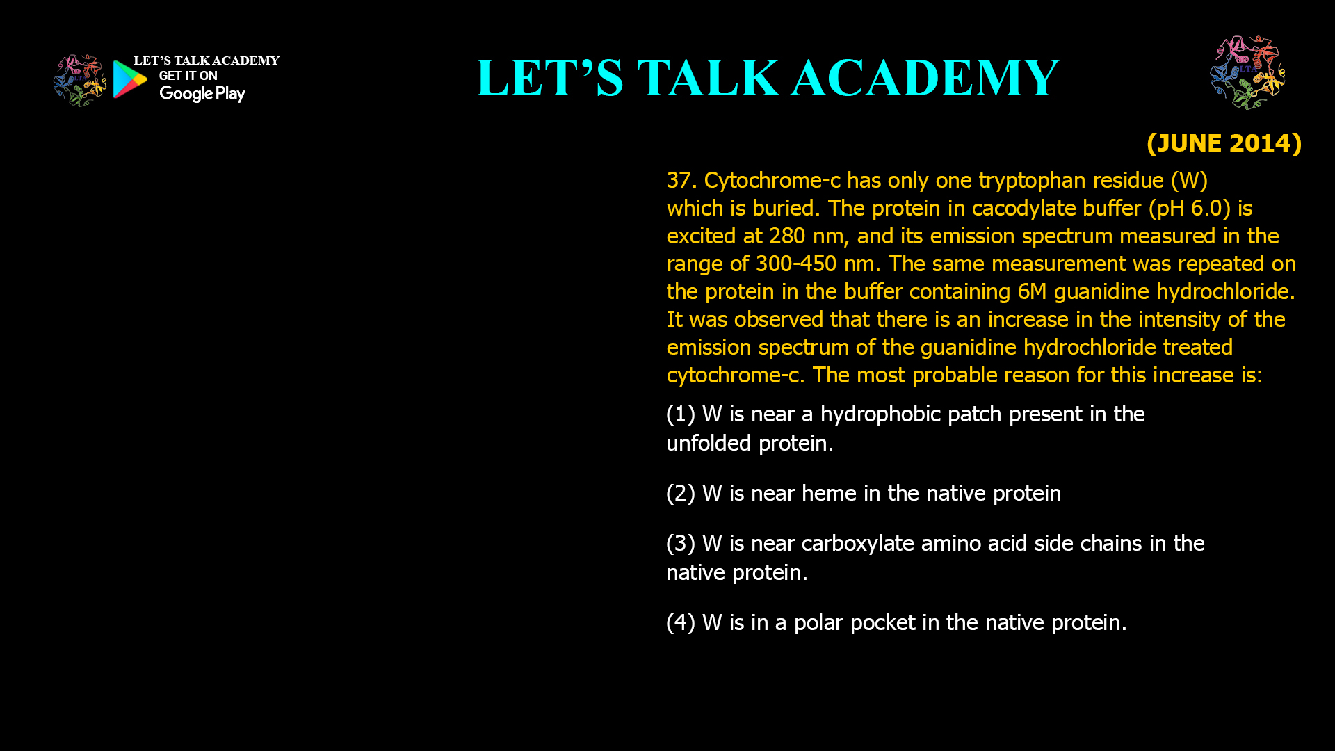
37. Cytochrome-c has only one tryptophan residue (W) which is buried. The protein in cacodylate buffer (pH 6.0) is excited at 280 nm, and its emission spectrum measured in the range of 300–450 nm. The same measurement was repeated on the protein in the buffer containing 6 M guanidine hydrochloride. It was observed that there is an increase in the intensity of the emission spectrum of the guanidine hydrochloride-treated cytochrome-c. The most probable reason for this increase is:
(A) W is near a hydrophobic patch present in the unfolded protein.
(B) W is near heme in the native protein.
(C) W is near carboxylate amino acid side chains in the native protein.
(D) W is in a polar pocket in the native protein.
Fluorescence Emission of Cytochrome-c After Guanidine Hydrochloride Denaturation
Correct Answer
Option (2): W is near heme in the native protein.
Explanation
Fluorescence spectroscopy is one of the most sensitive techniques for studying protein folding and conformational changes. Proteins containing aromatic amino acids such as tryptophan, tyrosine, and phenylalanine exhibit intrinsic fluorescence when excited with ultraviolet light. Among these residues, tryptophan is the strongest fluorophore because of its high quantum yield and strong absorption near 280 nm. Consequently, changes in tryptophan fluorescence are widely used to investigate the structural environment of proteins.
Cytochrome-c contains only a single tryptophan residue, making its fluorescence particularly easy to interpret. In the native folded protein, this tryptophan residue is located close to the heme prosthetic group. The heme functions as a highly efficient fluorescence quencher because the excited-state energy of tryptophan is transferred non-radiatively to the heme through electron transfer and resonance energy transfer mechanisms. As a result, the fluorescence intensity of native cytochrome-c is significantly reduced.
When the protein is treated with 6 M guanidine hydrochloride, the strong chaotropic agent disrupts hydrogen bonding, hydrophobic interactions, and other non-covalent forces that stabilize the native conformation. The protein unfolds, increasing the distance between the tryptophan residue and the heme group. Since fluorescence quenching decreases rapidly as the distance between the fluorophore and quencher increases, separation of tryptophan from the heme allows a much larger fraction of the absorbed energy to be emitted as fluorescence. Consequently, the emission intensity increases after denaturation.
The observed increase in fluorescence therefore does not result from the tryptophan becoming more hydrophobic or more polar. Instead, it arises because unfolding removes the quenching effect exerted by the nearby heme group in the native protein.
Why Option (1) is Incorrect
A hydrophobic patch in an unfolded protein would not explain the increase in fluorescence intensity observed here. During denaturation, hydrophobic regions generally become exposed to the solvent rather than forming new hydrophobic pockets. More importantly, the dominant factor controlling the fluorescence of cytochrome-c is quenching by the heme group, not the presence of hydrophobic regions.
Why Option (2) is Correct
The native structure of cytochrome-c places the single tryptophan residue close to the heme prosthetic group. The heme efficiently quenches tryptophan fluorescence through non-radiative energy transfer. Denaturation by guanidine hydrochloride unfolds the protein and separates the tryptophan from the heme, reducing quenching and producing a significant increase in fluorescence intensity. This explanation is fully consistent with the experimental observation.
Why Option (3) is Incorrect
Carboxylate-containing amino acid side chains such as aspartate and glutamate can influence the local environment of tryptophan, but they are not sufficiently efficient fluorescence quenchers to account for the dramatic increase observed after denaturation. The characteristic fluorescence behaviour of cytochrome-c is primarily governed by the nearby heme group.
Why Option (4) is Incorrect
If the tryptophan residue were located within a polar pocket in the native protein, unfolding would generally expose it to the aqueous environment, which often causes a decrease in fluorescence intensity together with a red shift in the emission maximum. This behaviour does not explain the increased fluorescence observed after treatment with guanidine hydrochloride.
Effect of Guanidine Hydrochloride on Protein Structure
Guanidine hydrochloride is a powerful protein denaturant that disrupts hydrogen bonds, electrostatic interactions, and hydrophobic packing within proteins. As these stabilizing interactions are lost, the compact tertiary structure unfolds and previously buried amino acid residues become exposed to the solvent. Denaturation also alters the spatial relationships between fluorophores and quenching groups, making fluorescence spectroscopy an effective method for monitoring protein unfolding.
Fluorescence Quenching by the Heme Group
The heme prosthetic group contains a conjugated porphyrin ring capable of accepting excitation energy from nearby fluorophores. When tryptophan is located close to the heme, excited-state energy is transferred efficiently to the porphyrin ring instead of being emitted as fluorescence. This process, known as fluorescence quenching, greatly reduces the observed emission intensity. Increasing the separation between tryptophan and the heme during unfolding diminishes this energy transfer and restores fluorescence.
Conclusion
The increase in fluorescence intensity observed after treatment of cytochrome-c with guanidine hydrochloride is caused by disruption of the native protein structure, which separates the single tryptophan residue from the nearby heme prosthetic group. As the quenching effect of the heme decreases, tryptophan fluorescence becomes significantly more intense. Therefore, the correct answer is Option (2): W is near heme in the native protein.


