
1. Introduction to the Complement System
The complement system is an essential component of the innate immune system that protects the body against invading microorganisms and helps eliminate damaged or abnormal cells. It consists of a large group of soluble proteins, membrane-associated proteins, receptors, and regulatory molecules that interact with one another in a highly organized cascade.
The term complement originated from the observation that certain heat-sensitive components of blood could enhance, or “complement,” the activity of antibodies in destroying microorganisms. Although complement was initially considered primarily an antibody-associated defense mechanism, it is now known to be an important part of both innate and adaptive immunity.
Complement proteins circulate predominantly in the blood and extracellular fluids in inactive forms. When an appropriate trigger is encountered, these proteins become sequentially activated. Each activated component can promote activation of the next component, resulting in rapid amplification of the response.
The major consequences of complement activation include:
- Recognition and elimination of microorganisms
- Opsonization of pathogens for efficient phagocytosis
- Direct destruction of susceptible target cells
- Recruitment and activation of inflammatory cells
- Removal of immune complexes and cellular debris
- Enhancement of adaptive immune responses
- Maintenance of tissue homeostasis
The complement system is therefore not simply a collection of proteins that destroy pathogens. It is a sophisticated surveillance and effector network that connects pathogen recognition, inflammation, phagocytosis, cell lysis, and immune regulation.
2. Components of the Complement System
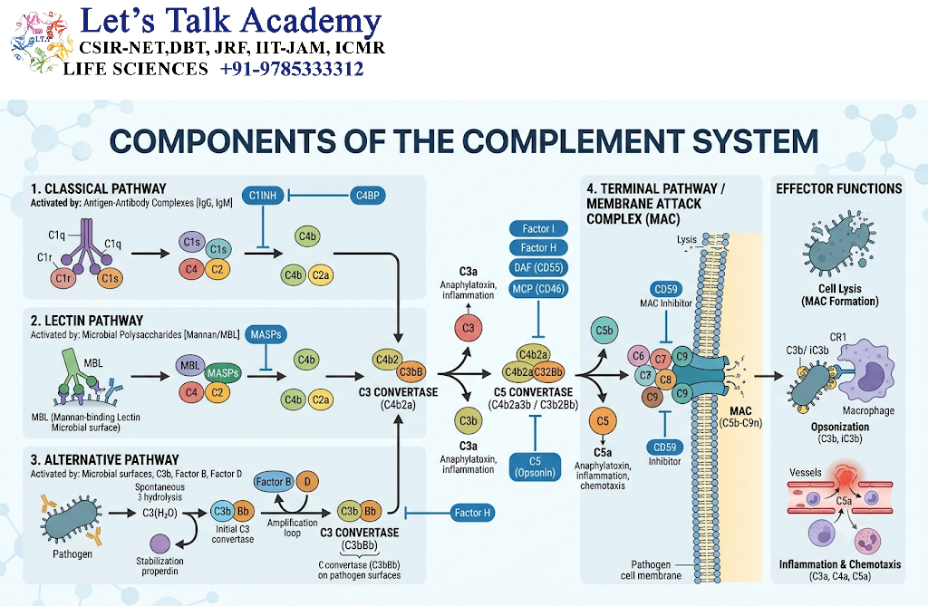
The complement system contains more than 30 proteins and protein fragments. These components are found either in the circulation, on cell surfaces, or within tissues.
Most complement proteins are synthesized by the liver, although important amounts are also produced by monocytes, macrophages, epithelial cells, endothelial cells, and other tissues.
Complement components are commonly designated by the letter C followed by a number, such as C1, C2, C3, C4, C5, C6, C7, C8, and C9.
Some complement proteins are designated by letters rather than numbers, including:
- Factor B
- Factor D
- Factor H
- Factor I
- Properdin
Complement receptors and regulatory proteins have separate names and functions.
2.1 Major Complement Components
The principal components include:
| Component | Major function |
|---|---|
| C1 | Initiates the classical pathway |
| C2 | Participates in formation of the classical/lectin pathway C3 convertase |
| C3 | Central component of complement activation |
| C4 | Participates in classical and lectin pathways |
| C5 | Initiates the terminal pathway after cleavage |
| C6–C9 | Form the membrane attack complex |
| Factor B | Component of the alternative pathway C3 convertase |
| Factor D | Cleaves Factor B during alternative pathway activation |
| Properdin | Stabilizes the alternative pathway C3 convertase |
| Factor H | Regulates the alternative pathway |
| Factor I | Inactivates C3b and related fragments |
Among these components, C3 is particularly important because all three major complement activation pathways converge at the level of C3 activation.
3. Complement Activation Pathways
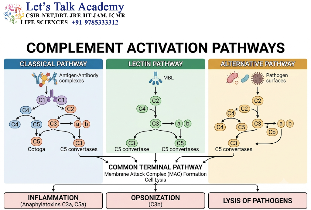
Complement can be activated through three major pathways:
- Classical pathway
- Lectin pathway
- Alternative pathway
Although these pathways differ in their initiating mechanisms, they ultimately generate enzymes called C3 convertases. These enzymes cleave C3 and initiate a common sequence of events that can result in formation of the membrane attack complex.
3.1 Classical Pathway
The classical pathway is generally initiated when antibodies bound to an antigen interact with the C1 complex.
The pathway is strongly associated with antibodies of the IgM and certain IgG subclasses.
The C1 complex consists of:
- C1q
- C1r
- C1s
C1q recognizes the Fc region of antibodies that have already bound to antigen. Binding of C1q causes activation of C1r and C1s, which are serine proteases.
Activated C1s cleaves C4 into:
- C4a
- C4b
C4b attaches to the nearby surface, while C4a is released.
C1s then cleaves C2 into C2a and C2b. The surface-associated C4b combines with C2a to form the classical pathway C3 convertase:
C4b2a
The C3 convertase cleaves C3 into:
- C3a
- C3b
C3b can attach to the microbial surface and participate in the formation of the C5 convertase.
The classical pathway therefore provides an important mechanism by which antibodies can recruit complement to the surface of a pathogen.
3.2 Lectin Pathway
The lectin pathway is activated without requiring antibodies.
It begins when soluble pattern-recognition molecules recognize specific carbohydrate structures present on microbial surfaces.
Important recognition molecules include:
- Mannose-binding lectin (MBL)
- Ficolins
MBL recognizes carbohydrate residues such as mannose and N-acetylglucosamine that are commonly present on microbial surfaces.
MBL is associated with enzymes called MBL-associated serine proteases, or MASPs.
Following recognition of microbial carbohydrates, MASPs become activated and cleave C4 and C2. This produces the same C3 convertase generated by the classical pathway:
C4b2a
Thus, the lectin pathway joins the complement cascade at the C3 activation stage.
The lectin pathway is particularly important because it provides antibody-independent recognition of conserved microbial structures.
3.3 Alternative Pathway
The alternative pathway differs substantially from the classical and lectin pathways because it can become activated directly on suitable microbial surfaces and does not require antibodies.
A small amount of C3 undergoes spontaneous hydrolysis in plasma. This process continuously provides a low level of complement surveillance.
When C3b becomes associated with an appropriate microbial surface, it can bind Factor B.
Factor D then cleaves Factor B, generating an active complex that functions as an alternative pathway C3 convertase:
C3bBb
Properdin can stabilize this enzyme complex and increase its activity.
The alternative pathway therefore provides an important amplification mechanism for complement activation.
3.4 Comparison of the Three Pathways
| Feature | Classical pathway | Lectin pathway | Alternative pathway |
|---|---|---|---|
| Major initiating mechanism | Antibody-antigen complexes | Microbial carbohydrates | Microbial surfaces and spontaneous C3 activation |
| Antibody requirement | Usually associated with antibodies | No | No |
| Recognition molecules | C1q | MBL and ficolins | C3b and associated factors |
| Major C3 convertase | C4b2a | C4b2a | C3bBb |
| Major biological outcome | Complement activation | Complement activation | Amplification and surveillance |
Despite their different initiation mechanisms, all three pathways converge on C3.
4. C3: The Central Component of Complement
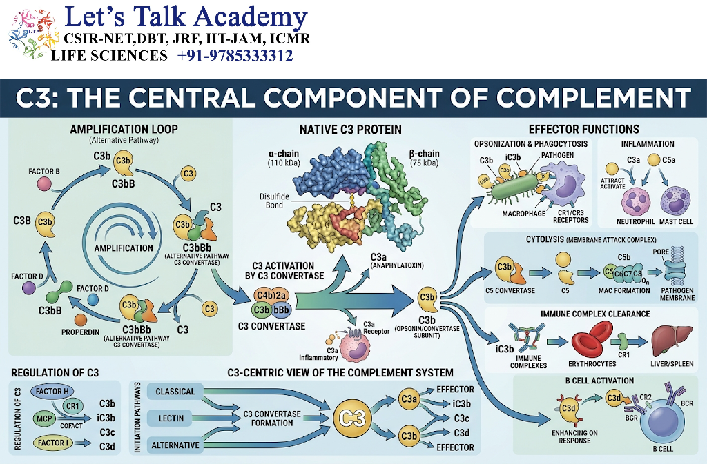
C3 is considered the central molecule of the complement system because activation of all three pathways ultimately leads to C3 cleavage.
C3 is cleaved by C3 convertases into:
C3 → C3a + C3b
These fragments have different biological functions.
4.1 Functions of C3a
C3a is an anaphylatoxin. It contributes to inflammatory responses by acting on cells such as mast cells and other immune cells.
Its effects can include:
- Promotion of inflammatory mediator release
- Increased vascular permeability
- Recruitment and activation of immune cells
- Contribution to local inflammatory responses
4.2 Functions of C3b
C3b is one of the most important opsonins of the complement system.
It attaches to microbial surfaces and makes those microorganisms easier for phagocytes to recognize and ingest.
C3b also participates in formation of C5 convertases.
Therefore, C3b connects complement activation with both opsonization and terminal complement activation.
4.3 iC3b and Other C3 Fragments
C3b can be further processed by complement regulatory mechanisms into iC3b.
Although iC3b no longer functions as a C5 convertase component, it remains an important opsonin.
Complement receptors on phagocytes can recognize C3-derived fragments and promote phagocytosis.
5. Formation of C5 Convertase
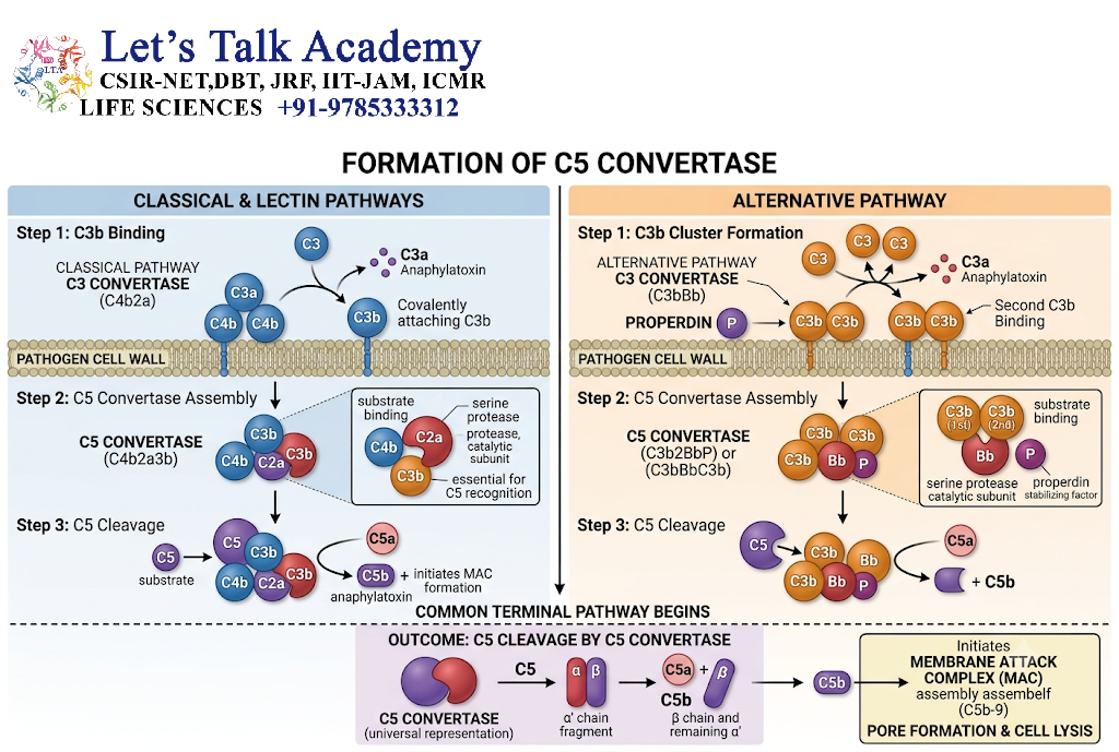
Once sufficient C3b has been deposited, C3 convertases can acquire additional C3b and become C5 convertases.
For the classical and lectin pathways, the C5 convertase is commonly represented as:
C4b2a3b
For the alternative pathway, it is commonly represented as:
C3bBb3b
C5 convertases cleave C5 into:
C5 → C5a + C5b
These two fragments have distinct functions.
5.1 C5a
C5a is one of the most potent inflammatory mediators generated by the complement system.
It promotes:
- Chemotaxis of leukocytes
- Activation of neutrophils
- Increased endothelial adhesion
- Release of inflammatory mediators
- Enhancement of inflammatory responses
C5a is therefore a major link between complement activation and acute inflammation.
5.2 C5b
C5b initiates the terminal complement pathway.
It binds sequentially to C6, C7, C8, and multiple molecules of C9 to form the membrane attack complex.
6. Membrane Attack Complex
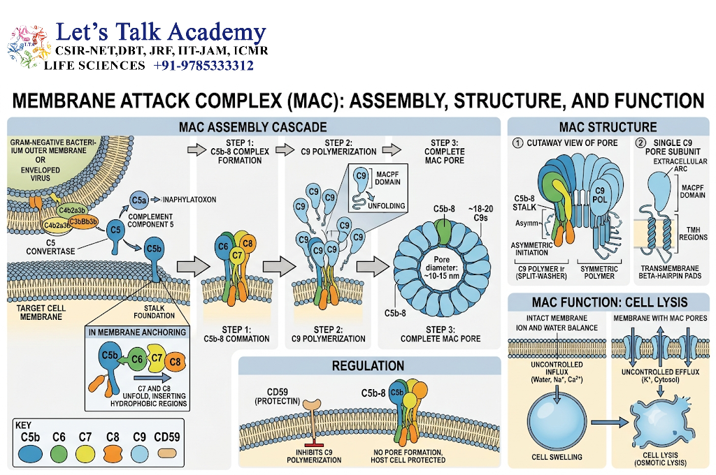
The membrane attack complex, or MAC, is the terminal lytic structure of the complement system.
It consists primarily of:
C5b-C6-C7-C8-C9
The complex inserts into the lipid bilayer of susceptible target cells and forms a transmembrane pore.
This pore disrupts membrane integrity and can lead to osmotic imbalance and cell lysis.
6.1 Steps in MAC Formation
The major sequence is:
- C5 is cleaved into C5a and C5b.
- C5b binds C6.
- The C5b6 complex binds C7.
- C5b67 associates with the target membrane.
- C8 binds to the complex and contributes to membrane insertion.
- Multiple C9 molecules polymerize.
- A membrane pore is formed.
The MAC is particularly effective against certain Gram-negative bacteria, especially Neisseria species.
7. Major Biological Functions of Complement
Complement performs several important functions in host defense.
7.1 Opsonization
Opsonization refers to the coating of a microorganism with molecules that facilitate its recognition and uptake by phagocytes.
C3b and iC3b are major complement-derived opsonins.
Phagocytes possess complement receptors that recognize these fragments.
The overall process can be summarized as:
Microbe → C3b deposition → complement receptor recognition → phagocytosis
Opsonization is particularly important when microorganisms are difficult for phagocytes to recognize directly.
7.2 Inflammation
Complement contributes strongly to inflammation.
C3a and C5a are called anaphylatoxins because they can promote inflammatory reactions.
C5a is especially important in:
- Neutrophil recruitment
- Neutrophil activation
- Endothelial activation
- Increased vascular permeability
- Release of inflammatory mediators
Complement therefore helps direct immune cells toward sites of infection or tissue injury.
7.3 Direct Cell Lysis
The membrane attack complex can directly damage susceptible microorganisms.
This mechanism is especially important for some Gram-negative bacteria.
However, not all microorganisms are equally susceptible to MAC-mediated killing.
7.4 Clearance of Immune Complexes
Complement contributes to the removal of antigen-antibody complexes from the circulation.
C3b deposited on immune complexes can interact with complement receptors on erythrocytes and other cells. These complexes can then be transported to organs such as the liver and spleen, where they are removed by phagocytic cells.
Efficient immune-complex clearance helps prevent excessive deposition of immune complexes in tissues.
7.5 Removal of Dead and Damaged Cells
Complement proteins also participate in recognition and clearance of apoptotic cells and cellular debris.
This function contributes to tissue homeostasis and helps prevent unnecessary persistence of cellular material that could promote inflammation.
7.6 Enhancement of Adaptive Immunity
Complement does not operate independently of adaptive immunity.
Complement fragments can enhance B-cell responses.
For example, complement fragment C3d can remain associated with antigen and interact with complement receptor 2 (CR2), also known as CD21, on B cells.
The interaction between antigen-associated C3d and CR2 can lower the threshold required for B-cell activation.
Thus, complement provides an important bridge between innate immune recognition and adaptive immune responses.
8. Complement Receptors
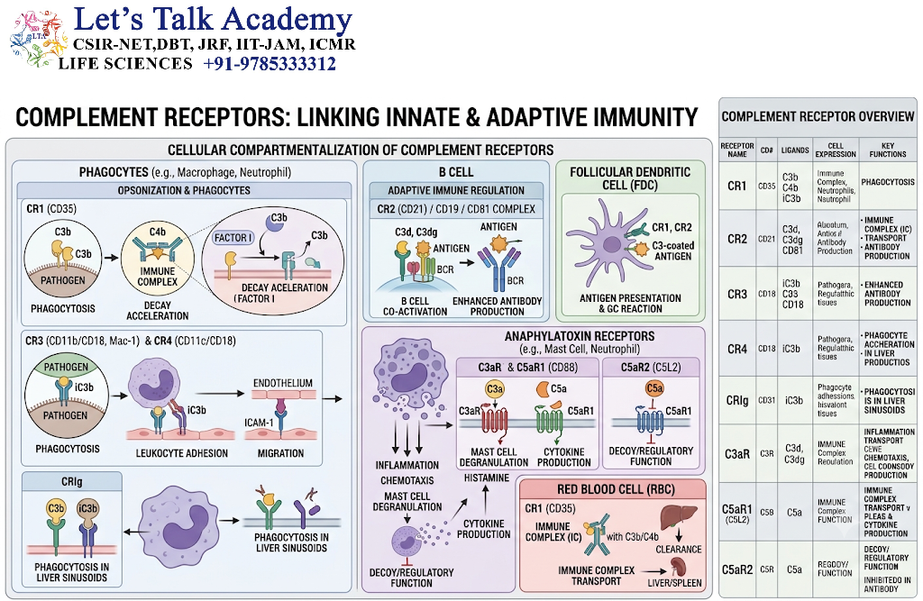
Complement receptors are found on various immune and non-immune cells.
Important receptors include:
8.1 CR1
CR1, also known as CD35, recognizes complement fragments including C3b and C4b.
It is expressed on:
- Erythrocytes
- B cells
- Neutrophils
- Monocytes
- Macrophages
CR1 contributes to immune-complex clearance and phagocytosis.
8.2 CR2
CR2, or CD21, is expressed prominently on B cells.
It recognizes complement fragments such as C3d.
CR2 plays an important role in enhancing B-cell activation.
8.3 CR3
CR3 is an integrin expressed on various leukocytes.
It recognizes iC3b and contributes to:
- Adhesion
- Phagocytosis
- Cell migration
- Immune-cell activation
8.4 C5a Receptor
The C5a receptor, particularly C5aR1, is expressed on several inflammatory cells.
C5a-C5aR signaling contributes to:
- Leukocyte chemotaxis
- Cell activation
- Inflammatory responses
9. Regulation of the Complement System
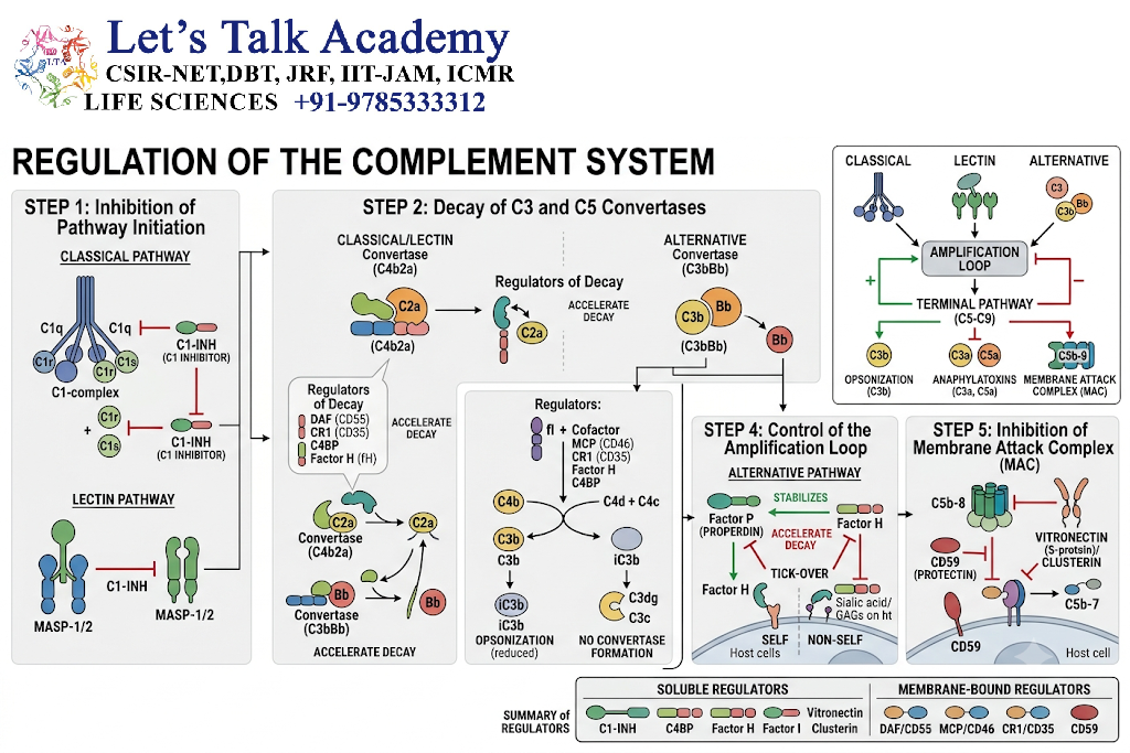
Because complement activation can cause inflammation and tissue damage, it must be tightly controlled.
Complement regulatory proteins prevent uncontrolled activation on host cells.
Important regulatory molecules include:
- C1 inhibitor
- Factor H
- Factor I
- CD46
- CD55
- CD59
9.1 C1 Inhibitor
C1 inhibitor, commonly called C1-INH, regulates the classical and lectin pathways.
It inhibits activated components associated with C1 and MASPs.
Loss or dysfunction of C1-INH can result in excessive complement and related protease-system activation.
9.2 Factor H
Factor H is an important regulator of the alternative pathway.
It preferentially protects host surfaces by promoting the inactivation of C3b and interfering with alternative pathway amplification.
9.3 Factor I
Factor I is a serine protease that inactivates C3b and C4b in cooperation with cofactors such as Factor H and membrane-associated regulatory proteins.
9.4 CD46
CD46, also known as membrane cofactor protein, acts as a cofactor for Factor I-mediated cleavage of C3b and C4b.
It helps protect host cells from complement-mediated damage.
9.5 CD55
CD55, also known as decay-accelerating factor, promotes the dissociation of C3 convertases.
This reduces complement amplification on host cell surfaces.
9.6 CD59
CD59 protects host cells from formation of the membrane attack complex.
It prevents effective polymerization of C9 and therefore limits MAC formation.
9.7 Importance of Complement Regulation
Complement regulation illustrates an important principle of immunology:
The immune system must eliminate pathogens while minimizing damage to host tissues.
Failure of this balance can contribute to inflammatory and autoimmune disease.
10. Complement Activation on Pathogens Versus Host Cells
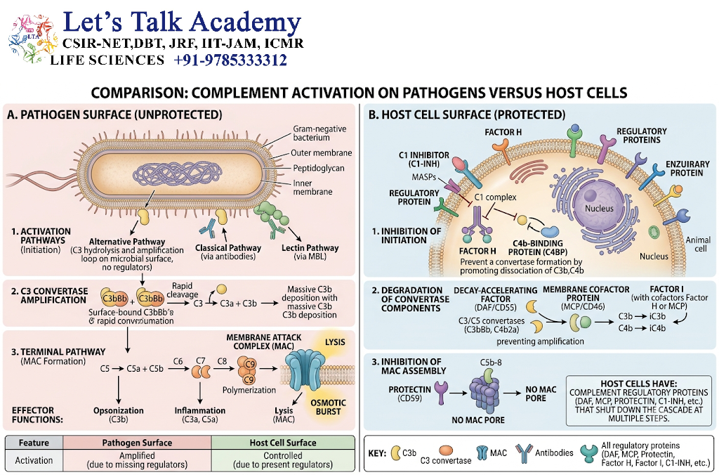
One of the central questions in complement biology is how the system distinguishes foreign surfaces from self-surfaces.
Microbial surfaces often lack the complement regulatory proteins that protect mammalian cells.
Host cells, in contrast, express regulators such as:
- CD46
- CD55
- CD59
Host plasma also contains regulatory proteins such as Factor H and Factor I.
Consequently, complement activation is generally amplified on susceptible microbial surfaces while being restricted on healthy host cells.
Some pathogens, however, have evolved mechanisms that interfere with complement activation.
These mechanisms include:
- Binding host complement regulatory proteins
- Blocking complement components
- Preventing complement deposition
- Altering their surface structures
- Shedding complement-bound molecules
This demonstrates the evolutionary competition between host defense mechanisms and microbial immune evasion.
11. Complement Deficiencies
Deficiencies in complement components can produce characteristic susceptibility to infections or immune disorders.
The clinical consequences depend on which component is deficient.
11.1 Early Classical Pathway Deficiencies
Deficiencies involving components such as C1, C2, or C4 can be associated with increased susceptibility to certain infections and immune-complex-mediated autoimmune manifestations.
Because the classical pathway participates in immune-complex handling and adaptive immune responses, deficiencies may affect more than antimicrobial defense.
11.2 C3 Deficiency
C3 deficiency is particularly significant because C3 is central to all three major activation pathways.
A severe deficiency can result in:
- Recurrent bacterial infections
- Increased susceptibility to encapsulated bacteria
- Impaired opsonization
- Reduced immune-complex clearance
11.3 C5–C9 Deficiencies
Deficiencies in the terminal complement components can impair membrane attack complex formation.
Individuals with deficiencies in terminal components have increased susceptibility to infections caused by Neisseria, particularly recurrent meningococcal disease.
11.4 Alternative Pathway Deficiencies
Deficiencies involving Factor B, Factor D, or properdin can interfere with alternative pathway activity.
Properdin deficiency is associated with increased susceptibility to certain bacterial infections, particularly infections caused by Neisseria.
11.5 Regulatory Protein Deficiencies
Defects in complement regulators can cause excessive complement activation.
A well-known example involves C1 inhibitor deficiency or dysfunction, which can lead to hereditary angioedema.
The underlying problem is dysregulated bradykinin-mediated vascular permeability rather than simply uncontrolled MAC-mediated cell lysis.
12. Clinical Significance of Complement
Complement is involved in numerous human diseases.
Its activity can be protective under normal circumstances but harmful when excessively activated or improperly regulated.
12.1 Autoimmune Diseases
Complement activation can contribute to tissue injury in autoimmune diseases.
In conditions involving immune complexes, complement may become activated after antibodies bind to self-antigens.
Complement consumption can also affect laboratory measurements of circulating complement proteins.
12.2 Systemic Lupus Erythematosus
Systemic lupus erythematosus is associated with immune-complex formation and complement activation.
Levels of components such as C3 and C4 may decrease during periods of active immune-complex-mediated disease because complement is being consumed.
Complement measurements can therefore provide useful information when interpreted together with clinical findings and other laboratory data.
12.3 Hereditary Angioedema
Hereditary angioedema is commonly associated with abnormalities involving C1 inhibitor.
Episodes can involve swelling of:
- Skin
- Gastrointestinal tissues
- Upper airway tissues
The disease illustrates the importance of complement-related regulatory mechanisms in controlling inflammatory and vascular pathways.
12.4 Paroxysmal Nocturnal Hemoglobinuria
Paroxysmal nocturnal hemoglobinuria is associated with an acquired defect in the synthesis of GPI-anchored proteins.
This results in reduced expression of complement regulatory proteins such as CD55 and CD59 on affected blood cells.
Consequently, red blood cells become more susceptible to complement-mediated destruction.
12.5 Complement in Kidney Disease
Complement activation has an important role in several kidney disorders.
Dysregulated complement activity may contribute to glomerular injury and inflammation.
Complement abnormalities are particularly relevant in diseases involving immune complexes and alternative pathway dysregulation.
12.6 Complement and Sepsis
During severe systemic infection, complement activation can become extensive.
Although complement is important for antimicrobial defense, excessive or dysregulated activation can contribute to:
- Systemic inflammation
- Endothelial dysfunction
- Vascular injury
- Organ dysfunction
This demonstrates the dual nature of complement as both a protective and potentially damaging system.
13. Complement and Pathogen Evasion
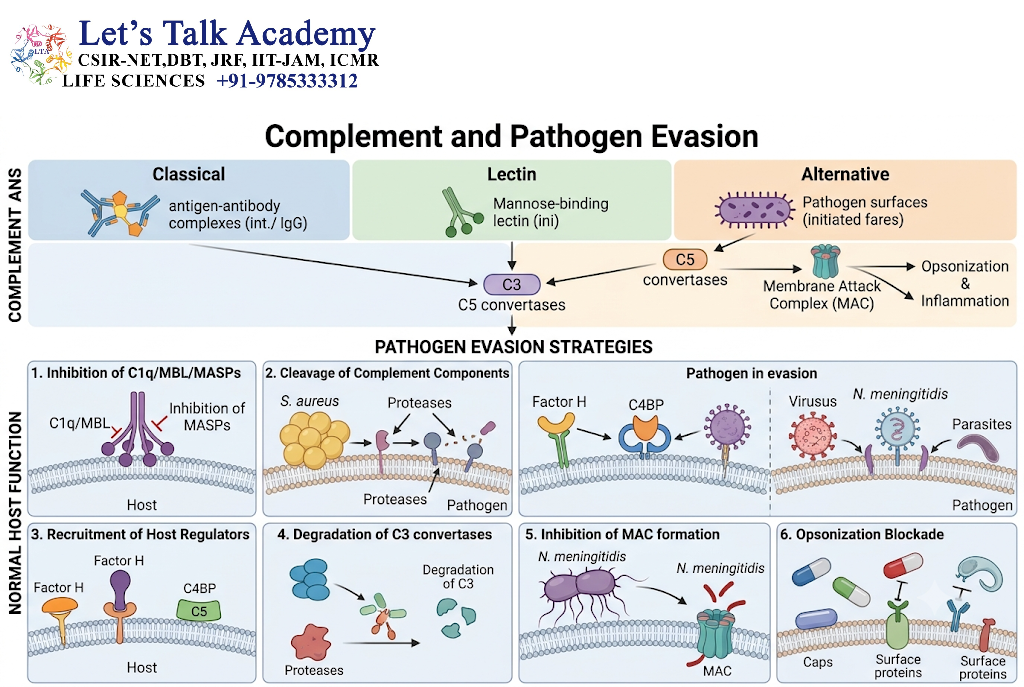
Microorganisms have evolved multiple strategies to escape complement-mediated destruction.
Some bacteria produce proteins that:
- Bind Factor H
- Inactivate complement components
- Prevent C3b deposition
- Recruit host regulatory proteins
- Reduce MAC insertion
- Modify surface structures
Certain pathogens can also recruit complement regulators to their surfaces.
These strategies allow microorganisms to survive despite complement activation.
Understanding complement evasion is important because it demonstrates how innate immune mechanisms influence host-pathogen interactions.
14. Complement and Bacterial Infections
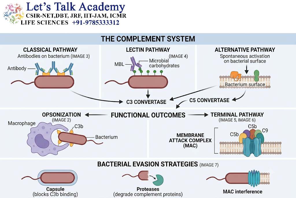
Complement is particularly important in defense against extracellular bacteria.
The major protective mechanisms include:
Recognition → C3 activation → C3b deposition → Opsonization → Phagocytosis
Complement also contributes to inflammation through C3a and C5a.
The terminal pathway is especially important for defense against some Gram-negative bacteria.
Encapsulated bacteria can be difficult for phagocytes to ingest without effective opsonization. Complement-derived C3b and iC3b can therefore be particularly important in their clearance.
15. Complement and Viral Infections
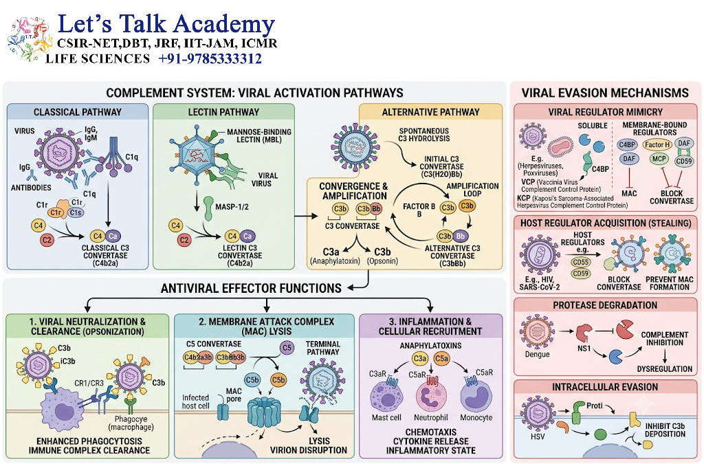
Complement can recognize and interact with viruses, viral particles, and infected cells.
Its functions can include:
- Opsonization of viral particles
- Promotion of phagocytic clearance
- Neutralization of certain viruses
- Recruitment of inflammatory cells
- Interaction with antibody-mediated immunity
However, viruses have also evolved mechanisms that interfere with complement activation.
Some enveloped viruses can acquire host complement regulatory proteins during budding, helping them avoid complement-mediated destruction.
16. Complement and Fungal Infections
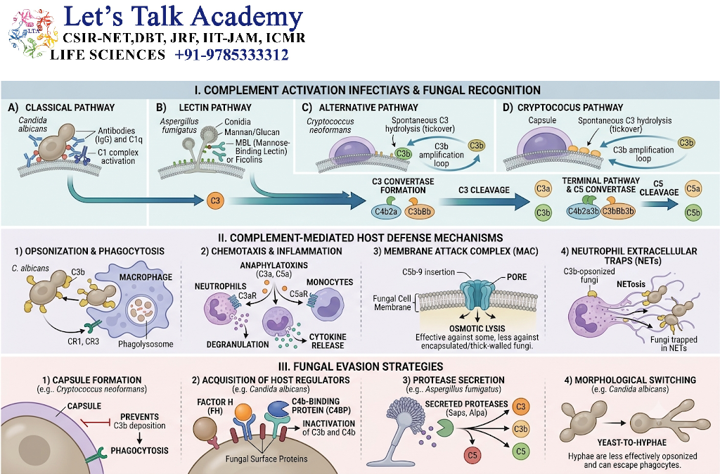
Complement also contributes to antifungal immunity.
Complement proteins can bind fungal surfaces and promote:
- Opsonization
- Phagocytosis
- Inflammatory responses
- Recruitment of immune cells
C3-derived fragments are particularly important for recognition and uptake of fungal organisms by phagocytes.
17. Complement and Parasitic Infections
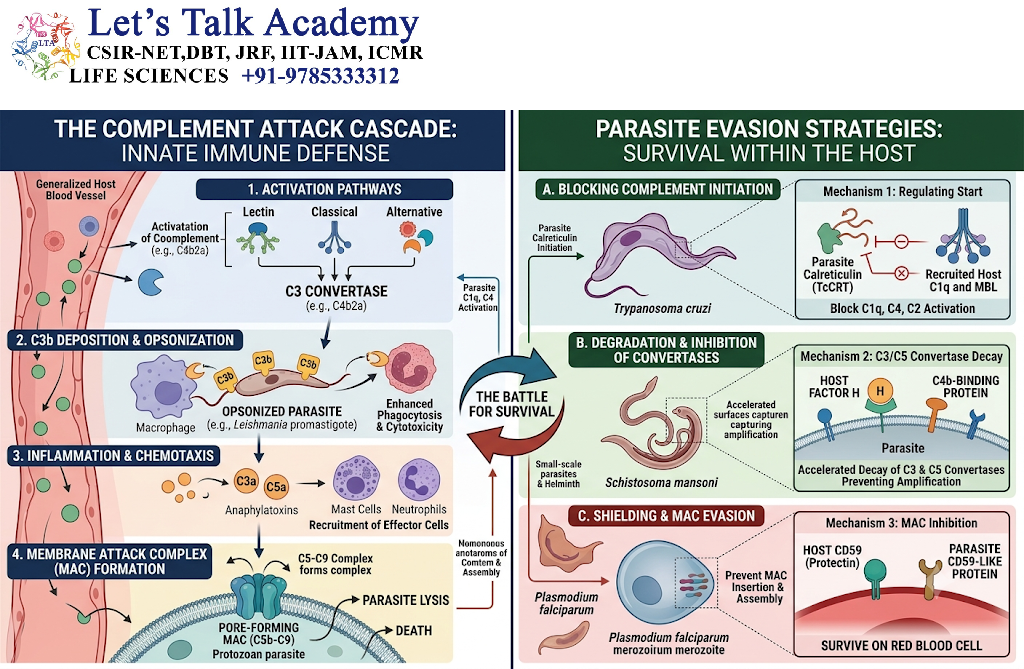
Complement can participate in defense against parasites, although the effectiveness of complement varies widely among different parasite species.
Some parasites are susceptible to complement-mediated damage, whereas others have developed mechanisms that resist complement activation.
The interaction between complement and parasites therefore represents another example of host-pathogen coevolution.
18. Laboratory Evaluation of the Complement System
Complement activity can be assessed through several laboratory methods.
18.1 Serum C3 and C4
Measurement of serum C3 and C4 can provide information about complement activity.
Low levels may occur because of:
- Increased consumption
- Reduced synthesis
- Genetic deficiency
- Certain inflammatory or immune-mediated conditions
Interpretation requires clinical context.
18.2 CH50 Assay
The CH50 test evaluates the functional activity of the classical complement pathway.
A markedly reduced CH50 can suggest a deficiency or significant dysfunction involving components of the classical or terminal pathway.
18.3 AH50 Assay
The AH50 test evaluates functional activity of the alternative pathway.
Abnormal results can help identify defects involving components of the alternative pathway or terminal pathway.
18.4 Importance of Functional Testing
Measuring the concentration of an individual complement protein does not always reveal whether the protein is functioning normally.
Functional assays therefore provide complementary information.
19. Classical Pathway Versus Alternative Pathway
The classical and alternative pathways differ in their mechanisms of activation but eventually converge on common downstream events.
The classical pathway is strongly associated with antibody-mediated recognition.
The alternative pathway provides continuous surveillance and amplification on appropriate surfaces.
A useful conceptual distinction is:
Classical pathway = antibody-associated activation
Lectin pathway = carbohydrate recognition
Alternative pathway = spontaneous activation and amplification
These descriptions are simplified because the pathways interact extensively in vivo.
20. Classical Pathway Versus Lectin Pathway
The classical and lectin pathways share many downstream components.
Both produce:
C4b2a → C3 activation → C5 activation → MAC formation
The major difference is how activation begins.
The classical pathway generally involves recognition of antibody-antigen complexes by C1q.
The lectin pathway begins when MBL or ficolins recognize microbial carbohydrate patterns.
Therefore, the lectin pathway provides an antibody-independent route that can produce the same major C3 convertase as the classical pathway.
21. Lectin Pathway Versus Alternative Pathway
Both pathways can operate independently of antibodies, but their initiation mechanisms differ.
The lectin pathway begins through recognition of specific carbohydrate structures by soluble pattern-recognition molecules.
The alternative pathway involves spontaneous C3 activation and strong amplification on suitable surfaces.
The alternative pathway also serves as an amplification loop for complement activation initiated through the classical or lectin pathways.
22. Important Complement Convertases
The concept of convertases is fundamental to understanding complement activation.
22.1 C3 Convertases
Classical pathway:
C4b2a
Lectin pathway:
C4b2a
Alternative pathway:
C3bBb
Their primary function is to cleave C3.
22.2 C5 Convertases
Classical and lectin pathways:
C4b2a3b
Alternative pathway:
C3bBb3b
Their primary function is to cleave C5 and initiate the terminal pathway.
23. Complement Cascade: A Simplified Sequence
The entire complement system can be understood through the following sequence:
Recognition
↓
Complement activation
↓
C3 convertase formation
↓
C3 cleavage
↓
C3b deposition and opsonization
↓
C5 convertase formation
↓
C5 cleavage
↓
C5a-mediated inflammation
↓
C5b-mediated terminal pathway
↓
C5b-C9 membrane attack complex formation
This sequence provides a useful framework for understanding the relationship between complement activation, inflammation, opsonization, and cell lysis.
24. Complement as a Bridge Between Innate and Adaptive Immunity
The complement system is traditionally classified as part of innate immunity, but its functions extend deeply into adaptive immunity.
Antibodies can activate the classical pathway.
Complement fragments can enhance B-cell activation.
Complement receptors participate in immune-complex processing.
Complement also influences the behavior of antigen-presenting cells and other immune cells.
Therefore, complement should be viewed as a functional bridge between innate and adaptive immunity rather than as an isolated innate immune mechanism.
25. Complement in Tissue Homeostasis
Complement activity is not restricted to infection.
Low-level complement activation contributes to normal tissue maintenance.
Complement participates in:
- Removal of apoptotic cells
- Clearance of cellular debris
- Immune-complex processing
- Regulation of inflammatory responses
- Interaction with tissue-resident immune cells
Controlled complement activity can therefore support tissue homeostasis.
However, excessive activation can transform a protective process into a source of tissue injury.
26. Regulation of Complement on Cell Surfaces
Healthy host cells are protected by several membrane-associated proteins.
The major mechanisms include:
CD46 → Cofactor for Factor I
CD55 → Accelerates decay of C3 convertases
CD59 → Prevents effective MAC formation
These regulatory proteins are strategically positioned on host cell membranes to prevent complement from damaging normal tissues.
This is an example of how the immune system uses multiple layers of regulation rather than relying on a single protective mechanism.
27. Complement and Inflammation
Complement-derived inflammatory signals must be carefully controlled.
C3a and C5a can activate inflammatory pathways, but excessive production may contribute to tissue damage.
C5a is especially powerful because it can activate neutrophils and promote their migration toward sites of complement activation.
Activated neutrophils can release:
- Reactive oxygen species
- Proteases
- Cytokines
- Other inflammatory mediators
Therefore, complement activation can initiate a broader inflammatory cascade extending beyond the complement system itself.
28. Important Conceptual Relationships
Several relationships are particularly important for understanding complement biology.
28.1 C3b and Opsonization
C3b → surface deposition → complement receptor recognition → enhanced phagocytosis
28.2 C3a and Inflammation
C3 cleavage → C3a → inflammatory effects
28.3 C5a and Chemotaxis
C5 cleavage → C5a → leukocyte recruitment and activation
28.4 C5b and MAC
C5 cleavage → C5b → C6 → C7 → C8 → C9 → MAC
28.5 Complement Regulation
Complement activation + regulatory proteins → controlled host defense
These relationships provide a conceptual framework for understanding the entire system.
29. Key Complement Proteins and Their Functions
| Protein | Major role |
|---|---|
| C1q | Recognition in classical pathway |
| C1r | Serine protease of C1 complex |
| C1s | Cleaves C4 and C2 |
| C3 | Central complement component |
| C3a | Anaphylatoxin |
| C3b | Opsonin and convertase component |
| C4 | Classical and lectin pathway component |
| C5 | Precursor of C5a and C5b |
| C5a | Potent inflammatory mediator |
| C5b | Initiates terminal pathway |
| C6 | Terminal pathway component |
| C7 | Helps associate terminal complex with membrane |
| C8 | Contributes to membrane insertion |
| C9 | Forms MAC pore |
| Factor B | Alternative pathway component |
| Factor D | Activates Factor B |
| Properdin | Stabilizes alternative pathway convertase |
| Factor H | Alternative pathway regulator |
| Factor I | Inactivates C3b and C4b |
| C1-INH | Regulates classical and lectin pathway initiation |
| CD46 | Cofactor for complement regulation |
| CD55 | Accelerates convertase decay |
| CD59 | Inhibits MAC formation |


