
1. Introduction
Cells constantly receive signals from their environment and from other cells. These signals may come from hormones, neurotransmitters, growth factors, cytokines, extracellular matrix components, or changes in nutrients and physical conditions.
For a cell to respond correctly, signaling pathways must be precisely regulated. A signal should become active when required, produce an appropriate response, and then be reduced or terminated when the stimulus disappears.
Without proper regulation, signaling may become:
- Too weak
- Too strong
- Too long-lasting
- Too short-lived
- Activated at the wrong location
- Activated at the wrong time
Such disturbances can alter normal cellular behavior and contribute to processes such as uncontrolled proliferation, abnormal metabolism, inflammation and cellular dysfunction.
A simplified principle is:
Signal → Receptor → Signal transduction → Cellular response → Regulation → Signal termination
Regulation therefore does not occur only at the end of a pathway. It can occur at almost every step.
2. Definition of Regulation of Signaling Pathways
2.1 Definition
Regulation of signaling pathways is the coordinated control of signal generation, transmission, amplification, integration, cellular response and termination to ensure an appropriate response to a stimulus.
The regulation determines:
- Whether a pathway is active or inactive
- How strongly it is activated
- How long it remains active
- Where signaling occurs
- How the cell adapts to repeated stimulation
3. Why Signaling Pathways Need Regulation
Signaling pathways can generate large responses through amplification.
For example:
One receptor
↓
Multiple signaling molecules
↓
Many second messengers
↓
Multiple protein kinases
↓
Many target proteins
This amplification is useful, but uncontrolled amplification can be harmful.
Therefore, cells use regulatory mechanisms to maintain signaling within an appropriate range.
3.1 Major Objectives of Signaling Regulation
- Maintain signal specificity
- Prevent excessive activation
- Control signal duration
- Control signal strength
- Prevent inappropriate responses
- Adapt to persistent stimulation
- Coordinate multiple pathways
- Terminate signaling efficiently
4. Levels of Signaling Regulation
Regulation can occur at multiple levels.
| Level | Major Regulatory Mechanisms |
|---|---|
| Ligand | Production, release and degradation |
| Receptor | Activation, inhibition, desensitization, internalization |
| G proteins | GTP hydrolysis and regulatory proteins |
| Second messengers | Synthesis, degradation and sequestration |
| Protein kinases | Activation and inhibition |
| Protein phosphatases | Dephosphorylation |
| Scaffold proteins | Spatial organization |
| Transcription factors | Activation and repression |
| Target proteins | Modification, localization and degradation |
| Entire pathway | Feedback and crosstalk |
Thus, signaling regulation is distributed throughout the signaling network.
5. Regulation at the Ligand Level
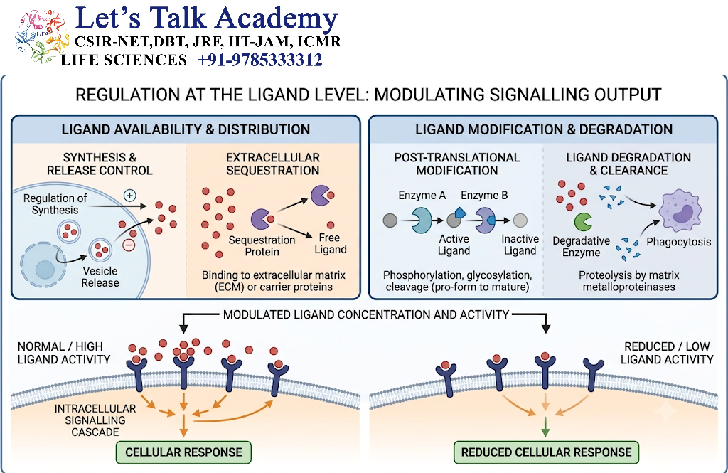
The first level of regulation often occurs before the receptor is activated.
Cells control:
- Ligand synthesis
- Ligand storage
- Ligand release
- Ligand transport
- Ligand degradation
For example, a signaling molecule may be stored inside secretory vesicles and released only after appropriate stimulation.
5.1 Ligand Degradation
Extracellular signaling molecules can be degraded by enzymes.
This reduces their concentration and therefore decreases receptor activation.
Ligand release → Receptor activation → Ligand degradation → Reduced signaling
6. Regulation at the Receptor Level
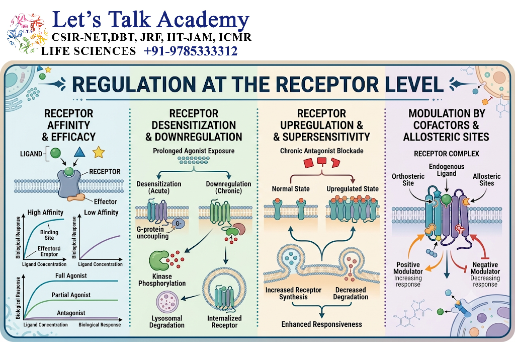
Receptors are major control points in signaling pathways.
Cells regulate:
- Receptor number
- Receptor localization
- Ligand-binding affinity
- Receptor activation
- Receptor phosphorylation
- Receptor internalization
- Receptor degradation
A cell can therefore change its sensitivity to a signal by changing receptor abundance.
7. Receptor Desensitization
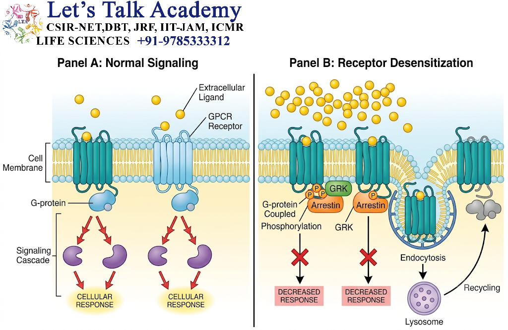
Desensitization refers to the reduction in receptor responsiveness despite continued presence of the signaling molecule.
This is particularly important when a cell is exposed to a signal for a long period.
For example:
Continuous ligand stimulation
↓
Receptor activation
↓
Receptor regulatory modification
↓
Reduced receptor responsiveness
Desensitization prevents excessive cellular responses.
7.1 GPCR Desensitization
GPCRs can be phosphorylated by receptor kinases.
Phosphorylated receptors may bind β-arrestins, which reduce further coupling to heterotrimeric G proteins.
This is an important mechanism for limiting GPCR signaling.
8. Receptor Internalization
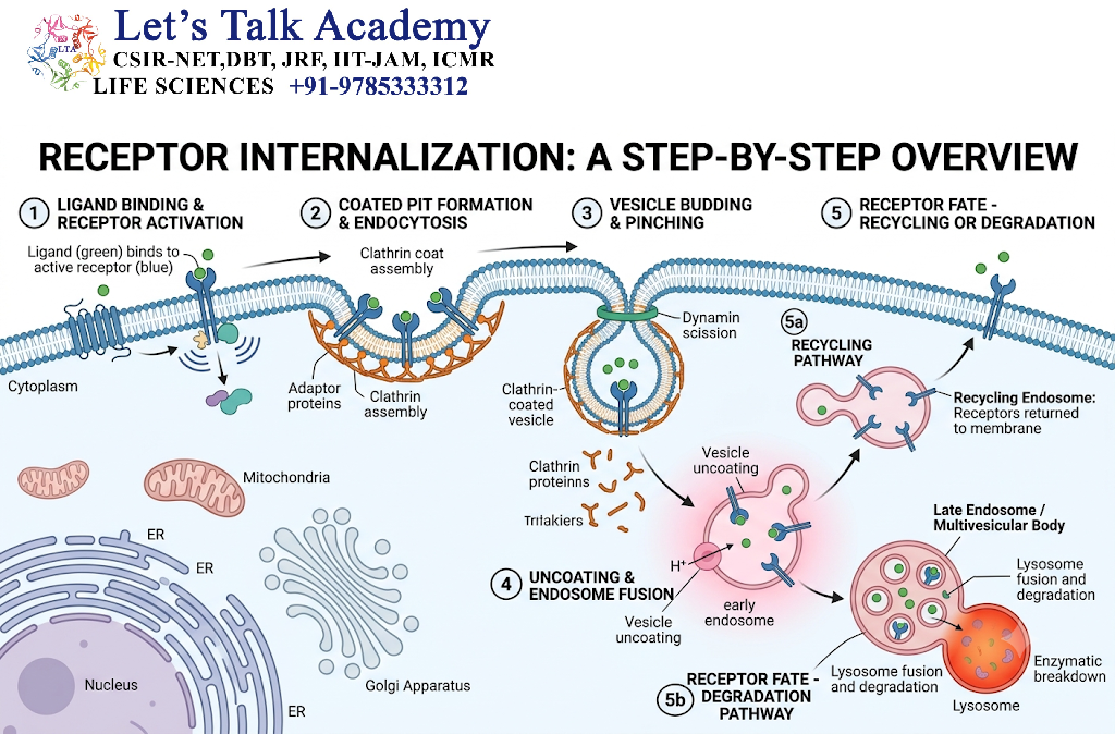
Activated receptors can be removed from the plasma membrane through endocytosis.
The process can be summarized as:
Receptor activation
↓
Receptor modification
↓
Recruitment of endocytic machinery
↓
Endocytosis
↓
Endosomal receptor
↓
Recycling or degradation
Internalization can reduce the number of receptors available at the cell surface.
However, internalized receptors do not always become inactive immediately. In some pathways, endosomes can continue to support signaling.
9. Receptor Recycling
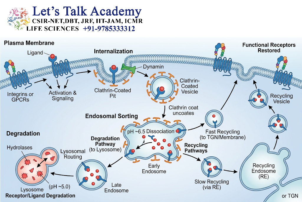
Some internalized receptors are returned to the plasma membrane.
Cell surface receptor
↓
Internalization
↓
Endosome
↓
Recycling
↓
Cell surface receptor
Recycling restores cellular sensitivity to extracellular signals.
This allows cells to dynamically adjust receptor availability.
10. Receptor Degradation and Downregulation
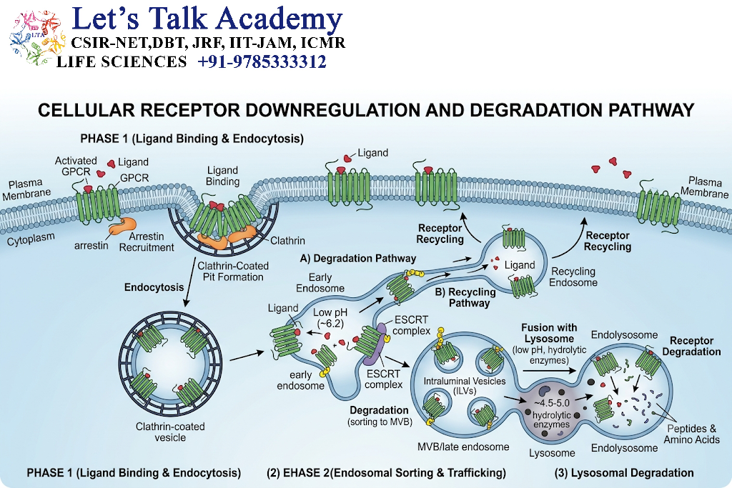
Some receptors are directed toward degradation.
For example:
Receptor
↓
Ubiquitination
↓
Endocytosis
↓
Lysosomal or proteasomal processing
↓
Reduced receptor abundance
Long-term reduction in receptor number is often referred to as downregulation.
11. Regulation of G Proteins
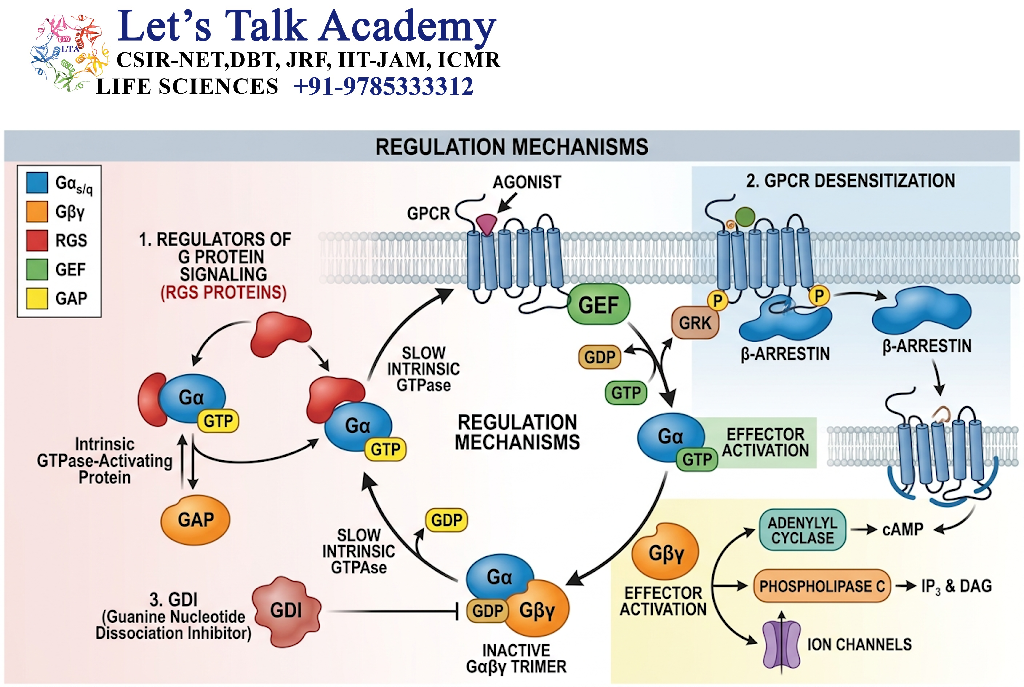
Heterotrimeric G proteins act as molecular switches.
The Gα subunit is:
GDP-bound → inactive
GTP-bound → active
The signal is terminated when GTP is hydrolyzed to GDP.
Gα-GTP → Gα-GDP
11.1 GTPase-Activating Proteins
Proteins known as RGS proteins (Regulators of G-protein Signaling) can accelerate GTP hydrolysis by Gα subunits.
Therefore:
RGS activity ↑ → GTP hydrolysis ↑ → G-protein signaling ↓
12. Regulation of Second Messengers
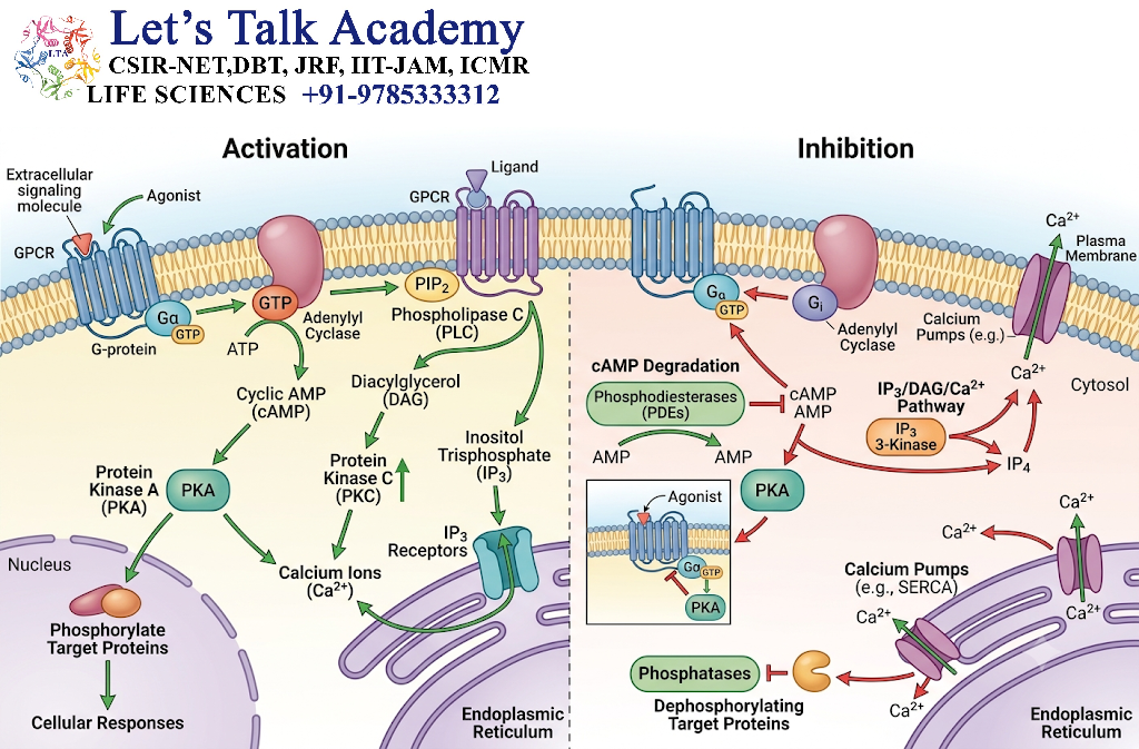
Second messengers must be tightly controlled.
Important examples include:
- cAMP
- cGMP
- IP₃
- DAG
- Ca²⁺
Their concentrations depend on the balance between production and removal.
12.1 cAMP Regulation
Adenylyl cyclase produces cAMP.
Phosphodiesterases degrade cAMP.
Adenylyl cyclase → cAMP ↑
Phosphodiesterase → cAMP ↓
This balance controls the strength and duration of cAMP signaling.
12.2 cGMP Regulation
Guanylyl cyclase produces cGMP, while phosphodiesterases degrade it.
GTP → cGMP → PKG
Termination:
cGMP → GMP
12.3 Calcium Regulation
Cytosolic Ca²⁺ is controlled by:
- Ca²⁺ pumps
- Ion exchangers
- ER uptake
- Mitochondrial buffering
- Calcium-binding proteins
Maintaining low resting cytosolic Ca²⁺ is essential for Ca²⁺ to function effectively as a second messenger.
13. Regulation by Protein Kinases
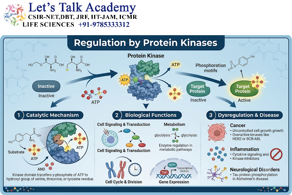
Protein kinases transfer phosphate groups from ATP to target proteins.
Protein + ATP → Phosphorylated protein + ADP
Phosphorylation can:
- Activate a protein
- Inhibit a protein
- Change its localization
- Change its stability
- Alter protein-protein interactions
Protein kinases therefore act as important regulatory switches.
14. Regulation by Protein Phosphatases
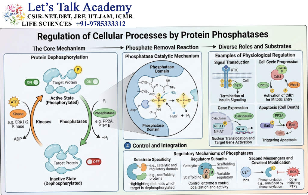
Protein phosphatases remove phosphate groups from proteins.
Phosphorylated protein → Dephosphorylated protein
Thus, signaling often depends on a balance:
Kinase activity ↔ Phosphatase activity
If kinase activity dominates, phosphorylation increases.
If phosphatase activity dominates, phosphorylation decreases.
This reversible modification allows rapid regulation.
15. Negative Feedback
Negative feedback occurs when activation of a signaling pathway eventually produces an effect that reduces the activity of that same pathway.
General mechanism:
Signal
↓
Pathway activation
↓
Response
↓
Inhibitory effect
↓
Reduced pathway activity
Negative feedback prevents excessive or prolonged signaling.
15.1 Example
A signaling pathway may activate a protein phosphatase that subsequently dephosphorylates and inhibits components of the same pathway.
Thus:
Pathway activation → inhibitory regulator → pathway suppression
16. Positive Feedback
Positive feedback occurs when activation of a pathway promotes further activation.
General mechanism:
Initial signal
↓
Pathway activation
↓
Amplification
↓
Further pathway activation
Positive feedback can produce rapid and strong cellular responses.
However, it usually requires additional mechanisms to prevent uncontrolled activation.
17. Feedback Loops
Feedback regulation can occur through:
- Receptor regulation
- Kinase inhibition
- Phosphatase activation
- Gene expression
- Protein degradation
- Second-messenger degradation
Feedback loops are important for maintaining signaling balance.
18. Regulation by Inhibitory Proteins
Cells contain proteins that directly inhibit signaling components.
Examples include:
- Protein phosphatases
- Kinase inhibitors
- G-protein regulators
- Receptor-associated inhibitory proteins
- Inhibitory adaptor proteins
These proteins act as molecular brakes.
19. Regulation by Scaffold Proteins
Scaffold proteins organize multiple signaling components into functional complexes.
For example:
Scaffold
↙ ↓ ↘
Kinase 1 → Kinase 2 → Kinase 3
This organization can:
- Increase signaling efficiency
- Improve specificity
- Reduce unwanted pathway interactions
- Control the location of signaling
Scaffolds are particularly important in kinase cascades such as MAPK signaling.
20. Spatial Regulation of Signaling
Signaling is often restricted to specific cellular regions.
Important locations include:
- Plasma membrane
- Cytoplasm
- Endosomes
- Mitochondria
- Endoplasmic reticulum
- Nucleus
For example, a signaling protein may need to be transported to the nucleus before it can regulate gene expression.
Thus:
Location of signaling component → accessibility of target → cellular response
21. Temporal Regulation
Signaling pathways are also controlled over time.
A signal may be:
- Transient
- Sustained
- Oscillatory
- Pulsatile
The same signaling molecule can produce different responses depending on the duration of activation.
For example:
Short ERK activation and sustained ERK activation can lead to different cellular outcomes in some cellular contexts.
Therefore, signaling information is encoded not only in signal intensity but also in timing.
22. Regulation Through Signal Amplification
Amplification increases the magnitude of a signal.
However, amplification must be controlled.
For example:
Receptor
↓
G protein
↓
Adenylyl cyclase
↓
Many cAMP molecules
↓
PKA
↓
Multiple target proteins
Phosphodiesterases, phosphatases and inhibitory proteins can limit this amplification.
23. Regulation of MAPK Pathways
MAPK pathways commonly involve a kinase cascade:
Ras → Raf → MEK → ERK
Each kinase activates the next component.
Regulation occurs through:
- Phosphatases
- Scaffold proteins
- Feedback inhibition
- Protein degradation
- Receptor regulation
- Crosstalk with other pathways
The duration and intensity of ERK activity can influence the final cellular response.
24. Regulation of PI3K-AKT Signaling
The PI3K-AKT pathway is regulated at several levels.
Simplified pathway:
Receptor
↓
PI3K
↓
PIP₃
↓
AKT
↓
Cellular responses
One important negative regulator is PTEN, which converts PIP₃ back toward the PIP₂ state.
Thus:
PI3K → PIP₃ formation
PTEN → PIP₃ reduction
This balance is important for controlling AKT signaling.
25. Regulation of JAK-STAT Signaling
JAK-STAT signaling can be regulated by inhibitory proteins such as SOCS (Suppressor of Cytokine Signaling) proteins.
General mechanism:
Cytokine
↓
Receptor
↓
JAK
↓
STAT
↓
Gene expression
↓
SOCS induction
↓
Reduced signaling
This represents a feedback mechanism in which pathway activation promotes production of its own inhibitors.
26. Regulation of TGF-β Signaling
TGF-β signaling is regulated through:
- Receptor activity
- SMAD phosphorylation
- SMAD degradation
- Inhibitory SMAD proteins
- Nuclear cofactors
- Feedback mechanisms
Inhibitory SMADs can limit signaling and help maintain pathway control.
27. Signal Crosstalk
Cells rarely use signaling pathways independently.
Different pathways can interact.
Examples include:
- MAPK and PI3K-AKT
- Ca²⁺ and protein kinase pathways
- cAMP and MAPK
- TGF-β and other transcriptional pathways
This is known as signal crosstalk.
Crosstalk allows the cell to integrate multiple signals.
28. Signal Integration
A cell may receive several signals simultaneously.
For example:
Growth factor
+
Hormone
+
Nutrient signal
The cell integrates these signals before producing a final response.
This prevents the response from depending on only one signaling pathway.
29. Regulation by Protein Degradation
Signaling proteins can be removed through controlled degradation.
Major mechanisms include:
- Ubiquitin-proteasome system
- Lysosomal degradation
- Autophagy-related mechanisms in appropriate contexts
Protein degradation changes the abundance of signaling components and therefore changes pathway activity.
30. Regulation by Ubiquitination
Ubiquitination involves attachment of ubiquitin molecules to target proteins.
Depending on the ubiquitin linkage and cellular context, ubiquitination can influence:
- Protein degradation
- Protein trafficking
- Receptor internalization
- Signaling complex formation
Therefore, ubiquitination is not simply a degradation signal; it can also directly regulate signaling.
31. Regulation by Subcellular Localization
A signaling protein may be active only when present in a particular cellular compartment.
For example:
Cytoplasm → nucleus
Movement into the nucleus can allow a signaling protein to interact with transcriptional regulators.
Similarly, membrane localization can bring signaling proteins close to receptors and phospholipid substrates.
32. Regulation by Protein-Protein Interactions
Signaling proteins frequently require interaction with other proteins.
Regulation can therefore occur by controlling:
- Protein binding
- Complex formation
- Adaptor recruitment
- Scaffold association
- Competitive inhibition
These interactions determine which downstream pathway becomes active.
33. Regulation Through Receptor Affinity
Changes in receptor structure can influence ligand binding.
A receptor with high ligand affinity may respond strongly even at relatively low ligand concentrations.
Conversely, lower affinity can reduce sensitivity.
Thus, receptor properties influence cellular responsiveness.
34. Cellular Adaptation
When cells are continuously exposed to a signal, they may adapt.
For example:
Persistent signal
↓
Receptor desensitization
↓
Reduced receptor availability
↓
Reduced cellular response
Adaptation prevents cells from remaining permanently in a highly activated state.
35. Signal Termination
Signal termination is the final major stage of pathway regulation.
Important mechanisms include:
- Ligand removal
- Receptor inactivation
- Receptor internalization
- GTP hydrolysis
- Second-messenger degradation
- Protein dephosphorylation
- Protein degradation
- Inhibitory feedback
General flow:
Signal
↓
Receptor activation
↓
Intracellular signaling
↓
Cellular response
↓
Negative regulation
↓
Signal termination
36. Regulation of Signaling: Integrated Flowchart
Extracellular signal
↓
Receptor activation
↓
Signal transduction
↓
Second messengers / kinase cascades
↓
Signal amplification
↓
Cellular response
↙ ↓ ↘
Feedback inhibition | Phosphatases | Protein degradation
↓
Receptor desensitization/internalization
↓
Second-messenger removal
↓
Signal termination
37. Major Regulatory Mechanisms
| Mechanism | Main Function |
|---|---|
| Desensitization | Reduces receptor responsiveness |
| Internalization | Removes receptors from cell surface |
| Recycling | Restores receptors to membrane |
| Degradation | Reduces protein abundance |
| Phosphorylation | Changes protein activity |
| Dephosphorylation | Reverses phosphorylation |
| GTP hydrolysis | Terminates G-protein signaling |
| PDE activity | Removes cAMP/cGMP |
| Feedback inhibition | Limits pathway activity |
| Scaffold proteins | Organize signaling components |
| Crosstalk | Integrates different pathways |
| Compartmentalization | Controls signaling location |
38. Regulation and Cellular Specificity
The same signal can produce different effects in different cells.
This occurs because cells differ in:
- Receptor expression
- Signaling proteins
- Kinases
- Phosphatases
- Scaffold proteins
- Transcription factors
- Second-messenger machinery
Therefore:
Same signal + different cellular machinery = different response
This is an important principle of cellular signaling.
39. Regulation of Signaling Pathways and Disease
Abnormal signaling regulation can contribute to disease.
39.1 Cancer
Persistent activation of pathways such as:
- Ras-MAPK
- PI3K-AKT-mTOR
- JAK-STAT
can promote inappropriate cell proliferation or survival when regulatory controls are disrupted.
39.2 Metabolic Disorders
Defective regulation of hormone signaling can alter glucose and lipid metabolism.
39.3 Inflammatory Disorders
Excessive or prolonged cytokine signaling can contribute to chronic inflammatory states.
39.4 Cardiovascular Disorders
Abnormal regulation of NO-cGMP signaling can affect vascular function.
40. Important Principles of Signaling Regulation
40.1 Balance
Signaling depends on a balance between activation and inhibition.
40.2 Reversibility
Many signaling modifications are reversible.
Kinase → phosphorylation
Phosphatase → dephosphorylation
40.3 Specificity
Cells restrict signals to particular pathways and locations.
40.4 Adaptation
Persistent stimulation can decrease cellular sensitivity.
40.5 Integration
Multiple signals can be combined to produce one cellular decision.
40.6 Termination
Every signaling pathway requires mechanisms to switch the signal off.


