
1. Introduction to Cell Surface Receptors
1.1 Overview of Cell Surface Receptors
Cells are the fundamental structural and functional units of all living organisms. Although cells operate as independent units, they must continuously communicate with their surrounding environment to survive, grow, divide, differentiate, and perform specialized functions. This communication is particularly important in multicellular organisms, where individual cells work together to maintain tissue organization and physiological balance.
Cells receive information from their surroundings through specialized proteins known as cell surface receptors. These receptors are generally embedded in the plasma membrane and act as molecular sensors that detect signals outside the cell and transmit information into the cell.
Cell surface receptors recognize a wide range of signaling molecules, including hormones, neurotransmitters, growth factors, cytokines, chemokines, and extracellular matrix components. When a signaling molecule binds to its specific receptor, the receptor undergoes a structural or conformational change or initiates a receptor-associated interaction. This event activates intracellular signaling pathways that alter cellular activities.
For example, insulin binds to the insulin receptor on the surface of target cells and stimulates signaling pathways involved in glucose uptake, metabolism, growth, and gene regulation. Similarly, neurotransmitters such as acetylcholine bind to specific receptors on nerve or muscle cells and influence electrical activity or cellular responses.
The importance of cell surface receptors extends beyond simple signal detection. They determine how cells interpret information, distinguish between different extracellular signals, and produce appropriate biological responses. A single signal can produce different effects in different cell types because the nature of the receptor, the associated signaling proteins, and the cellular environment may vary.
Cell surface receptors are therefore essential components of cellular communication, signal transduction, tissue development, immune responses, metabolism, and homeostasis.
1.2 Definition of Cell Surface Receptors
Cell surface receptors are specialized proteins, usually located within the plasma membrane, that recognize specific extracellular signaling molecules and convert the information carried by those molecules into intracellular biochemical, electrical, or mechanical responses.
These receptors are also called plasma membrane receptors or membrane receptors.
A typical cell surface receptor has an extracellular region that interacts with a signaling molecule, a membrane-associated region that anchors the receptor, and an intracellular region that either possesses signaling activity or interacts with proteins capable of initiating intracellular signaling.
However, not all receptors share the same structural organization. Some receptors contain a single transmembrane segment, whereas others form multipass membrane proteins or large receptor complexes. Their mechanisms of signaling also differ considerably.
General mechanism
The basic mechanism of cell surface receptor signaling can be represented as follows:
Extracellular signal → Receptor binding → Receptor activation → Intracellular signal transduction → Cellular response
The cellular response may include:
-
Activation or inhibition of enzymes.
-
Changes in ion concentrations.
-
Alterations in gene expression.
-
Changes in cellular metabolism.
-
Cell growth and proliferation.
-
Cell differentiation.
-
Cell movement and adhesion.
-
Regulation of cell survival or programmed cell death.
1.3 Historical Development of Receptor Biology
The concept of cellular receptors developed from the observation that certain hormones and drugs produce specific biological effects only in particular tissues. Early pharmacological studies suggested that cells possess selective binding sites for chemical messengers.
In the twentieth century, researchers provided experimental evidence for the existence of specific hormone-binding proteins. Studies of cell signaling subsequently established that receptors are not merely passive binding sites but active components of communication systems.
The development of biochemical, molecular, and structural techniques led to the identification of distinct receptor families, including G protein-coupled receptors, receptor tyrosine kinases, cytokine receptors, and ligand-gated ion channels.
Modern receptor biology integrates structural biology, biochemistry, genetics, cell biology, and systems biology to explain how extracellular information is detected, processed, amplified, and regulated.
1.4 Importance of Cell Surface Receptors in Cellular Communication
Cell surface receptors are essential because most hydrophilic signaling molecules cannot freely cross the hydrophobic interior of the plasma membrane. These molecules require receptors to communicate with the intracellular machinery.
The major functions of cell surface receptors include:
1. Signal recognition: Receptors detect specific molecules in the extracellular environment.
2. Signal transduction: They convert extracellular information into intracellular signals.
3. Signal amplification: A single receptor activation event can stimulate multiple downstream signaling molecules.
4. Signal integration: Receptors participate in networks that combine information from several signaling pathways.
5. Regulation of cellular activity: They control processes such as metabolism, proliferation, differentiation, and migration.
6. Adaptation to environmental changes: Receptors allow cells to respond to changing nutrient levels, stress, chemical signals, and interactions with neighboring cells.
7. Maintenance of homeostasis: Receptor-mediated signaling helps maintain stable physiological conditions within tissues and organisms.
1.5 Extracellular Signaling Molecules and Ligands
A molecule that binds to a receptor and influences its activity is called a ligand. In receptor biology, ligands may act as activators, inhibitors, or modulators of receptor function.
Common classes of extracellular signaling molecules include:
|
Ligand or signal |
Example |
Major biological function |
|---|---|---|
|
Hormones |
Insulin, glucagon |
Metabolism and physiological regulation |
|
Growth factors |
EGF, FGF |
Growth, survival, and proliferation |
|
Cytokines |
Interleukins, interferons |
Immune regulation |
|
Neurotransmitters |
Acetylcholine, glutamate |
Neuronal communication |
|
Chemokines |
CXCL8 |
Cell migration and immune signaling |
|
Extracellular matrix proteins |
Fibronectin, collagen |
Adhesion and mechanical signaling |
A ligand may bind to a receptor with high specificity, but binding alone does not always guarantee receptor activation. Some ligands inhibit receptor activity, while others alter the strength, duration, or nature of signaling.
The biological effect of a ligand depends on the receptor it binds to, the receptor’s cellular context, and the signaling machinery available within the cell.
2. General Structure and Organization of Cell Surface Receptors
2.1 Plasma Membrane as the Platform for Receptor Function
The plasma membrane is a selectively permeable biological membrane that separates the interior of a cell from its external environment. It consists primarily of a phospholipid bilayer containing proteins, cholesterol, and carbohydrates.
The phospholipid bilayer has a hydrophilic outer surface and a hydrophobic interior. This arrangement allows the membrane to act as a selective barrier between the extracellular and intracellular environments.
Cell surface receptors are embedded within this membrane or associated with its surface. Their position within the membrane allows them to interact with extracellular signaling molecules while communicating with intracellular signaling proteins.
The plasma membrane is not a static structure. It is dynamic and contains specialized regions known as membrane microdomains, which can influence receptor localization and signaling. Some receptors cluster into specific membrane regions, bringing signaling proteins into close proximity and facilitating efficient signal transmission.
The organization of the plasma membrane is particularly important for receptors involved in immune signaling, cell adhesion, and growth factor responses.
2.2 Basic Structural Organization of Cell Surface Receptors
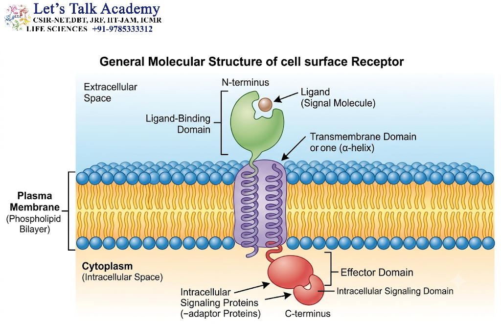
Although cell surface receptors differ greatly in structure, many have three functional regions:
2.2.1 Extracellular Domain
The extracellular domain extends into the space outside the cell. It is responsible for recognizing and binding signaling molecules, such as hormones, growth factors, neurotransmitters, or extracellular matrix proteins.
The extracellular domain may contain:
-
Ligand-binding sites.
-
Protein–protein interaction regions.
-
Disulfide bonds that stabilize the protein structure.
-
Glycosylation sites.
-
Regions that interact with other membrane proteins.
The three-dimensional structure of the extracellular domain determines which ligands can bind to the receptor. The binding interaction often depends on complementary shapes, electrostatic interactions, hydrogen bonds, and hydrophobic interactions.
Some receptors possess highly selective binding sites, whereas others recognize groups of structurally related ligands.
2.2.2 Transmembrane Domain
The transmembrane domain anchors the receptor in the plasma membrane.
In many receptors, this region consists of one or more hydrophobic stretches of amino acids that form α-helices across the lipid bilayer. These regions interact with the hydrophobic interior of the membrane.
The number of transmembrane segments varies among receptor families.
-
Receptor tyrosine kinases generally contain a single transmembrane segment.
-
G protein-coupled receptors contain seven transmembrane α-helices.
-
Ligand-gated ion channels possess multiple membrane-spanning regions arranged around an ion-conducting pore.
-
Integrins contain one transmembrane segment per subunit.
The transmembrane region can participate directly in receptor activation. In certain receptors, ligand binding changes the relative orientation or association of transmembrane helices, thereby influencing the intracellular signaling machinery.
2.2.3 Intracellular Domain
The intracellular domain faces the cytoplasm and is responsible for initiating or regulating intracellular signaling.
Depending on the receptor family, the intracellular region may:
-
Possess enzymatic activity.
-
Bind intracellular adaptor proteins.
-
Interact with heterotrimeric G proteins.
-
Regulate ion channels.
-
Recruit protein kinases or phosphatases.
-
Interact with the cytoskeleton.
-
Influence transcription factors through downstream signaling pathways.
For example, the intracellular domain of a receptor tyrosine kinase contains a protein kinase region that phosphorylates specific tyrosine residues. In contrast, cytokine receptors generally lack intrinsic enzymatic activity and recruit cytoplasmic kinases, such as Janus kinases.
2.3 Membrane Topology and Orientation
Membrane topology refers to the arrangement of a membrane protein within the lipid bilayer and the orientation of its functional domains.
The extracellular portion of a receptor is exposed to the external environment, while its intracellular portion interacts with cytoplasmic molecules.
The orientation of a receptor is established during protein synthesis and insertion into the endoplasmic reticulum. Specific sequences within the protein help determine how the protein is inserted into the membrane.
The orientation is maintained during transport through the secretory pathway and delivery to the plasma membrane.
This organization is essential because receptor signaling depends on the correct positioning of its ligand-binding and signaling domains.
2.4 Receptor Specificity and Ligand Recognition
Receptor specificity refers to the ability of a receptor to recognize particular ligands rather than unrelated molecules.
The interaction between a receptor and its ligand is influenced by the chemical and structural compatibility of the interacting molecules.
Two important concepts in receptor biology are affinity and specificity.
Affinity
Affinity describes the strength of the interaction between a receptor and its ligand. A ligand with high affinity can bind strongly to its receptor under suitable conditions.
Specificity
Specificity describes the ability of a receptor to preferentially recognize a particular ligand or group of related ligands.
A receptor may have high affinity for a ligand but still produce different biological responses depending on the cellular environment.
Receptor–ligand binding equilibrium
The reversible binding of a ligand to a receptor can be represented as:
Where:
-
RR = unbound receptor.
-
LL = ligand.
-
RLRL = receptor–ligand complex.
The dissociation constant is represented as:
A lower KdK_d generally indicates a higher binding affinity under the conditions being studied.
However, receptor affinity alone does not determine the strength of a cellular response. The number of receptors, downstream signaling efficiency, ligand concentration, and receptor regulation also influence the outcome.
2.5 Receptor Activation and Conformational Changes
Receptor activation occurs when ligand binding or another regulatory event changes the functional state of a receptor.
In many receptors, ligand binding induces a conformational change. This alteration may expose a binding site, promote receptor dimerization, rearrange transmembrane helices, or activate an intracellular enzyme.
For example, the binding of epidermal growth factor to the epidermal growth factor receptor promotes receptor dimerization and activation of its intracellular tyrosine kinase domains.
In some receptors, ligand binding does not cause a large structural change but instead stabilizes an active receptor conformation.
Receptor activation is therefore a structural and biochemical process that initiates a cascade of intracellular events.
2.6 Receptor Dimerization and Oligomerization
Dimerization is the association of two receptor molecules, whereas oligomerization refers to the formation of complexes containing multiple receptor molecules.
Many cell surface receptors require dimerization or higher-order assembly for activation.
For example, receptor tyrosine kinases often become activated when ligand binding promotes receptor dimerization. This brings the intracellular kinase domains close enough to phosphorylate one another.
Some receptors form stable dimers even in the absence of ligand, while others associate only after ligand binding.
Receptor assembly can influence:
-
Ligand-binding properties.
-
Enzymatic activity.
-
Signaling specificity.
-
Receptor trafficking.
-
Signal duration.
-
Interaction with other membrane proteins.
The formation of receptor complexes is an important mechanism for regulating the strength and nature of cellular responses.
3. Major Types of Cell Surface Receptors
3.1 Classification of Cell Surface Receptors
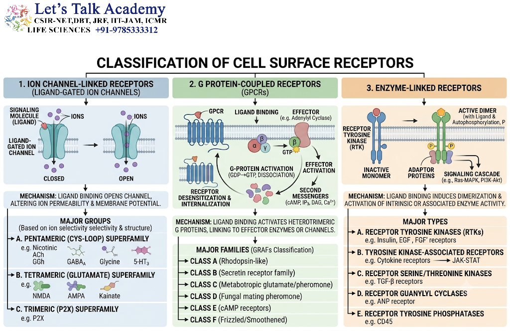
Cell surface receptors can be classified according to their structural organization, signaling mechanisms, and intracellular functions.
The major receptor families include:
-
G protein-coupled receptors.
-
Receptor tyrosine kinases.
-
Receptor serine/threonine kinases.
-
Cytokine receptors associated with cytoplasmic tyrosine kinases.
-
Receptor guanylyl cyclases.
-
Ligand-gated ion channel receptors.
-
Integrins and other cell adhesion receptors.
Each receptor family has a characteristic structure and activates specific intracellular pathways.
3.2 Overview of Receptor Families
|
Receptor family |
Structural feature |
Major signaling mechanism |
Example |
|---|---|---|---|
|
G protein-coupled receptors |
Seven transmembrane helices |
Activation of heterotrimeric G proteins |
β-adrenergic receptor |
|
Receptor tyrosine kinases |
Single transmembrane segment and kinase domain |
Tyrosine phosphorylation |
Insulin receptor, EGFR |
|
Cytokine receptors |
Usually single transmembrane segment |
Activation of JAK–STAT signaling |
Interferon receptor |
|
Receptor serine/threonine kinases |
Single transmembrane segment |
Serine/threonine phosphorylation |
TGF-β receptors |
|
Receptor guanylyl cyclases |
Membrane-spanning region and catalytic domain |
Production of cyclic GMP |
Natriuretic peptide receptor |
|
Ligand-gated ion channels |
Multiple membrane-spanning regions |
Opening or closing of ion channels |
Nicotinic acetylcholine receptor |
|
Integrins |
α and β subunit heterodimers |
Adhesion and bidirectional signaling |
Fibronectin receptors |
3.3 Enzyme-Linked Receptors
Enzyme-linked receptors are membrane receptors that either possess intrinsic enzymatic activity or associate with enzymes that become activated following ligand binding.
These receptors commonly contain a single transmembrane segment and an intracellular region involved in signal transduction.
Major examples include:
-
Receptor tyrosine kinases.
-
Receptor serine/threonine kinases.
-
Receptor guanylyl cyclases.
-
Cytokine receptors that associate with cytoplasmic tyrosine kinases.
Enzyme-linked receptors are particularly important in growth regulation, differentiation, metabolism, immune responses, and tissue development.
3.4 G Protein-Coupled Receptors
G protein-coupled receptors, commonly abbreviated as GPCRs, are a large family of cell surface receptors containing seven transmembrane α-helices.
These receptors transmit signals through heterotrimeric G proteins and regulate diverse physiological processes.
GPCRs participate in:
-
Vision.
-
Smell and taste.
-
Heart rate regulation.
-
Neurotransmission.
-
Hormone responses.
-
Smooth muscle contraction.
-
Immune signaling.
The β-adrenergic receptor, muscarinic acetylcholine receptor, and rhodopsin are well-known examples.
3.5 Ligand-Gated Ion Channel Receptors
Ligand-gated ion channels are membrane proteins that open or close in response to the binding of specific chemical ligands.
They regulate the movement of ions across the plasma membrane and are essential for rapid cellular communication.
These receptors are particularly important in the nervous system.
Examples include:
-
Nicotinic acetylcholine receptors.
-
GABAA_A receptors.
-
Ionotropic glutamate receptors.
-
Glycine receptors.
Their activation can rapidly change the membrane potential of a cell.
4. G Protein-Coupled Receptors
4.1 Introduction to G Protein-Coupled Receptors
G protein-coupled receptors are one of the most extensively studied families of cell surface receptors. They are also known as seven-transmembrane receptors or 7TM receptors because their polypeptide chains cross the plasma membrane seven times.
GPCRs convert extracellular chemical signals into intracellular responses through interactions with heterotrimeric G proteins.
They are involved in a wide variety of biological processes, including sensory perception, neurotransmission, hormonal regulation, cardiovascular control, and immune responses.
The functional versatility of GPCRs is partly due to their ability to activate different G protein subtypes and interact with several intracellular regulatory proteins.
4.2 Structure of G Protein-Coupled Receptors
A typical GPCR contains:
-
Seven transmembrane α-helices.
-
An extracellular N-terminal region.
-
An intracellular C-terminal region.
-
Three extracellular loops.
-
Three intracellular loops.
-
An intracellular ligand-dependent signaling interface.
The extracellular region may contain ligand-binding sites, glycosylation sites, or disulfide bonds that stabilize the receptor.
The intracellular loops and the C-terminal region interact with G proteins, arrestins, and other regulatory proteins.
Different GPCRs recognize different types of ligands. Some ligands bind within the transmembrane region, while others interact with extracellular domains or loops.
4.3 Heterotrimeric G Proteins
GPCRs typically signal through heterotrimeric G proteins, which consist of three subunits:
-
Gα
-
Gβ
-
Gγ
The Gα subunit binds guanine nucleotides, such as GDP and GTP, and has intrinsic GTPase activity.
The Gβ and Gγ subunits form a functional complex known as the Gβγ dimer.
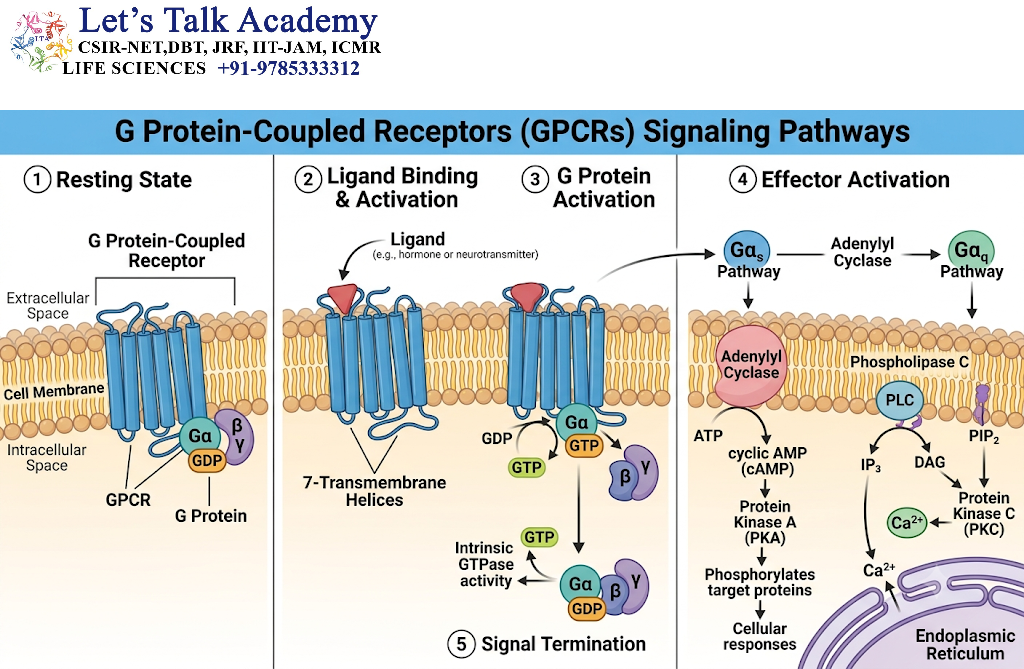
In the inactive state, Gα is bound to GDP and is associated with the Gβγ dimer.
When an activated GPCR interacts with the G protein, it promotes the exchange of GDP for GTP on the Gα subunit. The G protein then undergoes a conformational change that enables Gα-GTP and, in many cases, Gβγ to regulate downstream effectors.
G protein signaling is terminated when Gα hydrolyzes GTP to GDP. This process is often accelerated by regulatory proteins known as regulators of G protein signaling, or RGS proteins.
4.4 Mechanism of GPCR Activation
The general mechanism of GPCR signaling occurs through the following steps:
Step 1: Ligand Binding
A signaling molecule binds to the extracellular or transmembrane region of a GPCR.
Step 2: Receptor Conformational Change
Ligand binding stabilizes an active conformation of the receptor.
Step 3: G Protein Activation
The activated receptor interacts with a heterotrimeric G protein and promotes the exchange of GDP for GTP on the Gα subunit.
Step 4: Dissociation or Rearrangement of G Protein Components
Gα-GTP and Gβγ can regulate downstream signaling proteins.
Step 5: Activation of Effector Proteins
The activated G protein components influence effectors such as adenylyl cyclase, phospholipase C, or ion channels.
Step 6: Generation of Intracellular Signals
Effector proteins generate second messengers, including cyclic AMP, inositol trisphosphate, and diacylglycerol.
Step 7: Cellular Response
Second messengers activate protein kinases, alter ion concentrations, or regulate gene expression.
Step 8: Signal Termination
GTP hydrolysis, receptor phosphorylation, arrestin recruitment, and other regulatory mechanisms reduce or terminate signaling.
4.5 Major G Protein Signaling Pathways
Different Gα subfamilies activate distinct signaling pathways.
4.5.1 Gs-Mediated Signaling
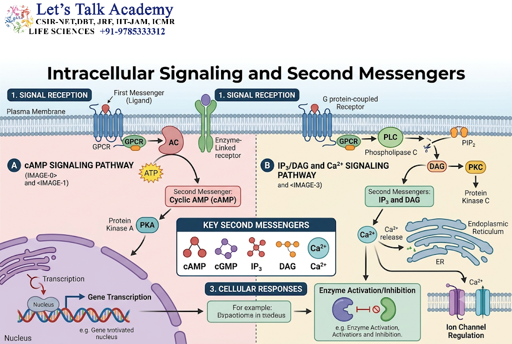
The Gs protein stimulates adenylyl cyclase, which catalyzes the conversion of ATP into cyclic AMP.
The increase in cAMP activates protein kinase A, also known as PKA.
PKA phosphorylates specific target proteins and can influence metabolism, ion transport, and gene expression.
For example, activation of β-adrenergic receptors in certain cells stimulates the Gs–adenylyl cyclase–cAMP pathway.
The effects of cAMP depend on the cell type and the proteins expressed within the cell.
4.5.2 Gi-Mediated Signaling
Gi proteins generally inhibit adenylyl cyclase and reduce cAMP production.
However, Gi-associated signaling is more complex than simple inhibition of cAMP synthesis. The Gβγ subunits can also regulate ion channels and other signaling proteins.
Gi-coupled receptors participate in neurotransmitter signaling, immune regulation, and the control of cellular excitability.
4.5.3 Gq-Mediated Signaling
Gq proteins activate phospholipase C-β, an enzyme that hydrolyzes a membrane phospholipid known as phosphatidylinositol 4,5-bisphosphate, or PIP₂.
The reaction produces two important second messengers:
-
Inositol 1,4,5-trisphosphate, or IP₃.
-
Diacylglycerol, or DAG.
IP₃ diffuses through the cytoplasm and binds to IP₃ receptors on the endoplasmic reticulum, promoting calcium release.
DAG remains associated with the plasma membrane and, together with calcium in many settings, activates protein kinase C.
The Gq–PLCβ pathway is involved in smooth muscle contraction, secretion, metabolism, and other cellular responses.
4.6 Second Messengers in GPCR Signaling
GPCRs regulate several important second messengers.
Cyclic AMP
Cyclic AMP is produced from ATP by adenylyl cyclase. It activates PKA and can regulate ion channels and transcriptional regulators such as CREB.
Calcium Ions
Calcium ions function as intracellular signaling molecules. Their concentration in the cytoplasm is tightly controlled.
Calcium may enter the cell through plasma membrane channels or be released from intracellular stores.
IP₃
IP₃ is generated through the hydrolysis of PIP₂ and promotes calcium release from the endoplasmic reticulum.
DAG
DAG is a lipid second messenger that activates protein kinase C and other signaling proteins.
The combined action of these second messengers allows GPCRs to regulate multiple intracellular processes.
4.7 Desensitization of GPCRs
Continuous or repeated stimulation of a GPCR can reduce the cellular response to a ligand. This process is known as receptor desensitization.
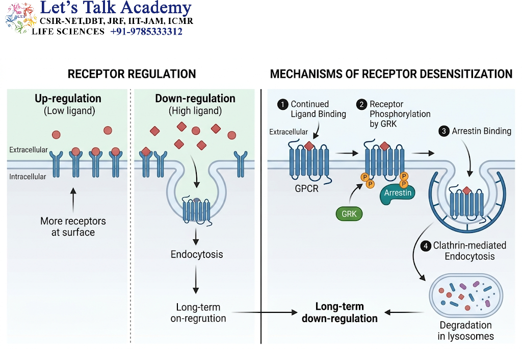
A major mechanism involves phosphorylation of the activated receptor by GPCR kinases, or GRKs.
The phosphorylated receptor can bind arrestin proteins. Arrestin binding interferes with further G protein coupling and may promote receptor internalization.
Internalized receptors may be recycled to the plasma membrane or directed toward degradation.
Desensitization prevents excessive cellular responses and allows cells to adapt to persistent signals.
4.8 Physiological and Medical Significance of GPCRs
GPCRs regulate many important physiological processes, including:
-
Heart rate and cardiac function.
-
Blood pressure regulation.
-
Vision.
-
Smell.
-
Taste.
-
Hormone secretion.
-
Neurotransmission.
-
Immune cell migration.
-
Smooth muscle contraction.
Because GPCRs are involved in many biological processes, they are major targets of pharmacological research. Drugs may activate receptors, inhibit them, or modify their signaling activity.
However, the effect of a receptor-targeting drug depends on the receptor subtype, tissue distribution, and downstream signaling pathways.
5. Receptor Tyrosine Kinases
5.1 Introduction to Receptor Tyrosine Kinases
Receptor tyrosine kinases, commonly abbreviated as RTKs, are cell surface receptors that possess an intracellular protein tyrosine kinase domain.
They are activated by a wide range of extracellular signaling molecules, particularly growth factors, hormones, and other regulatory proteins.
RTKs play essential roles in:
-
Cell growth.
-
Cell proliferation.
-
Cell differentiation.
-
Cell survival.
-
Cell migration.
-
Embryonic development.
-
Tissue repair.
-
Metabolic regulation.
Important examples include the epidermal growth factor receptor, insulin receptor, platelet-derived growth factor receptor, and vascular endothelial growth factor receptors.
5.2 Structure of Receptor Tyrosine Kinases
A typical RTK consists of the following structural regions:
-
An extracellular ligand-binding domain.
-
A single transmembrane α-helix.
-
An intracellular juxtamembrane region.
-
An intracellular tyrosine kinase domain.
-
A C-terminal tail containing regulatory tyrosine residues.
The extracellular domain recognizes the appropriate ligand. The transmembrane region anchors the receptor within the plasma membrane.
The intracellular kinase domain transfers phosphate groups from ATP to specific tyrosine residues on protein substrates.
The phosphorylated C-terminal tail can serve as a docking platform for intracellular signaling proteins.
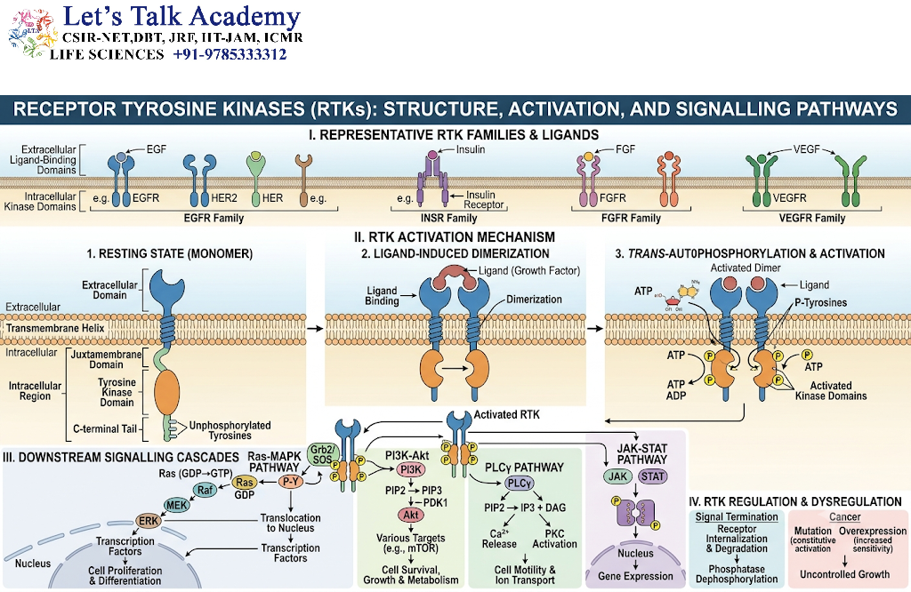
5.3 Activation Mechanism of Receptor Tyrosine Kinases
The activation of many RTKs involves ligand-induced receptor dimerization or rearrangement of pre-existing receptor complexes.
The mechanism can be explained as follows.
Step 1: Ligand Recognition
A growth factor or another extracellular ligand binds to the receptor’s extracellular domain.
Step 2: Receptor Association
Ligand binding promotes the association of two receptor molecules or changes the arrangement of receptors within a complex.
Step 3: Activation of Kinase Domains
The intracellular kinase domains become appropriately positioned and activated.
Step 4: Autophosphorylation
The kinase domains phosphorylate tyrosine residues on the receptor. In many RTKs, this involves phosphorylation between receptor molecules, known as trans-autophosphorylation.
Step 5: Recruitment of Signaling Proteins
Proteins containing phosphotyrosine-recognition domains, such as SH2 or PTB domains, bind to specific phosphorylated receptor sites.
Step 6: Activation of Downstream Pathways
Signaling complexes activate pathways such as Ras–MAPK, PI3K–AKT, and PLCγ.
Step 7: Cellular Response
The resulting signals influence gene expression, metabolism, growth, survival, migration, and differentiation.
5.4 Autophosphorylation and Phosphotyrosine Docking Sites
Autophosphorylation is a critical mechanism in RTK signaling.
When tyrosine residues on the receptor become phosphorylated, they can function as docking sites for intracellular proteins.
Different receptors possess different phosphorylation sites, and each site may recruit a distinct signaling protein.
For example, phosphorylated receptor regions can recruit proteins involved in:
-
Ras activation.
-
PI3K signaling.
-
Phospholipase C-γ activation.
-
Cytoskeletal regulation.
-
Adaptor protein assembly.
This mechanism allows a single receptor to activate several signaling pathways simultaneously.
The specific combination of phosphorylated sites and recruited proteins contributes to signaling specificity.
5.5 The Ras–MAPK Signaling Pathway
The Ras–MAPK pathway is one of the major signaling pathways activated by many receptor tyrosine kinases.
It is involved in cell proliferation, differentiation, development, and changes in gene expression.
The general pathway is:
5.5.1 Activation of Ras
Ras is a small GTP-binding protein located on the inner surface of the plasma membrane.
In its inactive state, Ras is bound to GDP.
Activated RTKs recruit adaptor proteins, such as Grb2, which help bring the guanine nucleotide exchange factor SOS near Ras.
SOS promotes the exchange of GDP for GTP.
Ras-GTP is the active form of Ras and can activate downstream signaling proteins.
5.5.2 Activation of the MAP Kinase Cascade
Active Ras stimulates Raf, a serine/threonine protein kinase.
Raf phosphorylates and activates MEK.
MEK phosphorylates and activates ERK.
ERK can phosphorylate cytoplasmic proteins and transcriptional regulators, thereby influencing gene expression.
Depending on the cellular context, this pathway can promote proliferation, differentiation, or other biological responses.
5.6 The PI3K–AKT Signaling Pathway
The PI3K–AKT pathway is another major signaling route activated by certain RTKs.
It regulates:
-
Cell survival.
-
Glucose metabolism.
-
Protein synthesis.
-
Cell growth.
-
Nutrient responses.
-
Metabolic adaptation.
Mechanism
Activated receptors recruit or activate phosphoinositide 3-kinase, or PI3K.
PI3K phosphorylates membrane phosphoinositides, including the conversion of PIP₂ to phosphatidylinositol 3,4,5-trisphosphate, or PIP₃.
PIP₃ recruits proteins containing pleckstrin homology domains, including AKT and PDK1.
AKT is activated through phosphorylation by upstream kinases, including PDK1 and mTORC2-associated mechanisms.
Activated AKT regulates several target proteins involved in cell survival, metabolism, and growth.
One important downstream target is the mTOR signaling system, which regulates protein synthesis and cellular growth.
5.7 PLCγ Signaling
Some receptor tyrosine kinases activate phospholipase C-γ, or PLCγ.
PLCγ hydrolyzes PIP₂ to generate IP₃ and DAG.
The resulting second messengers regulate calcium signaling and protein kinase C.
This pathway is involved in cell proliferation, secretion, cytoskeletal regulation, and other cellular responses.
5.8 The Insulin Receptor as a Specialized RTK
The insulin receptor is a receptor tyrosine kinase that regulates metabolism and growth.
Unlike many RTKs, the mature insulin receptor exists as a disulfide-linked receptor complex containing two extracellular α subunits and two membrane-spanning β subunits.
Insulin binding induces structural changes that activate the intracellular kinase domains.
The receptor phosphorylates insulin receptor substrate proteins, including IRS proteins, which then recruit downstream signaling components.
Two important pathways activated by insulin signaling are:
-
The PI3K–AKT pathway, which regulates glucose transport, metabolism, and other metabolic effects.
-
The Ras–MAPK pathway, which contributes to growth-related responses.
In skeletal muscle and adipose tissue, insulin signaling promotes the translocation of GLUT4-containing vesicles to the plasma membrane, increasing glucose uptake.
5.9 Regulation of RTK Signaling
RTK signaling must be tightly regulated to prevent excessive or prolonged activation.
Important regulatory mechanisms include:
-
Protein tyrosine phosphatases that remove phosphate groups from signaling proteins.
-
Receptor internalization through endocytosis.
-
Receptor recycling to the plasma membrane.
-
Lysosomal receptor degradation.
-
Inhibitory adaptor proteins.
-
Negative-feedback phosphorylation.
-
Regulation of ligand availability.
Disruption of these regulatory processes can lead to abnormal cell growth and other pathological effects.
6. Cytokine Receptors and the JAK–STAT Signaling Pathway
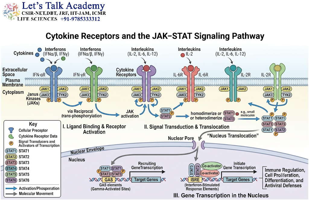
6.1 Introduction to Cytokine Receptors
Cytokine receptors are cell surface receptors that recognize cytokines, which are signaling proteins involved in immune regulation, inflammation, cell growth, and communication between cells.
Many cytokine receptors do not possess intrinsic enzymatic activity. Instead, they associate with cytoplasmic protein kinases that initiate intracellular signaling.
A major signaling mechanism used by several cytokine receptor families is the Janus kinase–signal transducer and activator of transcription pathway, commonly called the JAK–STAT pathway.
Cytokine signaling is essential for immune responses, hematopoiesis, tissue repair, and communication between immune and non-immune cells.
6.2 Structural Organization of Cytokine Receptors
Many cytokine receptors contain:
-
An extracellular ligand-binding region.
-
A single transmembrane segment.
-
An intracellular domain that associates with signaling proteins.
Their cytoplasmic regions generally lack intrinsic tyrosine kinase activity.
Instead, they associate with Janus kinases, including JAK1, JAK2, JAK3, and TYK2, depending on the receptor complex and cell type.
Cytokine receptors may form homodimeric or heteromeric complexes. Ligand binding can promote receptor association or rearrangement, allowing the associated kinases to activate one another.
6.3 The JAK–STAT Signaling Mechanism
The JAK–STAT pathway provides a relatively direct connection between extracellular cytokine binding and changes in gene transcription.
The major steps are as follows:
Step 1: Cytokine Binding
A cytokine binds to the extracellular domain of its receptor.
Step 2: Receptor Rearrangement
Ligand binding changes the arrangement of receptor subunits and their associated JAK proteins.
Step 3: JAK Activation
The associated JAK proteins become activated and phosphorylate one another or undergo other activating structural changes.
Step 4: Receptor Phosphorylation
Activated JAKs phosphorylate tyrosine residues on the intracellular region of the receptor.
Step 5: STAT Recruitment
Signal transducers and activators of transcription, known as STAT proteins, bind to phosphorylated receptor sites through their SH2 domains.
Step 6: STAT Phosphorylation
JAKs phosphorylate the recruited STAT proteins.
Step 7: STAT Dimerization
Phosphorylated STAT proteins form dimers through reciprocal phosphotyrosine–SH2 domain interactions.
Step 8: Nuclear Translocation
STAT dimers enter the nucleus.
Step 9: Regulation of Gene Expression
STAT dimers bind regulatory DNA sequences and influence the transcription of target genes.
The genes activated depend on the cytokine, receptor complex, STAT proteins involved, and the cellular context.
6.4 Biological Functions of the JAK–STAT Pathway
The JAK–STAT pathway regulates several important processes:
-
Antiviral defense.
-
Immune cell differentiation.
-
Inflammation.
-
Hematopoiesis.
-
Cell proliferation.
-
Cell survival.
-
Communication between immune cells.
-
Responses to extracellular cytokines.
Interferons, for example, can activate JAK–STAT signaling and induce the expression of genes involved in antiviral defense.
6.5 Negative Regulation of JAK–STAT Signaling
The JAK–STAT pathway is controlled by several inhibitory mechanisms.
Suppressors of Cytokine Signaling
Suppressors of cytokine signaling, or SOCS proteins, inhibit cytokine signaling through mechanisms that may involve direct kinase inhibition, competition for receptor-binding sites, and recruitment of protein degradation machinery.
Protein Tyrosine Phosphatases
Protein tyrosine phosphatases can remove phosphate groups from JAKs, receptors, or STAT proteins, reducing signaling activity.
Protein Inhibitors of Activated STATs
PIAS proteins can inhibit STAT-dependent transcription through several mechanisms.
The coordinated action of these regulatory systems prevents excessive cytokine signaling.
7. Receptor Serine/Threonine Kinases
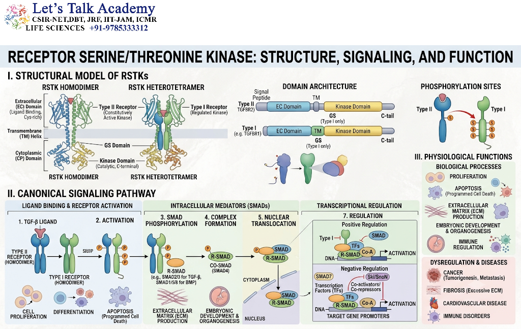
7.1 Introduction to Receptor Serine/Threonine Kinases
Receptor serine/threonine kinases are cell surface receptors that contain intracellular kinase domains capable of phosphorylating serine and threonine residues on target proteins.
They are particularly important in signaling by the transforming growth factor beta, or TGF-β, superfamily.
These receptors regulate:
-
Cell proliferation.
-
Cell differentiation.
-
Embryonic development.
-
Extracellular matrix production.
-
Tissue remodeling.
-
Immune regulation.
-
Cell fate determination.
7.2 Structure of Receptor Serine/Threonine Kinases
These receptors generally contain:
-
An extracellular ligand-binding domain.
-
A single transmembrane region.
-
An intracellular serine/threonine kinase domain.
TGF-β family signaling commonly involves two receptor classes:
-
Type II receptors.
-
Type I receptors.
Type II receptors possess constitutive kinase activity in many signaling contexts and activate type I receptors after ligand-dependent receptor complex formation.
7.3 TGF-β Signaling Mechanism
The general mechanism of TGF-β receptor signaling involves the following steps:
-
TGF-β or another related ligand binds to a receptor complex.
-
Type II receptors interact with and phosphorylate type I receptors.
-
Activated type I receptors phosphorylate receptor-regulated SMAD proteins.
-
Phosphorylated SMAD proteins associate with a common mediator SMAD.
-
The SMAD complex enters the nucleus.
-
The complex regulates the transcription of target genes with the help of other transcriptional regulators.
Different TGF-β family ligands activate different receptor combinations and SMAD proteins.
7.4 SMAD Proteins and Gene Regulation
SMAD proteins are intracellular signaling molecules that transmit signals from activated TGF-β family receptors to the nucleus.
They can be divided into functional groups:
-
Receptor-regulated SMADs.
-
Common mediator SMADs.
-
Inhibitory SMADs.
For example, TGF-β signaling commonly involves SMAD2 and SMAD3, which associate with SMAD4 after activation.
Bone morphogenetic protein signaling commonly involves other receptor-regulated SMAD proteins, such as SMAD1, SMAD5, and SMAD8, with SMAD9 also functioning in certain pathways.
SMAD complexes do not generally act alone. They interact with transcription factors, chromatin regulators, and other nuclear proteins to control gene expression.
7.5 Biological Importance of TGF-β Signaling
TGF-β signaling contributes to:
-
Embryonic development.
-
Cell fate specification.
-
Regulation of immune responses.
-
Extracellular matrix synthesis.
-
Wound healing.
-
Tissue homeostasis.
-
Regulation of epithelial and mesenchymal cell behavior.
Abnormal TGF-β signaling can contribute to fibrosis, developmental abnormalities, and cancer-associated changes, depending on the biological context.
8. Receptor Guanylyl Cyclases
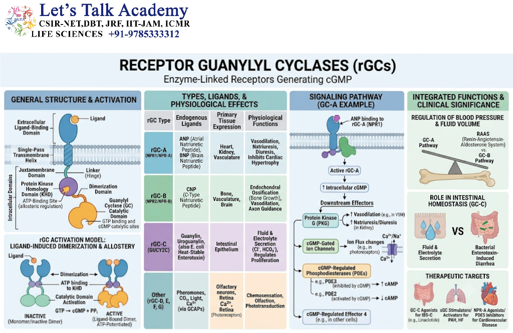
8.1 Introduction to Receptor Guanylyl Cyclases
Receptor guanylyl cyclases are membrane-associated receptors that possess an intracellular catalytic domain capable of producing cyclic guanosine monophosphate, or cGMP.
cGMP is an important intracellular second messenger involved in vascular regulation, sensory signaling, fluid balance, and other physiological processes.
Some receptor guanylyl cyclases are activated by peptide hormones, while other guanylyl cyclase systems are regulated by intracellular or membrane-associated signaling proteins.
8.2 Structural Organization
Many receptor guanylyl cyclases contain:
-
An extracellular ligand-binding domain.
-
A single transmembrane segment.
-
An intracellular kinase-homology region.
-
A dimerization region.
-
A catalytic guanylyl cyclase domain.
The catalytic domain converts GTP into cGMP.
The enzyme activity depends on the functional assembly of the receptor complex.
8.3 Signaling Through cGMP
cGMP regulates cellular processes through several downstream effectors.
These include:
-
cGMP-dependent protein kinase.
-
Cyclic nucleotide-gated ion channels.
-
Certain phosphodiesterases.
In vascular smooth muscle, cGMP signaling contributes to relaxation through mechanisms involving protein kinase G and the regulation of intracellular calcium and contractile proteins.
Receptor guanylyl cyclases activated by natriuretic peptides also participate in the regulation of blood volume and vascular physiology.
8.4 Physiological Importance
Receptor guanylyl cyclases contribute to:
-
Regulation of blood pressure.
-
Control of fluid and electrolyte balance.
-
Cardiovascular homeostasis.
-
Sensory signaling.
-
Regulation of intestinal fluid transport.
The activity of cGMP is controlled by phosphodiesterases, which hydrolyze cGMP and thereby help terminate the signal.
9. Integrins and Cell Adhesion Receptors
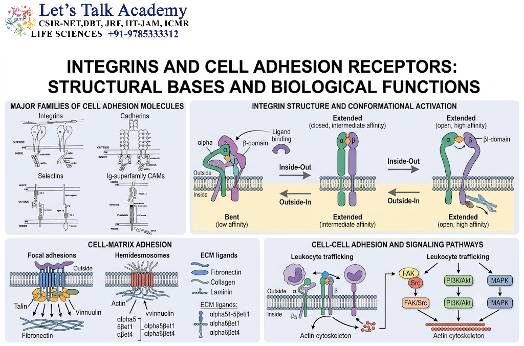
9.1 Introduction to Integrins
Integrins are transmembrane cell adhesion receptors that connect cells to the extracellular matrix or to other cells.
They are essential for maintaining tissue architecture, regulating cell movement, and transmitting mechanical and biochemical signals.
Unlike receptors that primarily detect soluble signaling molecules, integrins often recognize extracellular matrix proteins such as fibronectin, collagen, and laminins.
Integrins function as both adhesion molecules and signaling receptors.
9.2 Structure of Integrins
Integrins are heterodimeric proteins composed of two non-covalently associated subunits:
-
An α subunit.
-
A β subunit.
Mammals possess multiple α and β subunits that combine to form different integrin receptors with distinct ligand specificities.
Each subunit contains:
-
A large extracellular domain.
-
A single transmembrane segment.
-
A relatively short cytoplasmic tail.
The extracellular domains recognize specific extracellular matrix ligands, while the cytoplasmic tails interact with intracellular adaptor proteins and cytoskeletal components.
9.3 Bidirectional Integrin Signaling
Integrins transmit signals in two directions.
Inside-Out Signaling
Inside-out signaling occurs when intracellular signals alter the conformation and ligand-binding activity of integrins.
For example, signals from chemokine receptors or other intracellular pathways can increase the affinity or avidity of integrins for their ligands.
This mechanism is particularly important in the regulation of leukocyte adhesion.
Outside-In Signaling
Outside-in signaling occurs when extracellular matrix binding activates intracellular signaling pathways.
Ligand binding can promote integrin clustering and the assembly of signaling complexes known as focal adhesions.
These complexes contain proteins such as:
-
Talin.
-
Kindlin.
-
Focal adhesion kinase.
-
Paxillin.
-
Vinculin.
-
Src family kinases.
The resulting signals regulate cytoskeletal organization, cell migration, proliferation, and survival.
9.4 Focal Adhesions and Mechanical Signaling
Focal adhesions are dynamic multiprotein structures that connect integrins to the actin cytoskeleton.
They function as platforms for both biochemical signaling and mechanical force transmission.
Mechanical forces can alter the conformation of adhesion-associated proteins, influence signaling activity, and affect gene expression.
This process is known as mechanotransduction.
Mechanotransduction allows cells to respond to the stiffness, tension, and physical organization of their surrounding environment.
9.5 Biological Importance of Integrins
Integrins participate in:
-
Cell migration.
-
Wound healing.
-
Embryonic development.
-
Immune cell trafficking.
-
Blood clot formation.
-
Tissue organization.
-
Angiogenesis.
-
Cancer cell invasion.
-
Maintenance of tissue integrity.
Their ability to integrate chemical and mechanical information makes them essential regulators of cell behavior.
10. Ligand-Gated Ion Channel Receptors
10.1 Introduction to Ligand-Gated Ion Channels
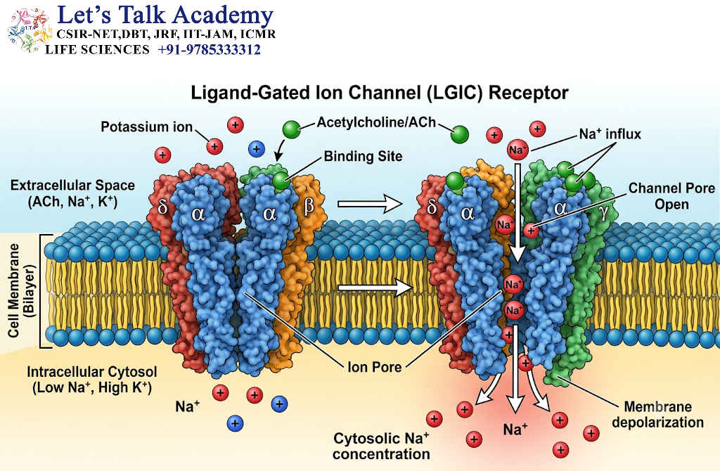
Ligand-gated ion channels are membrane proteins that respond to the binding of chemical messengers by changing their permeability to ions.
They are also called ionotropic receptors.
Unlike many enzyme-linked receptors and GPCRs, ligand-gated ion channels can produce electrical responses within milliseconds. This makes them particularly important in nervous system signaling.
Their activation can alter the membrane potential of a cell, thereby influencing neuronal excitability and muscle contraction.
10.2 Structural Organization
Ligand-gated ion channels contain multiple transmembrane regions that form an ion-conducting pore.
Depending on the receptor family, the channel may be assembled from several subunits.
The binding of a ligand changes the conformation of the channel, causing the pore to open or close.
The ions that pass through the channel depend on the channel’s selectivity properties.
Commonly transported ions include:
-
Sodium ions.
-
Potassium ions.
-
Calcium ions.
-
Chloride ions.
10.3 Mechanism of Ion Channel Activation
The signaling mechanism occurs through the following steps:
-
A neurotransmitter or other ligand is released into the extracellular space.
-
The ligand binds to the extracellular region of an ion channel receptor.
-
The receptor changes conformation.
-
The ion-conducting pore opens or closes.
-
Ions move across the membrane according to their electrochemical gradients.
-
The membrane potential changes.
-
The cell produces a physiological response.
The direction and magnitude of the electrical response depend on the ions involved, their concentration gradients, and the membrane potential.
10.4 Excitatory and Inhibitory Responses
Some ligand-gated ion channels produce depolarizing responses, while others promote hyperpolarization or stabilization of the membrane potential.
For example, many glutamate-gated channels promote excitatory signaling in neurons.
GABAA_A receptors are chloride-permeable ligand-gated ion channels that commonly mediate inhibitory responses in mature neurons, although the effect of chloride conductance depends on the chloride gradient across the membrane.
Thus, the functional effect of a receptor depends on both its ion selectivity and the physiological conditions of the cell.
10.5 Physiological Significance
Ligand-gated ion channels are essential for:
-
Synaptic transmission.
-
Muscle contraction.
-
Neuronal communication.
-
Sensory processing.
-
Learning and memory.
-
Regulation of neuronal excitability.
Abnormal activity of these receptors can interfere with nervous system function.
11. Intracellular Signaling and Second Messengers
11.1 Introduction to Signal Transduction
Signal transduction is the process by which a cell converts an extracellular signal into an intracellular response.
Cell surface receptors initiate this process by detecting extracellular ligands and activating signaling proteins.
Signal transduction pathways commonly involve protein phosphorylation, second messengers, changes in ion concentrations, protein–protein interactions, and alterations in gene expression.
The response may be rapid, such as the opening of an ion channel, or relatively slow, such as the activation of a gene expression program.
11.2 Protein Phosphorylation and Dephosphorylation
Protein phosphorylation is the addition of a phosphate group to a protein.
Protein kinases transfer phosphate groups from ATP to specific amino acid residues, commonly serine, threonine, or tyrosine.
Protein phosphatases remove phosphate groups from phosphorylated proteins.
These reversible reactions regulate protein activity, localization, stability, and interactions with other proteins.
Phosphorylation is a central mechanism of receptor signaling because it allows cells to rapidly alter the activity of signaling proteins.
11.3 Second Messengers
Second messengers are intracellular signaling molecules produced or released in response to receptor activation.
They transmit and amplify signals from activated receptors to intracellular targets.
Important second messengers include:
-
Cyclic AMP.
-
Cyclic GMP.
-
Calcium ions.
-
IP₃.
-
DAG.
-
Certain phosphoinositide lipids.
Second messengers often have short lifetimes and are regulated by enzymes, pumps, transporters, or binding proteins.
11.4 Signal Amplification
Signal amplification occurs when one activated receptor leads to the activation of many downstream signaling molecules.
For example, one receptor can activate an enzyme that produces many second messenger molecules. Each second messenger can activate multiple downstream targets.
Amplification allows cells to respond to very low concentrations of signaling molecules.
However, amplification must be regulated carefully because excessive signaling can produce inappropriate or harmful cellular responses.
11.5 Signal Termination
Cell signaling must eventually be reduced or terminated.
Important mechanisms include:
-
Ligand removal or degradation.
-
Receptor inactivation.
-
GTP hydrolysis.
-
Dephosphorylation of signaling proteins.
-
Second messenger degradation.
-
Calcium sequestration.
-
Receptor internalization.
-
Protein degradation.
-
Activation of inhibitory feedback pathways.
Signal termination is as important as signal activation because it prevents continuous stimulation and allows cells to respond to new signals.
12. Regulation and Desensitization of Cell Surface Receptors
12.1 Importance of Receptor Regulation
Cell surface receptors must be regulated to maintain an appropriate balance between signal sensitivity and cellular protection.
If receptors remain active for too long, cells may experience abnormal proliferation, metabolic disturbances, inflammation, or other harmful effects.
Receptor regulation controls the intensity, duration, location, and timing of signaling.
12.2 Receptor Phosphorylation
Receptor phosphorylation can either activate or inhibit receptor signaling, depending on the receptor and the specific amino acid residues involved.
For example, receptor tyrosine kinases are often activated through tyrosine phosphorylation.
In contrast, phosphorylation of GPCRs can promote receptor desensitization and arrestin binding.
The effect of phosphorylation is therefore context-dependent.
12.3 Receptor Internalization
Receptor internalization is the process by which receptors are removed from the plasma membrane through endocytosis.
Internalization can reduce the number of receptors available to bind extracellular ligands.
Once internalized, receptors may be:
-
Recycled back to the plasma membrane.
-
Transported to intracellular compartments.
-
Degraded in lysosomes.
-
Retained in signaling endosomes.
The fate of an internalized receptor influences the duration and location of receptor signaling.
12.4 Receptor Recycling
Receptor recycling allows internalized receptors to return to the plasma membrane.
This process restores receptor availability and can help maintain cellular sensitivity to extracellular signals.
Recycling is particularly important for receptors that undergo repeated cycles of activation and internalization.
12.5 Receptor Downregulation
Downregulation refers to a reduction in the number or functional availability of receptors.
It may occur through:
-
Decreased receptor synthesis.
-
Increased receptor degradation.
-
Enhanced internalization.
-
Reduced transport to the plasma membrane.
-
Changes in receptor stability.
Downregulation can occur in response to prolonged exposure to a ligand.
12.6 Receptor Upregulation
Upregulation refers to an increase in receptor abundance or functional availability.
Cells may increase receptor expression when signaling activity is persistently low or when additional sensitivity to a signal is required.
The precise response depends on the receptor type, ligand, cell type, and physiological conditions.
12.7 Receptor Desensitization
Desensitization is the reduction in cellular responsiveness to a ligand despite its continued presence.
Desensitization may be rapid or develop over a longer period.
It can involve receptor phosphorylation, arrestin recruitment, inhibitory proteins, changes in downstream signaling, and receptor trafficking.
Desensitization allows cells to adapt to persistent environmental stimuli.
13. Receptor Crosstalk and Signal Integration
13.1 Concept of Receptor Crosstalk
Receptor crosstalk occurs when signaling pathways activated by different receptors interact with one another.
Cells are exposed to multiple extracellular signals simultaneously. Their responses depend on the integration of these signals rather than on the activity of a single receptor in isolation.
For example, a growth factor receptor and an integrin may activate overlapping signaling proteins and jointly influence cell proliferation or migration.
13.2 Mechanisms of Receptor Crosstalk
Receptor crosstalk may occur through:
-
Shared intracellular signaling proteins.
-
Direct receptor–receptor interactions.
-
Convergent phosphorylation pathways.
-
Regulation of receptor trafficking.
-
Changes in gene expression.
-
Alterations in cytoskeletal organization.
-
Activation of common second messengers.
Crosstalk can enhance, reduce, or redirect a cellular response.
13.3 Signal Integration in Cellular Decision-Making
Cells often receive signals that promote different outcomes.
For example, one signal may promote cell proliferation, while another may promote differentiation or quiescence.
The cell integrates these signals through complex signaling networks.
The final response depends on:
-
Signal strength.
-
Signal duration.
-
Receptor abundance.
-
Cellular location.
-
Availability of signaling proteins.
-
Activity of inhibitory pathways.
-
Gene expression state.
This integration enables cells to make coordinated decisions.
13.4 Spatial Regulation of Receptor Signaling
Cell signaling is influenced by the location of receptors and signaling proteins.
Some signaling events occur at the plasma membrane, whereas others continue in endosomes or other intracellular compartments.
The same receptor may generate different effects depending on where it signals.
Spatial organization helps control the specificity of signaling and prevents inappropriate activation of unrelated cellular processes.
14. Biological Significance of Cell Surface Receptors
14.1 Role in Cell Growth and Proliferation
Cell surface receptors regulate cell division by detecting growth factors and other extracellular signals.
Receptor tyrosine kinases can activate pathways such as Ras–MAPK and PI3K–AKT, which influence cell cycle progression and cellular growth.
Growth factor signaling must be carefully controlled because excessive activation can contribute to abnormal tissue growth.
14.2 Role in Cell Differentiation
Cell differentiation is the process through which cells acquire specialized structures and functions.
Cell surface receptors help cells detect developmental signals that regulate gene expression and cell fate.
TGF-β family receptors, receptor tyrosine kinases, and other receptor systems contribute to differentiation in various tissues.
14.3 Role in Immune Responses
Immune cells use numerous cell surface receptors to recognize cytokines, chemokines, antigens, adhesion molecules, and danger signals.
These receptors regulate:
-
Immune cell activation.
-
Leukocyte migration.
-
Inflammatory responses.
-
Antigen recognition.
-
Communication between immune cells.
-
Development and maturation of immune cells.
Receptor signaling helps coordinate the immune response while preventing unnecessary tissue damage.
14.4 Role in Metabolism
Hormonal receptors regulate metabolic processes in response to changes in nutrient availability.
The insulin receptor is particularly important for glucose metabolism.
Other receptor systems regulate lipid metabolism, energy balance, and nutrient-responsive gene expression.
Metabolic signaling is influenced by both receptor activation and the integration of multiple intracellular pathways.
14.5 Role in Cell Adhesion and Migration
Integrins and other adhesion receptors help cells attach to their surroundings and regulate movement.
Cell migration is essential for embryonic development, wound healing, immune surveillance, and tissue repair.
Adhesion receptors coordinate the cytoskeleton, cell polarity, and mechanical forces required for movement.
14.6 Role in Tissue Homeostasis
Cell surface receptors contribute to tissue homeostasis by regulating cell survival, proliferation, differentiation, and communication.
The coordinated action of receptor signaling pathways helps maintain the balance between cell production, function, and removal.
Disturbances in these pathways can disrupt tissue organization.
15. Receptors in Human Diseases and Therapeutic Applications
15.1 Abnormal Receptor Signaling and Disease
Cell surface receptors play important roles in many diseases because they regulate essential biological processes.
Disease-associated receptor abnormalities may involve:
-
Genetic mutations.
-
Excessive receptor expression.
-
Reduced receptor expression.
-
Abnormal ligand production.
-
Defective receptor trafficking.
-
Persistent receptor activation.
-
Impaired signal termination.
The consequences depend on the receptor type and the affected tissue.
15.2 Receptor Tyrosine Kinases and Cancer
Some cancers are associated with abnormal activity of receptor tyrosine kinases.
Mechanisms may include receptor overexpression, gene amplification, activating mutations, or abnormal ligand production.
Persistent activation of RTK signaling can promote cell proliferation, survival, migration, and changes in tissue behavior.
Examples of clinically relevant receptor systems include EGFR, HER2, and certain receptor tyrosine kinases involved in blood vessel formation.
The presence of a receptor alteration does not automatically determine the clinical outcome. Its significance depends on the specific disease, molecular alteration, and other biological factors.
15.3 Receptors in Metabolic Disorders
Alterations in insulin receptor signaling can contribute to impaired glucose regulation.
Insulin resistance involves reduced biological responsiveness to insulin and may be associated with changes in receptor signaling, intracellular signaling proteins, inflammation, and metabolism.
The insulin receptor itself is only one component of the broader insulin signaling network.
15.4 Receptors in Immune and Inflammatory Disorders
Cytokine receptors and other immune receptors are involved in inflammatory signaling.
Excessive or dysregulated cytokine signaling can contribute to tissue inflammation and immune-mediated disease.
Therapeutic approaches may target cytokines, receptors, receptor-associated kinases, or downstream signaling components.
15.5 Receptors as Drug Targets
Cell surface receptors are important targets for drug development.
Drugs may act as:
-
Agonists that activate receptors.
-
Antagonists that block receptor activation.
-
Partial agonists that produce a limited response.
-
Allosteric modulators that alter receptor activity.
-
Antibodies that bind receptors or their ligands.
-
Inhibitors of receptor-associated enzymes.
The clinical effect of a drug depends on receptor distribution, signaling mechanisms, dose, pharmacokinetics, and tissue-specific responses.
15.6 Monoclonal Antibodies and Receptor-Based Therapy
Monoclonal antibodies are laboratory-produced antibodies that recognize specific molecular targets.
Some therapeutic antibodies bind cell surface receptors and inhibit their signaling. Others activate receptors or influence immune-cell interactions.
For example, certain antibodies target growth factor receptors or receptor-associated signaling systems.
Receptor-based therapy is an important area of modern biomedical research.
16. Experimental Approaches for Studying Cell Surface Receptors
16.1 Importance of Experimental Receptor Biology
Understanding cell surface receptors requires the integration of structural, biochemical, cellular, and molecular techniques.
Researchers study receptor expression, ligand binding, activation, trafficking, intracellular signaling, and physiological function.
Different experimental methods provide different types of information.
16.2 Ligand-Binding Assays
Ligand-binding assays are used to measure the interaction between a receptor and its ligand.
These assays can help determine:
-
Receptor affinity.
-
Binding capacity.
-
Receptor abundance.
-
Ligand specificity.
-
Binding kinetics.
Radiolabeled, fluorescent, or other tagged ligands may be used depending on the experimental design.
16.3 Western Blotting
Western blotting is used to detect specific proteins in a biological sample.
It can be used to study:
-
Receptor expression.
-
Receptor phosphorylation.
-
Changes in receptor abundance.
-
Activation of downstream signaling proteins.
For example, antibodies specific to phosphorylated tyrosine residues can be used to investigate receptor tyrosine kinase activation.
16.4 Immunofluorescence Microscopy
Immunofluorescence microscopy uses antibodies linked to fluorescent labels to visualize proteins in cells.
This technique can help determine:
-
Receptor localization.
-
Distribution on the plasma membrane.
-
Receptor internalization.
-
Colocalization with signaling proteins.
-
Changes in receptor distribution after ligand stimulation.
16.5 Flow Cytometry
Flow cytometry is used to analyze individual cells based on physical and fluorescent characteristics.
Fluorescent antibodies can be used to measure receptor expression on the surface of cells.
This technique is especially useful for studying immune cell populations and comparing receptor abundance between different cell types.
16.6 Co-Immunoprecipitation
Co-immunoprecipitation is a biochemical technique used to investigate protein–protein interactions.
It can help identify proteins associated with receptors, such as adaptor proteins, kinases, or cytoskeletal components.
The interpretation of results requires appropriate controls because biochemical association does not always demonstrate a direct physical interaction.
16.7 Mutational Analysis
Mutational analysis involves changing specific amino acids or regions of a receptor to investigate their functional importance.
Researchers may use this approach to study:
-
Ligand-binding sites.
-
Kinase activity.
-
Phosphorylation sites.
-
Receptor trafficking signals.
-
Protein interaction regions.
-
Structural elements involved in activation.
16.8 Live-Cell Imaging
Live-cell imaging allows researchers to monitor receptor behavior in living cells.
Fluorescently tagged receptors or signaling proteins can be used to study receptor movement, internalization, recycling, and interactions with other cellular structures.
These experiments provide information about the dynamic nature of receptor signaling.


