
1. Introduction
Cell division is one of the fundamental processes of life. It allows organisms to grow, replace damaged cells, reproduce, and maintain genetic continuity.
In eukaryotic organisms, two major forms of nuclear division are mitosis and meiosis.
Mitosis generally produces daughter cells that retain the chromosome number of the parent cell, whereas meiosis is a specialized division that reduces chromosome number and generates genetically diverse reproductive cells.
The basic relationship can be represented as:
Cell growth → DNA replication → Chromosome segregation → Cell division
Although both processes involve chromosome condensation, spindle formation, and chromosome segregation, their biological purposes are different.
Mitosis → Growth, tissue maintenance, cell replacement
Meiosis → Gamete formation, chromosome-number reduction, genetic variation
2. Cell Cycle and Cell Division
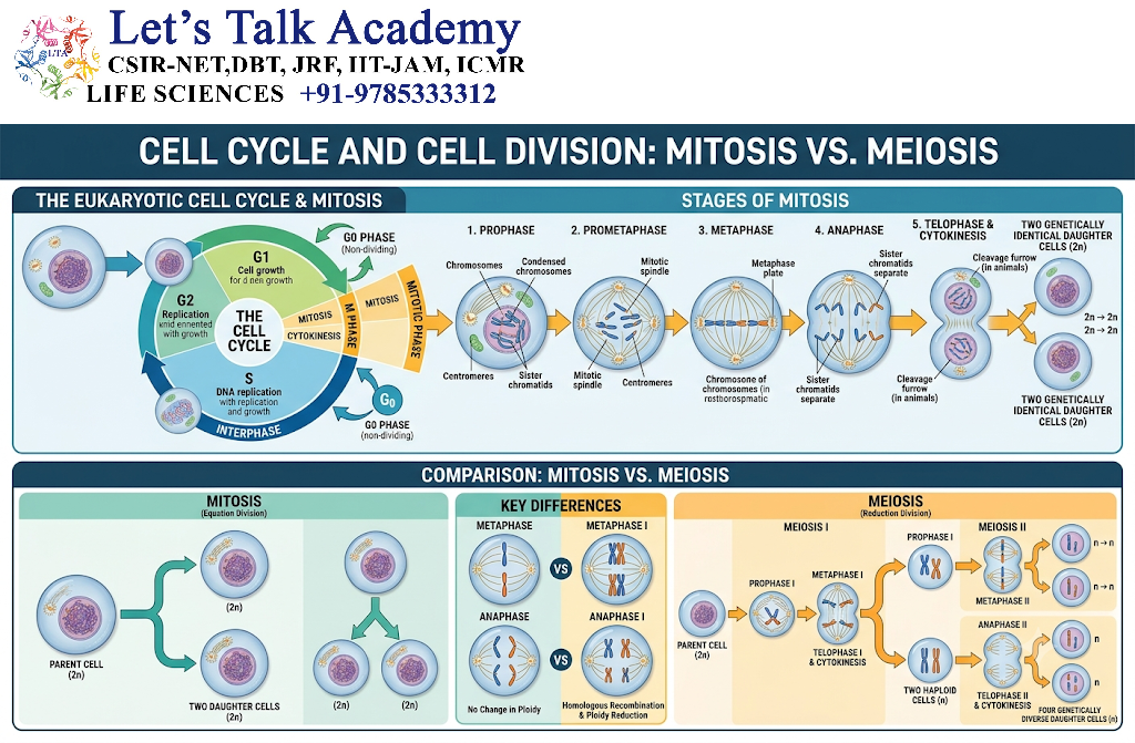
Before understanding mitosis and meiosis, it is important to understand the cell cycle.
The cell cycle consists broadly of:
- G1 phase
- S phase
- G2 phase
- M phase
During G1, the cell grows and performs normal metabolic functions.
During S phase, DNA is replicated.
During G2, the cell prepares for division.
The M phase includes chromosome segregation and cytokinesis.
A simplified sequence is:
G1 → S → G2 → M → G1
Meiosis also begins after a period of DNA replication, but the replicated chromosomes undergo two successive nuclear divisions.
3. DNA Replication Before Division
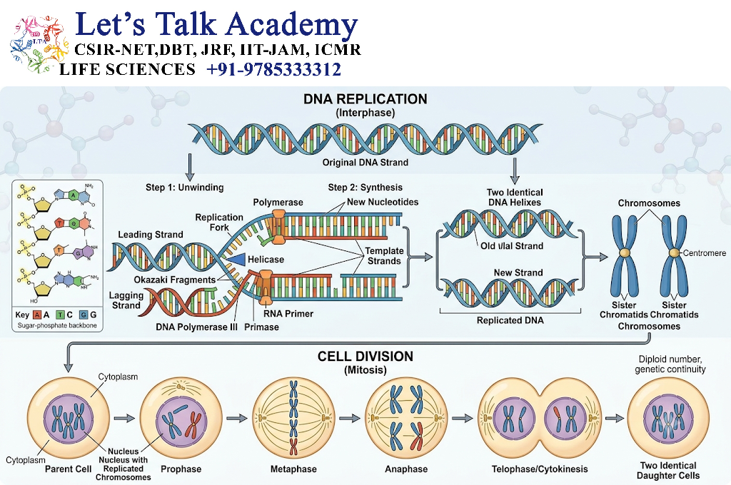
DNA replication occurs before mitosis and before meiosis.
During S phase, each chromosome is replicated to produce two sister chromatids.
Thus:
One chromosome → DNA replication → two sister chromatids
The sister chromatids remain physically associated until they are separated during chromosome segregation.
A crucial difference is:
- In mitosis, sister chromatids separate during the single chromosome-segregation division.
- In meiosis I, homologous chromosomes separate while sister chromatids remain together.
- In meiosis II, sister chromatids separate.
4. Mitosis: Definition
Mitosis is a form of nuclear division in which duplicated chromosomes are segregated so that each daughter nucleus generally receives one copy of each chromosome.
Mitosis is important for:
- Growth
- Development
- Tissue maintenance
- Cell replacement
- Asexual reproduction in some organisms
- Genome stability
The overall outcome is usually:
One parent cell → Two genetically similar daughter cells
5. Major Stages of Mitosis
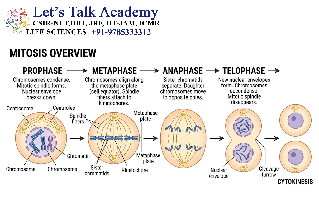
Mitosis is conventionally divided into:
- Prophase
- Prometaphase
- Metaphase
- Anaphase
- Telophase
Cytokinesis usually follows or overlaps with the final stages of mitosis.
6. Prophase
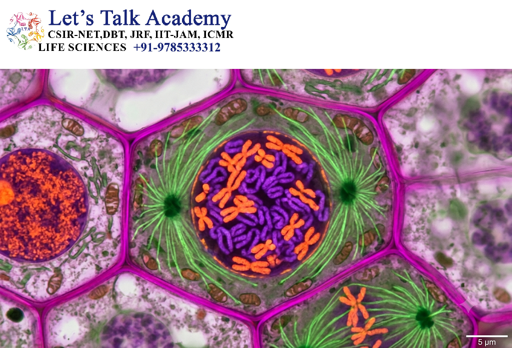
During prophase:
- Chromatin begins to condense.
- Chromosomes become increasingly visible.
- The mitotic spindle begins to form.
- Centrosomes move apart in animal cells.
- The nucleolus becomes less prominent.
Chromosome condensation is essential because compact chromosomes are easier to move and segregate without becoming excessively entangled.
7. Prometaphase
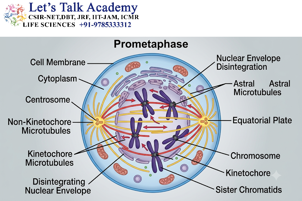
Prometaphase begins with breakdown of the nuclear envelope.
The spindle microtubules gain access to chromosomes.
Specialized protein structures called kinetochores assemble at centromeric regions.
Spindle microtubules attach to kinetochores.
Thus:
Nuclear-envelope breakdown → Kinetochore exposure → Microtubule attachment
8. Kinetochore Attachment
Each replicated chromosome contains two sister chromatids.
Their kinetochores are positioned so that sister chromatids can attach to spindle microtubules originating from opposite spindle poles.
This arrangement is called bipolar attachment.
Correct bipolar attachment is essential for accurate chromosome segregation.
9. Metaphase
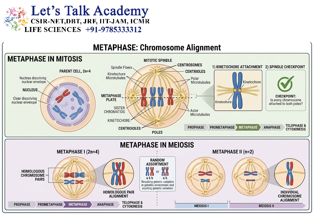
During metaphase, chromosomes become highly organized near the center of the spindle.
They form the metaphase plate.
At this stage, cells use surveillance mechanisms to ensure that chromosomes are appropriately attached to spindle microtubules.
The major regulatory system involved is the spindle assembly checkpoint.
10. Spindle Assembly Checkpoint
The spindle assembly checkpoint prevents premature chromosome segregation.
It monitors whether kinetochores have established appropriate interactions with spindle microtubules.
If chromosomes are not properly attached, the cell delays anaphase.
This provides time for correct spindle attachment to be established.
11. Anaphase
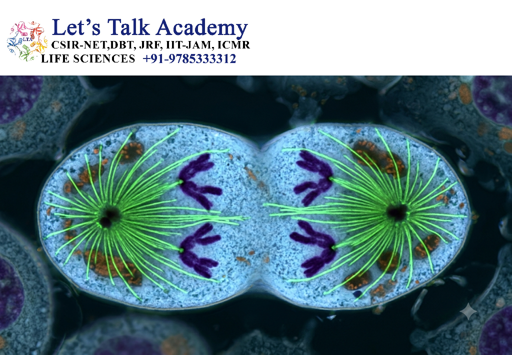
Anaphase begins when sister chromatids separate.
The sister chromatids are then considered individual daughter chromosomes.
They move toward opposite spindle poles.
A simplified sequence is:
Sister chromatid cohesion → Cohesion removal → Chromatid separation → Poleward movement
12. Telophase
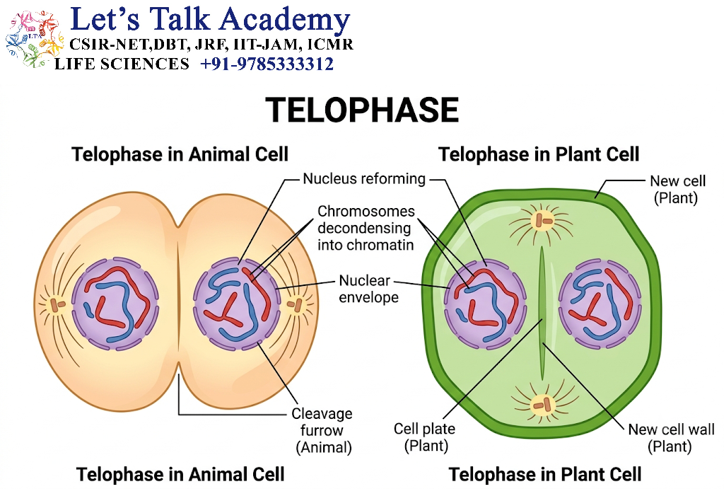
During telophase:
- Chromosomes reach opposite poles.
- Chromosomes begin to decondense.
- Nuclear envelopes reform.
- Nucleoli reappear.
- The mitotic spindle disassembles.
Two daughter nuclei are thereby established.
13. Cytokinesis
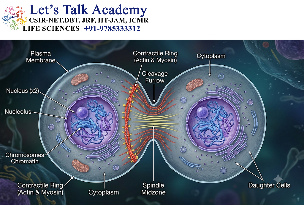
Cytokinesis is the physical division of the cytoplasm.
In animal cells, cytokinesis occurs through formation of a contractile ring containing:
- Actin
- Myosin
The contractile ring produces a cleavage furrow that divides the cell.
In plant cells, cytokinesis occurs through formation of a cell plate.
14. Outcome of Mitosis
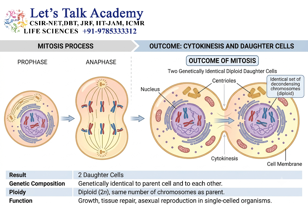
The general outcome of mitosis is:
One parent cell → Two daughter cells
The daughter cells generally maintain the same chromosome number as the parent cell.
For example:
Diploid cell → mitosis → two diploid daughter cells
Mitosis therefore maintains chromosome number across successive cell generations.
15. Biological Importance of Mitosis
Mitosis is essential for:
15.1 Growth
Multicellular organisms increase cell number through repeated cell divisions.
15.2 Tissue Repair
Damaged cells can be replaced through cell proliferation.
15.3 Development
Embryonic development depends on repeated mitotic divisions.
15.4 Tissue Maintenance
Many tissues continuously replace old or damaged cells.
15.5 Asexual Reproduction
Some organisms use mitotic division as part of asexual reproduction.
16. Meiosis: Definition
Meiosis is a specialized form of cell division that reduces chromosome number by half and generates genetically diverse reproductive cells.
Meiosis consists of two successive nuclear divisions:
- Meiosis I
- Meiosis II
DNA replication occurs once before meiosis I, but there is no second round of DNA replication between meiosis I and meiosis II.
The overall process can be represented as:
DNA replication → Meiosis I → Meiosis II → Four haploid products
17. Purpose of Meiosis
The major functions of meiosis include:
- Reduction of chromosome number.
- Production of haploid reproductive cells.
- Generation of genetic variation.
- Separation of homologous chromosomes.
- Facilitation of sexual reproduction.
Meiosis therefore connects chromosome inheritance with genetic diversity.
18. Meiosis I
Meiosis I is called the reductional division because homologous chromosomes separate from one another.
The major stages are:
- Prophase I
- Metaphase I
- Anaphase I
- Telophase I
19. Prophase I
Prophase I is the longest and most complex stage of meiosis.
It is characterized by:
- Homologous chromosome pairing.
- Synapsis.
- Formation of the synaptonemal complex.
- Crossing over.
- Recombination.
- Formation of chiasmata.
- Chromosome condensation.
Prophase I is divided into:
- Leptotene
- Zygotene
- Pachytene
- Diplotene
- Diakinesis
20. Leptotene
During leptotene:
- Chromosomes begin to condense.
- Chromosomal axes become visible.
- DNA has already been replicated.
- Homologous chromosomes begin the process of pairing.
Each chromosome consists of two sister chromatids, although these chromatids may not yet be individually distinguishable under the light microscope.
21. Zygotene
During zygotene, homologous chromosomes begin to pair closely.
This process is called synapsis.
The synaptonemal complex develops between homologous chromosomes and facilitates close alignment.
22. Synapsis
Synapsis is the pairing of homologous chromosomes during prophase I.
The paired homologues form a structure known as a bivalent or tetrad.
A tetrad contains:
2 homologous chromosomes × 2 sister chromatids = 4 chromatids
Synapsis is essential for proper meiotic recombination and chromosome segregation.
23. Synaptonemal Complex
The synaptonemal complex is a proteinaceous structure that forms between paired homologous chromosomes.
Its major functions include:
- Promoting homolog alignment.
- Supporting synapsis.
- Facilitating meiotic recombination.
- Organizing chromosome interactions.
The synaptonemal complex is especially prominent during the synapsed stages of prophase I.
24. Pachytene
During pachytene:
- Homologous chromosomes are fully synapsed.
- Crossing-over events are established through meiotic recombination.
- Genetic exchange occurs between non-sister chromatids.
Pachytene is therefore an important stage for generating genetic diversity.
25. Crossing Over
Crossing over is the reciprocal exchange of DNA between homologous chromosomes.
It usually occurs between non-sister chromatids of homologous chromosomes.
The simplified concept is:
Maternal chromosome + paternal chromosome
↓
DNA exchange
↓
Recombinant chromosomes
Crossing over creates new combinations of genetic variants.
26. Chiasmata
The visible manifestations of crossover-associated connections between homologues are called chiasmata.
Chiasmata help maintain physical connections between homologous chromosomes after the synaptonemal complex begins to disassemble.
They are important for:
- Proper homolog alignment.
- Accurate segregation.
- Genetic recombination.
27. Diplotene
During diplotene:
- The synaptonemal complex begins to disassemble.
- Homologous chromosomes begin to separate.
- Chiasmata become clearly visible.
The homologues remain connected at chiasmata.
28. Diakinesis
Diakinesis is the final stage of prophase I.
During this stage:
- Chromosomes become highly condensed.
- Chiasmata move toward chromosome ends in a process called terminalization.
- The nuclear envelope breaks down.
- Spindle formation progresses.
The cell is now prepared for metaphase I.
29. Metaphase I
During metaphase I, homologous chromosome pairs align at the metaphase plate.
The orientation of each homologous pair is random.
This phenomenon contributes to independent assortment.
Maternal and paternal homologues can therefore be distributed into daughter cells in different combinations.
30. Independent Assortment
Independent assortment refers to the random orientation and segregation of homologous chromosome pairs during meiosis I.
For a species with multiple chromosome pairs, different combinations of maternal and paternal chromosomes can enter gametes.
Independent assortment is therefore an important source of genetic variation.
31. Anaphase I
During anaphase I:
- Homologous chromosomes separate.
- Sister chromatids remain associated.
- Homologous chromosomes move toward opposite poles.
This is the major chromosome-number-reducing step of meiosis.
The key principle is:
Homologues separate, sister chromatids remain together.
32. Telophase I
During telophase I:
- Homologous chromosomes reach opposite poles.
- Nuclear envelopes may reform depending on organism and cell type.
- Chromosomes may partially decondense.
Cytokinesis can occur.
The result is two cells containing one chromosome from each homologous pair.
33. Interkinesis
The period between meiosis I and meiosis II is sometimes called interkinesis.
An important feature is:
No DNA replication occurs during interkinesis.
The chromosomes remain replicated and proceed directly toward meiosis II.
34. Meiosis II
Meiosis II resembles mitosis in several respects because sister chromatids are separated.
Its stages are:
- Prophase II
- Metaphase II
- Anaphase II
- Telophase II
35. Prophase II
During prophase II:
- Chromosomes condense if necessary.
- Spindle formation occurs.
- Nuclear envelopes, if present, break down.
- Kinetochores interact with spindle microtubules.
36. Metaphase II
Chromosomes align individually at the metaphase plate.
Each chromosome contains two sister chromatids.
The kinetochores of sister chromatids become attached to opposite spindle poles.
37. Anaphase II
During anaphase II:
- Sister chromatids separate.
- Each chromatid becomes an independent chromosome.
- Chromosomes move toward opposite poles.
Thus, meiosis II completes the separation of sister chromatids.
38. Telophase II
During telophase II:
- Chromosomes reach the poles.
- Nuclear envelopes reform.
- Chromosomes decondense.
- Cytokinesis occurs.
The final result is generally:
Four haploid cells
39. Outcome of Meiosis
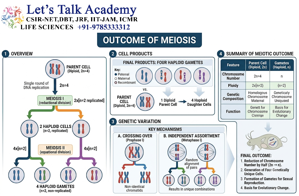
The general sequence is:
One diploid cell
↓
DNA replication
↓
Meiosis I
↓
Two haploid cells with replicated chromosomes
↓
Meiosis II
↓
Four haploid cells
These products are genetically different because of crossing over and independent assortment.
40. Mitosis and Meiosis: Major Comparison
| Feature | Mitosis | Meiosis |
|---|---|---|
| Number of divisions | One | Two |
| DNA replication | One round before division | One round before meiosis I |
| Daughter cells | Usually 2 | Usually 4 |
| Chromosome number | Generally maintained | Reduced by half |
| Homolog pairing | Absent | Present |
| Crossing over | Not a normal feature | Characteristic of prophase I |
| Genetic similarity | Generally similar | Genetically diverse |
| Homolog separation | Not the defining event | Occurs in meiosis I |
| Sister chromatid separation | Anaphase | Meiosis II |
| Major function | Growth and maintenance | Sexual reproduction and variation |
41. Regulation of Cell Division
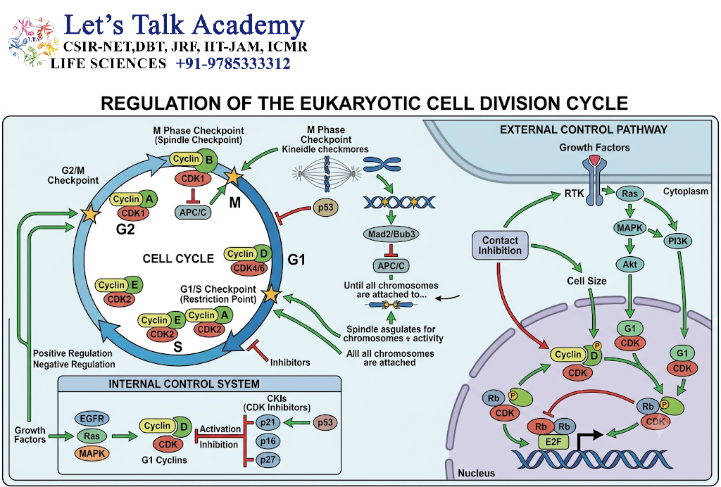
Cell division must be tightly regulated to prevent:
- DNA damage transmission.
- Incorrect chromosome segregation.
- Excessive cell proliferation.
- Chromosome-number abnormalities.
Major regulators include:
- Cyclins
- Cyclin-dependent kinases
- APC/C
- Spindle assembly checkpoint
- Cohesin
- Separase
- Condensin
- DNA-damage checkpoints
42. Cyclins and Cyclin-Dependent Kinases

Cyclin-dependent kinases (CDKs) are protein kinases that regulate progression through the cell cycle.
CDKs become active when associated with specific cyclins.
Major examples include:
- Cyclin D–CDK4/6
- Cyclin E–CDK2
- Cyclin A–CDK2
- Cyclin A/B–CDK1
The precise combinations and timing differ among organisms and cell types.
43. CDK1 and Entry into Mitosis
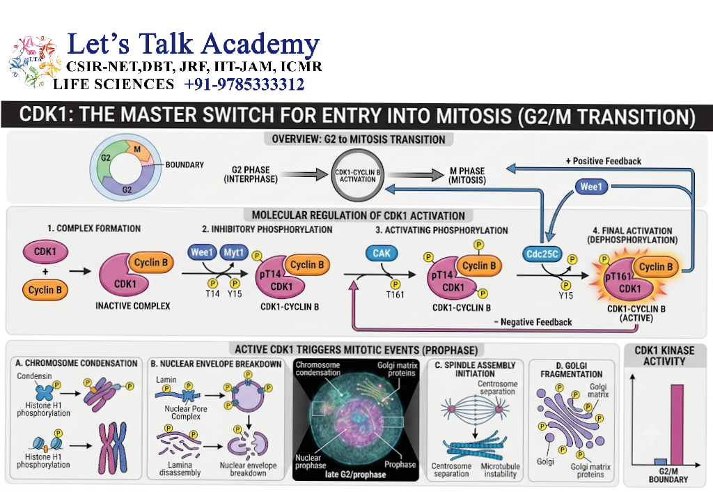
CDK1 is a central regulator of entry into mitosis.
Its association with mitotic cyclins promotes processes such as:
- Chromosome condensation.
- Nuclear-envelope breakdown.
- Spindle formation.
- Reorganization of cellular structures.
The activity of CDK1 is controlled through phosphorylation and dephosphorylation.
44. Cyclin B–CDK1 Complex

The Cyclin B–CDK1 complex is a major driver of mitotic entry.
Its activation triggers the transition from G2 phase to M phase.
When Cyclin B–CDK1 activity rises:
Chromosome condensation + spindle assembly + nuclear-envelope breakdown
are promoted.
45. Regulation of CDK Activity
CDKs are regulated by:
- Cyclin binding
- Activating phosphorylation
- Inhibitory phosphorylation
- CDK inhibitors
- Protein degradation
This provides multiple levels of control over cell-cycle progression.
46. CDK Inhibitors
CDK inhibitors prevent inappropriate CDK activity.
Important families include:
- INK4 proteins
- CIP/KIP proteins
These proteins can slow or stop cell-cycle progression under conditions such as:
- DNA damage
- Differentiation
- Growth-factor withdrawal
- Cellular stress
47. DNA Damage Checkpoint
Before a cell enters or progresses through division, it must ensure that its DNA is sufficiently intact.
DNA damage activates signaling pathways involving proteins such as:
- ATM
- ATR
- CHK1
- CHK2
- p53
These pathways can delay cell-cycle progression and promote DNA repair.
48. p53 and Cell-Cycle Control
p53 is an important tumor-suppressor protein involved in cellular responses to DNA damage.
When DNA damage is detected, p53 can promote expression of cell-cycle inhibitory proteins such as p21.
The resulting CDK inhibition can temporarily stop cell-cycle progression.
If damage is severe, p53-dependent pathways may also contribute to cellular senescence or apoptosis.
49. Spindle Assembly Checkpoint Regulation
The spindle assembly checkpoint prevents the cell from entering anaphase until chromosomes are correctly attached to the spindle.
Important checkpoint components include:
- MAD proteins
- BUB proteins
- Kinetochore-associated factors
The checkpoint inhibits premature activation of the machinery that triggers sister chromatid separation.
50. APC/C
The anaphase-promoting complex/cyclosome (APC/C) is a large ubiquitin ligase complex that promotes the destruction of specific cell-cycle proteins.
APC/C activation contributes to:
- Anaphase initiation
- Cyclin degradation
- Mitotic exit
It is therefore a major regulator of the transition from metaphase to anaphase and subsequent exit from mitosis.
51. Securin and Separase
Before anaphase, the enzyme separase is kept inactive by a protein called securin.
When the APC/C becomes appropriately activated:
Securin degradation → Separase activation → Cohesin cleavage → Sister chromatid separation
This is one of the central molecular switches controlling anaphase.
52. Cohesin
Cohesin is a protein complex that holds sister chromatids together after DNA replication.
It is essential for:
- Sister chromatid cohesion.
- Chromosome organization.
- Accurate segregation.
In mitosis, cohesin is removed from chromosome arms earlier, while centromeric cohesion is protected until anaphase.
53. Separase
Separase is a protease that cleaves a key cohesin component.
Its activation results in loss of sister chromatid cohesion.
Thus:
Separase activation → Cohesin cleavage → Chromatid separation
This mechanism ensures that sister chromatids remain together until the correct stage of division.
54. Shugoshin and Centromeric Protection
During meiosis I, sister chromatids must remain together while homologous chromosomes separate.
A protein system involving shugoshin protects centromeric cohesin from premature removal.
This is a crucial difference between meiosis I and mitosis.
55. Regulation of Meiosis I
Meiosis I requires mechanisms that are different from ordinary mitosis.
During meiosis I:
- Homologous chromosomes pair.
- Recombination occurs.
- Cohesin is protected at centromeres.
- Homologues separate.
- Sister chromatids remain together.
Thus, meiosis I requires specialized regulation of chromosome cohesion and spindle attachment.
56. Meiotic Recombination Regulation
Meiotic recombination begins with programmed DNA double-strand breaks.
These breaks are generated by specialized meiotic machinery, including the protein Spo11.
The breaks are repaired through homologous chromosome interactions, producing either:
- Crossovers
- Non-crossovers
Crossovers create chiasmata that contribute to proper homolog segregation.
57. Meiotic Double-Strand Breaks
Programmed DNA double-strand breaks may initially appear harmful, but they are essential for normal meiotic recombination.
The general sequence is:
Spo11-mediated break
↓
DNA-end processing
↓
Homology search
↓
Strand invasion
↓
DNA synthesis and repair
↓
Crossover or non-crossover product
58. Crossover Formation
Crossover formation is tightly regulated because too few or improperly positioned crossovers can interfere with chromosome segregation.
Crossovers are generally distributed along chromosome arms rather than occurring randomly without constraints.
Their number and distribution are influenced by:
- Chromosome structure
- Recombination machinery
- Cellular regulation
- Chromatin environment
59. Regulation of Meiosis II
Meiosis II is more similar to mitosis than meiosis I.
During meiosis II:
- Chromosomes align individually.
- Sister kinetochores become attached to opposite spindle poles.
- Centromeric cohesion is removed.
- Sister chromatids separate.
Thus, meiosis II completes the reduction process initiated in meiosis I.
60. Differences in Chromosome Behavior
A central distinction between mitosis and meiosis is the behavior of homologous chromosomes.
Mitosis
Sister chromatids separate
Meiosis I
Homologous chromosomes separate
Meiosis II
Sister chromatids separate
This can be summarized as:
Mitosis → sister separation
Meiosis I → homolog separation
Meiosis II → sister separation
61. Regulation of Mitosis vs Meiosis
| Regulatory feature | Mitosis | Meiosis |
|---|---|---|
| DNA replication | Once | Once before meiosis I |
| Number of divisions | One | Two |
| Homolog pairing | No | Yes |
| Recombination | Not characteristic | Essential feature |
| Centromeric cohesion | Removed at anaphase | Protected during meiosis I |
| APC/C regulation | Controls mitotic transitions | Controls meiotic transitions |
| Separase | Separates sister chromatids | Functions at different stages to separate homolog-associated and sister cohesion |
| Spindle attachment | Sister kinetochores orient oppositely | Meiosis I uses homolog-oriented attachment |
62. Checkpoints in Cell Division
Cell-cycle checkpoints help prevent errors.
Major checkpoints include:
- G1/S checkpoint
- G2/M checkpoint
- Spindle assembly checkpoint
These checkpoints assess:
- DNA integrity
- DNA replication
- Chromosome attachment
- Cellular conditions
63. G1/S Checkpoint
The G1/S checkpoint determines whether the cell is ready to enter DNA replication.
It responds to:
- Growth signals
- Nutrient availability
- DNA damage
- Cellular stress
If conditions are unfavorable, cell-cycle progression can be delayed.
64. G2/M Checkpoint
The G2/M checkpoint ensures that DNA replication has been completed properly before the cell enters mitosis.
If DNA remains damaged or incompletely replicated, entry into mitosis can be delayed.
65. Spindle Assembly Checkpoint
The spindle assembly checkpoint operates during mitosis and meiosis.
It prevents chromosome separation until appropriate kinetochore-spindle attachments are established.
This checkpoint is essential for preventing chromosome missegregation.
66. Chromosome Missegregation
Failure of accurate chromosome segregation can produce cells with abnormal chromosome numbers.
This condition is known as aneuploidy.
Aneuploidy can arise through:
- Nondisjunction
- Improper kinetochore attachment
- Checkpoint failure
- Cohesion defects
Such abnormalities can have major biological consequences.
67. Nondisjunction
Nondisjunction occurs when chromosomes fail to separate properly during cell division.
It can involve:
- Homologous chromosomes during meiosis I.
- Sister chromatids during meiosis II.
- Sister chromatids during mitosis.
Nondisjunction can produce daughter cells with abnormal chromosome numbers.
68. Mitosis and Genetic Stability
Accurate mitosis ensures that daughter cells receive appropriate chromosome complements.
This depends on:
- Accurate DNA replication.
- Correct spindle formation.
- Proper kinetochore attachment.
- Functional checkpoints.
- Controlled cohesin removal.
Defects in these mechanisms can lead to genomic instability.
69. Meiosis and Genetic Variation
Meiosis generates genetic diversity through at least two major mechanisms:
69.1 Crossing Over
DNA exchange between homologous chromosomes creates recombinant chromosomes.
69.2 Independent Assortment
Homologous chromosomes segregate into gametes in different combinations.
These processes create genetically diverse reproductive cells.
70. Meiosis in Spermatogenesis
In males of many animal species, meiosis occurs during spermatogenesis.
A simplified sequence is:
Spermatogonium → Primary spermatocyte → Meiosis I → Secondary spermatocytes → Meiosis II → Spermatids
The spermatids subsequently differentiate into sperm cells.
71. Meiosis in Oogenesis
In females of many animal species, meiosis occurs during oogenesis.
A simplified sequence is:
Oogonium → Primary oocyte → Meiosis I → Secondary oocyte + polar body → Meiosis II
The details of meiotic timing and completion differ substantially between species.
In humans, for example, oocytes begin meiosis during fetal development and progress through specific arrest points.
72. Regulation of Meiotic Arrest
Oocytes can remain arrested at particular stages of meiosis for extended periods.
The maintenance and release of these arrests involve complex signaling networks involving:
- Cyclins
- CDKs
- Protein kinases
- Phosphatases
- Calcium signaling
This specialized regulation distinguishes oocyte meiosis from the more continuously proliferative divisions of many somatic cells.
73. Mitotic Exit
Mitotic exit requires coordinated reduction of mitotic CDK activity.
A major mechanism is degradation of mitotic cyclins by APC/C-dependent ubiquitination.
This allows:
- Chromosome decondensation.
- Nuclear-envelope reformation.
- Spindle disassembly.
- Cytokinesis.
- Return to interphase organization.
74. Meiotic Exit
Meiotic progression also depends on regulated changes in CDK activity and protein degradation.
The two meiotic divisions must be coordinated so that:
One DNA replication event → Two chromosome segregation events
This unusual cell-cycle architecture is a defining characteristic of meiosis.
75. Mitosis and Meiosis in the Life Cycle
Mitosis primarily maintains chromosome number during organismal growth and tissue development.
Meiosis reduces chromosome number before sexual reproduction.
Fertilization subsequently restores the diploid chromosome complement.
The cycle can be represented as:
Diploid organism
↓
Meiosis
↓
Haploid gametes
↓
Fertilization
↓
Diploid zygote
↓
Mitosis
↓
Multicellular organism
76. Evolutionary Significance of Meiosis
Meiosis provides an important mechanism for generating genetic variation.
Through:
- Recombination
- Independent assortment
- Random combination of gametes during fertilization
meiosis contributes to genetic diversity within populations.
This variation provides raw material on which evolutionary processes can act.
77. Functional Significance of Mitosis
Mitosis provides genetic continuity within multicellular organisms.
It allows:
- Embryonic development.
- Tissue growth.
- Tissue repair.
- Cell replacement.
- Maintenance of cell populations.
Its high accuracy is essential because errors can be propagated to many descendant cells.
78. Mitosis, Meiosis and Chromatin
Chromatin must undergo dramatic structural changes during cell division.
During mitosis and meiosis:
- Chromosomes condense.
- Histone modifications change.
- Chromatin-associated proteins reorganize.
- Spindle attachment sites become accessible.
- Chromosome architecture becomes specialized for segregation.
Thus, chromosome structure and cell-cycle regulation are tightly interconnected.
79. Conceptual Flowchart of Mitosis
G1
↓
DNA replication
↓
G2
↓
Prophase
↓
Prometaphase
↓
Metaphase
↓
Anaphase
↓
Telophase
↓
Cytokinesis
↓
Two daughter cells
80. Conceptual Flowchart of Meiosis
DNA replication
↓
Prophase I
↓
Synapsis + Recombination
↓
Metaphase I
↓
Homolog separation
↓
Telophase I
↓
Meiosis II
↓
Sister chromatid separation
↓
Four haploid products


