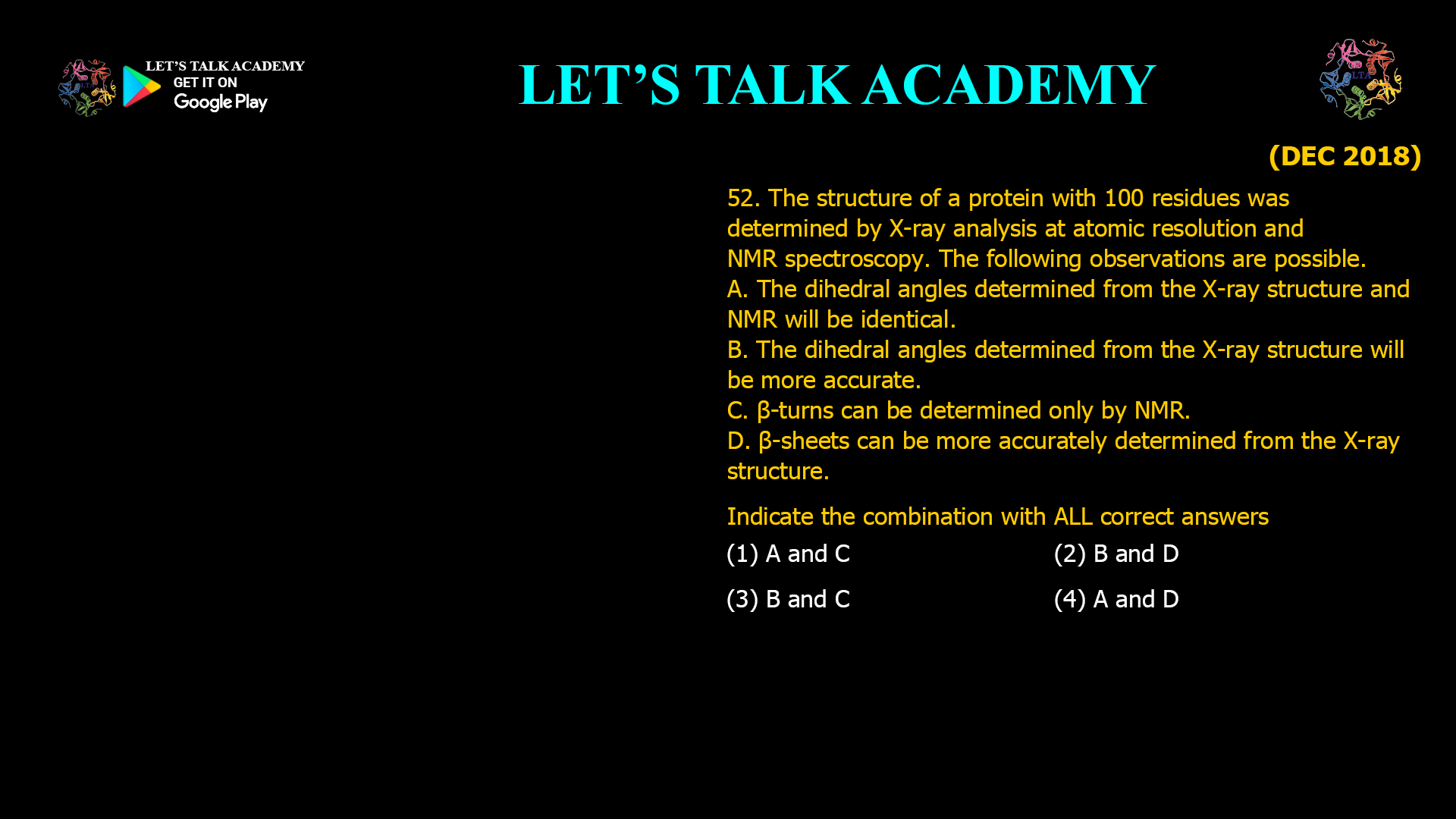
52. The structure of a protein with 100 residues was determined by X-ray analysis at atomic resolution and NMR spectroscopy. The following observations are possible.
A. The dihedral angles determined from the X-ray structure and NMR will be identical.
B. The dihedral angles determined from the X-ray structure will be more accurate.
C. β-turns can be determined only by NMR.
D. β-sheets can be more accurately determined from the X-ray structure.
Indicate the combination with ALL correct answers.
(A) A and C
(B) B and D
(C) B and C
(D) A and D
Comparison of X-ray Crystallography and NMR Spectroscopy for Protein Structure Determination
Correct Answer
Option (2): B and D
Explanation
X-ray crystallography and Nuclear Magnetic Resonance (NMR) spectroscopy are two of the most powerful techniques used to determine the three-dimensional structures of proteins. Although both techniques ultimately describe the arrangement of atoms within a protein, they are based on fundamentally different physical principles. X-ray crystallography determines electron density from protein crystals, whereas NMR spectroscopy derives structural information from magnetic interactions between atomic nuclei in proteins dissolved in solution. Consequently, the structural models obtained from the two techniques are usually very similar but are rarely identical.
When X-ray diffraction data are collected at atomic resolution, individual atoms can be localized with extremely high precision. The electron density maps allow accurate positioning of backbone and side-chain atoms, resulting in highly reliable measurements of bond lengths, bond angles, and backbone dihedral angles. In contrast, NMR structures are calculated from experimentally derived distance restraints, scalar coupling constants, residual dipolar couplings, and other spectroscopic parameters. Because NMR structures are represented as an ensemble of conformations that satisfy these restraints, individual dihedral angles generally exhibit greater variability than those obtained from atomic-resolution crystal structures.
β-sheets are stabilized by regular backbone hydrogen-bonding patterns and produce well-defined electron density in high-quality protein crystals. At atomic resolution, X-ray crystallography can define the positions of individual strands, hydrogen-bonding geometry, strand registry, and sheet orientation with exceptional accuracy. Therefore, β-sheet architecture is generally determined more precisely by X-ray crystallography than by solution NMR.
Analysis of Statement A
Statement A is incorrect.
Although the overall fold obtained by X-ray crystallography and NMR spectroscopy is usually highly similar, the backbone dihedral angles are not expected to be exactly identical. X-ray structures represent a single crystallographic model derived from electron density, whereas NMR structures consist of an ensemble of conformations representing the range of structures compatible with experimental restraints in solution. Small differences in molecular flexibility, crystal packing effects, and structural averaging make identical dihedral angles unlikely.
Analysis of Statement B
Statement B is correct.
Atomic-resolution X-ray crystallography provides highly accurate atomic coordinates from which backbone dihedral angles can be calculated with excellent precision. Because the coordinates are directly supported by well-resolved electron density, the resulting dihedral angles are generally more accurate than those derived from NMR restraint calculations.
Analysis of Statement C
Statement C is incorrect.
β-turns are not exclusive to NMR spectroscopy. Both X-ray crystallography and NMR can identify β-turns by examining the geometry of the peptide backbone and the arrangement of successive amino acid residues. High-resolution crystal structures clearly reveal β-turn conformations, while NMR identifies them through characteristic NOE patterns, scalar coupling constants, and backbone torsion angles. Therefore, β-turns can be determined using either technique.
Analysis of Statement D
Statement D is correct.
β-sheets contain highly ordered arrays of hydrogen-bonded β-strands that generate well-defined electron density in crystal structures. Atomic-resolution X-ray diffraction accurately establishes strand orientation, hydrogen-bonding patterns, and the detailed geometry of the β-sheet. Although NMR can also identify β-sheet structures, the greater positional precision of atomic-resolution crystallography generally provides a more accurate structural description.
Why Option (1) is Incorrect
Statement A is incorrect because X-ray and NMR structures do not produce identical dihedral angles, and Statement C is incorrect because β-turns can also be identified by X-ray crystallography. Therefore, this option contains two incorrect statements.
Why Option (2) is Correct
Statement B correctly states that atomic-resolution X-ray crystallography determines backbone dihedral angles with greater accuracy than NMR, and Statement D correctly states that β-sheet structures are more accurately defined in high-resolution crystal structures. Since both statements are scientifically correct, this is the correct option.
Why Option (3) is Incorrect
Although Statement B is correct, Statement C is incorrect because β-turns are readily identified using both X-ray crystallography and NMR spectroscopy. Consequently, this combination cannot be accepted.
Why Option (4) is Incorrect
Statement D is correct, but Statement A is incorrect because structural differences between crystal and solution environments prevent the two methods from producing identical dihedral angles. Therefore, this option contains one incorrect statement.
Comparison of X-ray Crystallography and NMR Spectroscopy
X-ray crystallography provides a high-resolution static view of proteins within a crystal lattice and is particularly powerful for determining atomic coordinates, side-chain orientations, hydrogen-bonding networks, and secondary structural elements. NMR spectroscopy, on the other hand, determines protein structures in solution and additionally provides valuable information about molecular flexibility, conformational exchange, and dynamic structural fluctuations that cannot be observed directly in crystal structures.
Determination of Secondary Structure by Structural Techniques
Both X-ray crystallography and NMR spectroscopy are capable of identifying α-helices, β-sheets, β-turns, and loop regions. X-ray crystallography determines these structures directly from atomic coordinates obtained from electron density maps, whereas NMR derives them from characteristic NOE patterns, scalar coupling constants, chemical shifts, hydrogen-deuterium exchange experiments, and other spectroscopic restraints. The combination of these complementary methods provides a comprehensive understanding of protein structure and dynamics.
Conclusion
Atomic-resolution X-ray crystallography generally provides more precise backbone dihedral angles and more accurate β-sheet geometry than solution NMR spectroscopy. β-turns can be identified by both techniques, and the structures obtained by X-ray crystallography and NMR are similar but not identical. Therefore, the correct answer is Option (2): B and D.


