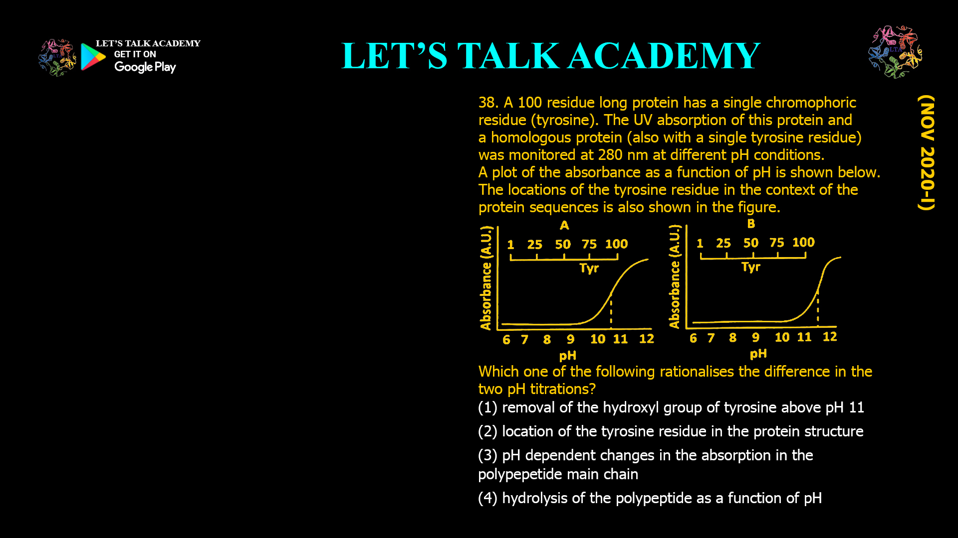
38. A 100 residue long protein has a single chromophoric residue (tyrosine). The UV absorption of this protein and a homologous protein (also with a single tyrosine residue) was monitored at 280 nm at different pH conditions. A plot of the absorbance as a function of pH is shown below. The locations of the tyrosine residue in the context of the protein sequence is also shown in the figure.
Which one of the following rationalises the difference in the two pH titrations?
(A) Removal of the hydroxyl group of tyrosine above pH 11.
(B) Location of the tyrosine residue in the protein structure.
(C) pH dependent changes in the absorption in the polypeptide main chain.
(D) Hydrolysis of the polypeptide as a function of pH.
Effect of Tyrosine Environment on UV Absorption and pH Titration Curves in Proteins
Correct Answer
Option (2): Location of the tyrosine residue in the protein structure.
Explanation
Tyrosine is an aromatic amino acid that exhibits a characteristic ultraviolet absorption maximum near 280 nm because of its phenolic aromatic ring. The absorbance of tyrosine changes with pH because its phenolic hydroxyl group can lose a proton at alkaline pH to form a negatively charged phenolate ion. The phenolate ion possesses a different electronic distribution from the neutral phenol group, resulting in an increase in absorbance at 280 nm. Consequently, monitoring absorbance as a function of pH provides information about the ionization behaviour of tyrosine.
The two proteins shown in the figure contain the same number of amino acid residues and each possesses only a single tyrosine residue. Since the chromophore is identical in both proteins, any difference in the pH titration curves cannot arise from differences in the chemical properties of tyrosine itself. Instead, the difference must originate from the environment surrounding the tyrosine residue within each protein.
In Protein A, the tyrosine residue is positioned close to the C-terminal region, whereas in Protein B it is located near the middle of the polypeptide chain. The local three-dimensional environment surrounding these residues is therefore different. One tyrosine may be exposed to the solvent, while the other may be partially buried within the protein interior or involved in hydrogen bonding and electrostatic interactions. These differences alter the apparent pKa of the phenolic hydroxyl group and shift the pH at which ionization occurs. As a result, the absorbance-versus-pH curves are displaced relative to one another.
This phenomenon is commonly observed in proteins because the microenvironment surrounding an ionizable amino acid strongly influences its acid-base behaviour. Hydrogen bonds, nearby charged residues, solvent accessibility, and local polarity can either stabilize or destabilize the ionized form, thereby shifting the titration curve without altering the chemical identity of the amino acid.
Why Option (1) is Incorrect
This option incorrectly suggests that the hydroxyl group of tyrosine is removed above pH 11. Under alkaline conditions, the hydroxyl group is not removed. Instead, it undergoes deprotonation, losing only its hydrogen ion to form a phenolate ion. The oxygen atom remains part of the tyrosine side chain throughout the process. Therefore, this statement is chemically incorrect.
Why Option (2) is Correct
The only meaningful difference between the two homologous proteins is the structural environment surrounding the tyrosine residue. Differences in solvent exposure, hydrogen bonding, electrostatic interactions, and local folding alter the apparent pKa of the phenolic hydroxyl group. Consequently, tyrosine ionizes at different pH values in the two proteins, producing different absorbance titration curves. Therefore, the location of the tyrosine residue within the protein structure correctly explains the observed behaviour.
Why Option (3) is Incorrect
The peptide backbone absorbs strongly in the far-ultraviolet region, generally below 230 nm. At 280 nm, the observed absorption arises almost entirely from aromatic amino acid residues, particularly tyrosine and tryptophan. The polypeptide main chain contributes negligibly at this wavelength and therefore cannot explain the difference between the two titration curves.
Why Option (4) is Incorrect
Hydrolysis of peptide bonds requires harsh chemical conditions and is not responsible for the gradual changes in absorbance observed during a pH titration experiment. The smooth sigmoidal curves shown in the figure represent reversible ionization of the tyrosine side chain rather than irreversible degradation of the protein.
Tyrosine Ionization and UV Absorption
The phenolic hydroxyl group of tyrosine behaves as a weak acid. At lower pH values it remains protonated, whereas at alkaline pH it loses a proton to form the phenolate ion. This ionization changes the electronic structure of the aromatic ring, producing an increase in absorbance at 280 nm. The pH at which this transition occurs depends not only on the intrinsic properties of tyrosine but also on the surrounding protein environment.
Influence of the Protein Microenvironment
The apparent pKa of an ionizable amino acid residue is rarely identical to its value in free solution because neighbouring amino acids influence its chemical behaviour. Hydrogen bonds, nearby acidic or basic residues, solvent accessibility, hydrophobic packing, and electrostatic interactions all affect the stability of the protonated and deprotonated forms. As a result, identical amino acid residues located at different positions within homologous proteins may exhibit different titration curves despite having the same chemical composition.
Conclusion
The difference between the two absorbance-versus-pH titration curves is caused by the different structural environments surrounding the tyrosine residue in the two proteins. Variations in solvent accessibility and local molecular interactions alter the apparent pKa of the phenolic hydroxyl group, shifting the ionization profile observed at 280 nm. Therefore, the correct answer is Option (2): Location of the tyrosine residue in the protein structure.


