
1. Introduction to Haematopoiesis
1.1 Definition of Haematopoiesis
Haematopoiesis, also spelled hematopoiesis, is the highly regulated biological process through which all mature blood cells are produced from a small population of undifferentiated cells called haematopoietic stem cells (HSCs). These stem cells possess two fundamental properties: self-renewal and multilineage differentiation.
Self-renewal enables haematopoietic stem cells to produce daughter cells that retain stem cell characteristics, thereby maintaining the stem cell pool throughout an individual’s lifetime. Differentiation allows these cells to generate specialized blood cell lineages, including red blood cells, white blood cells, and platelets.
Blood cell production is essential for maintaining oxygen transport, immune defense, haemostasis, tissue repair, and the overall internal balance of the body. Since mature blood cells have limited life spans, the body must continuously replace them through haematopoiesis.
The production of blood cells is not a random process. It is controlled by a complex interaction of intrinsic cellular programs, transcription factors, epigenetic mechanisms, extracellular signaling molecules, bone marrow stromal cells, metabolic pathways, and systemic physiological signals.
Central concept: Haematopoiesis maintains a balance between the preservation of stem cells and the generation of sufficient mature blood cells to meet the body’s changing needs.
1.2 Importance of Haematopoiesis
Haematopoiesis is essential for survival because mature blood cells perform specialized functions that cannot be maintained without continuous production.
1.2.1 Oxygen transport
Red blood cells contain haemoglobin, which transports oxygen from the lungs to tissues and assists in the transport of carbon dioxide back to the lungs. The production of red blood cells, known as erythropoiesis, must be adjusted according to the oxygen requirements of the body.
1.2.2 Immune defense
White blood cells, including neutrophils, lymphocytes, monocytes, eosinophils, and basophils, protect the body against pathogens and participate in immune surveillance. Haematopoiesis regulates the production of these cells according to developmental requirements and inflammatory conditions.
1.2.3 Blood clotting
Platelets are produced from large bone marrow cells called megakaryocytes. They are essential for the formation of blood clots and the prevention of excessive blood loss.
1.2.4 Maintenance of tissue homeostasis
Blood cells participate in inflammation, tissue repair, removal of damaged cells, and communication between different organs. Continuous blood cell production ensures that these functions remain operational.
1.2.5 Adaptation to physiological stress
During infection, bleeding, hypoxia, or inflammation, the body can modify blood cell production. This process is known as stress haematopoiesis and helps meet increased physiological demands.
1.3 General Characteristics of Haematopoiesis
Haematopoiesis is characterized by several important features:
-
Continuous activity: Blood cell production occurs throughout life.
-
Hierarchical organization: Stem cells give rise to progenitor cells, which subsequently generate more specialized cell populations.
-
Self-renewal: A portion of the stem cell population is preserved to maintain long-term blood production.
-
Multilineage differentiation: A single HSC can generate multiple blood cell lineages.
-
Regulated proliferation: Cell division is controlled to prevent excessive or insufficient blood cell production.
-
Microenvironmental dependence: Stem cells and progenitor cells interact with supporting cells and extracellular matrix components.
-
Feedback regulation: The production of certain blood cells is adjusted in response to physiological signals and the demand for mature cells.
-
Dynamic adaptability: Haematopoiesis changes in response to age, infection, inflammation, and tissue injury.
2. Developmental Sites of Haematopoiesis
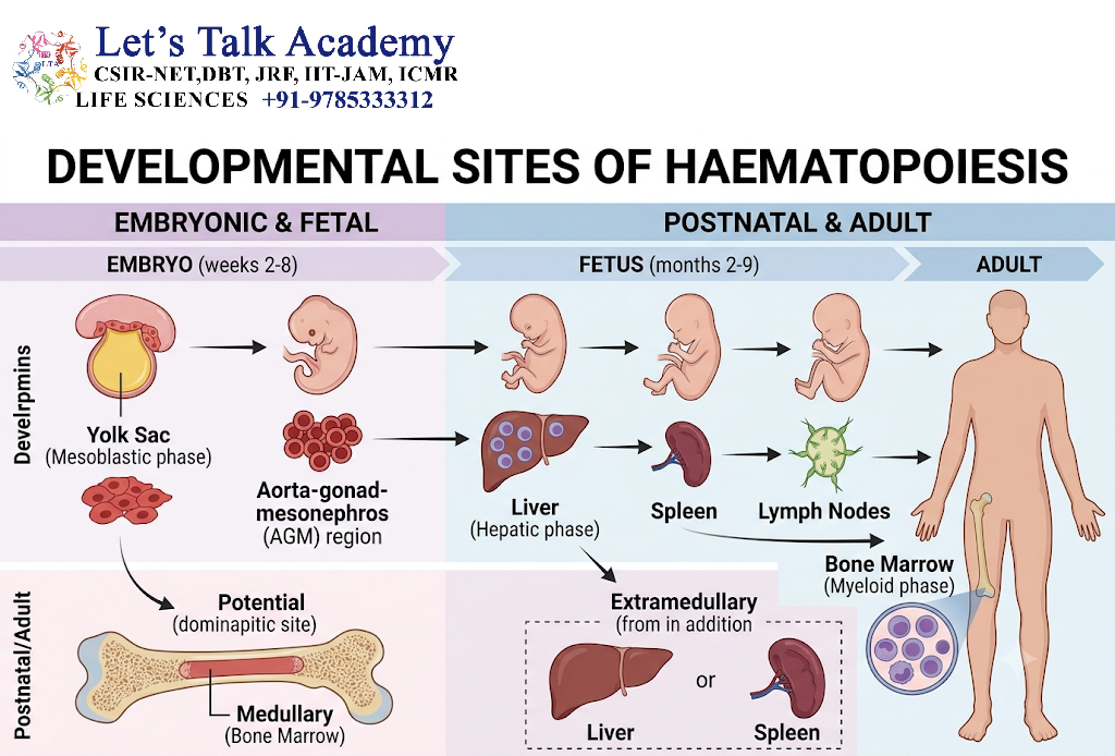
2.1 Embryonic Haematopoiesis
During embryonic development, haematopoiesis takes place at different anatomical sites. These changes reflect the increasing requirements of the developing organism.
The earliest blood-forming activity in mammals begins in the yolk sac, where primitive blood cells and early erythroid cells are generated. This initial phase provides blood cells needed during early development.
Subsequently, definitive haematopoietic stem cells emerge through a process associated with specialized endothelial cells in the embryonic aorta, particularly within the aorta–gonad–mesonephros region. This process is commonly described as endothelial-to-haematopoietic transition.
Definitive HSCs then migrate to other sites, where they expand and establish the capacity for lifelong blood production.
2.2 Foetal Liver Haematopoiesis
The foetal liver becomes the principal site of blood cell formation during much of embryonic and foetal development.
The foetal liver provides a supportive environment for the expansion and differentiation of haematopoietic stem and progenitor cells. Stromal cells, extracellular matrix components, and soluble growth factors help regulate this activity.
During this stage, erythropoiesis is particularly important because the developing foetus requires large numbers of red blood cells.
The foetal liver also contributes to the production of other blood cell lineages, although the relative contribution of different tissues changes during development.
2.3 Bone Marrow Haematopoiesis
Near birth and throughout adult life, the bone marrow becomes the primary site of haematopoiesis.
In adults, active haematopoiesis is concentrated in the marrow of bones such as the pelvis, vertebrae, sternum, ribs, and parts of the skull and proximal long bones.
The bone marrow contains a complex cellular environment consisting of:
-
Haematopoietic stem and progenitor cells.
-
Mesenchymal stromal cells.
-
Endothelial cells.
-
Macrophages.
-
Osteolineage cells.
-
Adipocytes.
-
Nerve-associated structures.
-
Extracellular matrix components.
-
Blood vessels and specialized vascular regions.
These components collectively form the haematopoietic microenvironment, which provides signals that regulate stem cell maintenance, proliferation, differentiation, migration, and survival.
2.4 Extramedullary Haematopoiesis
Extramedullary haematopoiesis refers to blood cell production outside the bone marrow.
It may occur during foetal development or in certain pathological conditions in adults, particularly when the bone marrow cannot meet the body’s demand for blood cell production.
Common sites of pathological extramedullary haematopoiesis include the spleen and liver.
It may be associated with conditions such as chronic haemolytic anaemias, marrow fibrosis, or disorders that impair normal marrow function.
3. Haematopoietic Stem Cells: The Foundation of Blood Cell Formation
3.1 Definition of Haematopoietic Stem Cells
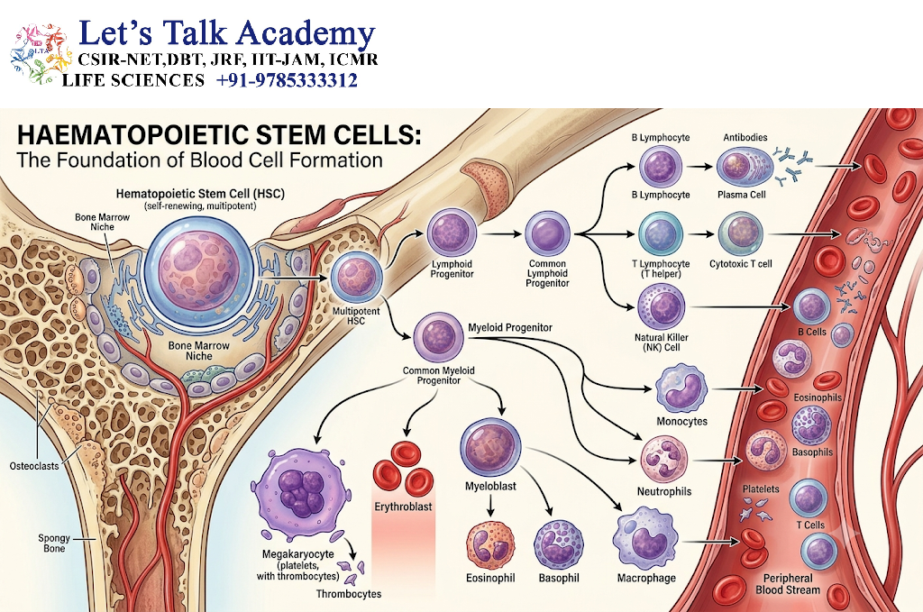
Haematopoietic stem cells are rare, self-renewing cells that can generate all major blood cell lineages.
They are responsible for sustaining blood production throughout life. Unlike mature blood cells, which have limited life spans, HSCs can remain in the body for decades under appropriate physiological conditions.
The ability of HSCs to produce both identical stem cells and differentiated progeny makes them the foundation of the haematopoietic system.
3.2 Fundamental Properties of HSCs
3.2.1 Self-renewal
Self-renewal is the process by which a stem cell produces daughter cells that retain stem cell properties.
Self-renewal can occur through different division patterns:
-
Symmetric self-renewal: One HSC produces two daughter cells that retain stem cell characteristics.
-
Asymmetric division: One daughter cell retains stem cell characteristics, while the other becomes more committed to differentiation.
-
Symmetric differentiation: Both daughter cells become more differentiated.
The balance between these division patterns is essential. Excessive self-renewal may contribute to abnormal stem cell accumulation, whereas excessive differentiation may deplete the stem cell pool.
3.2.2 Multipotency
Multipotency is the ability of an HSC to produce different types of mature blood cells.
These include:
-
Erythrocytes.
-
Megakaryocytes and platelets.
-
Neutrophils.
-
Monocytes and macrophages.
-
Dendritic cell populations.
-
B lymphocytes.
-
T lymphocytes.
-
Natural killer cells.
The precise developmental pathways and lineage relationships are more flexible than a strictly fixed tree in some models of adult haematopoiesis. Different HSC and progenitor populations may exhibit distinct lineage biases.
3.2.3 Quiescence
Many HSCs remain in a relatively inactive, non-dividing state known as quiescence.
Quiescence helps preserve the long-term regenerative capacity of HSCs and may reduce the accumulation of replication-associated damage.
However, quiescent cells are not completely inactive. They maintain essential metabolic processes, respond to environmental signals, and can enter the cell cycle when necessary.
3.2.4 Long-term and short-term repopulating activity
HSC populations can be described experimentally according to their ability to sustain blood production over different periods.
-
Long-term repopulating HSCs: Demonstrate durable, multilineage reconstitution in appropriate experimental systems.
-
Short-term repopulating cells: Support blood production for a limited period.
-
Multipotent progenitors: Retain broad differentiation potential but generally have less durable self-renewal capacity than long-term HSCs.
These functional categories are important for understanding the organization of the haematopoietic system.
3.3 Surface Markers of Haematopoietic Stem Cells
In experimental haematology, HSCs are identified using combinations of cell surface markers rather than a single universal marker.
In mice, a commonly studied enriched HSC population is characterized by a lineage-negative, Sca-1-positive, c-Kit-positive phenotype, often abbreviated as LSK, with additional markers used to identify more specific populations.
In humans, HSC-enriched populations commonly express CD34 and CD38-negative or low phenotypes, although these markers are not exclusive to HSCs.
Other markers used in research include CD90, CD45RA, CD49f, and additional phenotypic features depending on the experimental strategy.
Important distinction: A cell surface phenotype is an operational method for enriching or identifying a population. It does not automatically establish that every cell in that population has identical stem cell behavior.
3.4 Balance Between Stem Cell Maintenance and Differentiation
The maintenance of the HSC pool requires a carefully coordinated balance between:
-
Self-renewal.
-
Quiescence.
-
Differentiation.
-
Apoptosis.
-
Cellular metabolism.
-
DNA repair.
-
Interactions with the bone marrow microenvironment.
Intrinsic transcriptional programs determine how HSCs respond to signals. Extrinsic signals from the surrounding niche influence whether an HSC remains quiescent, divides, migrates, or differentiates.
The regulation of this balance is a central principle of haematopoietic homeostasis.
4. Organization of the Haematopoietic System
4.1 The Haematopoietic Hierarchy
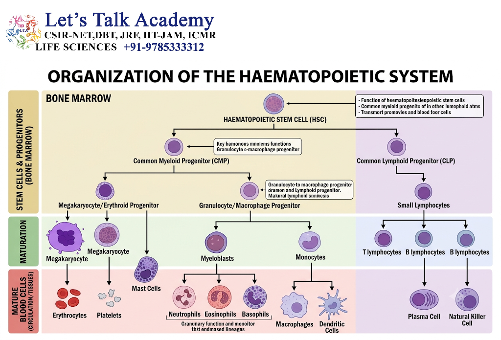
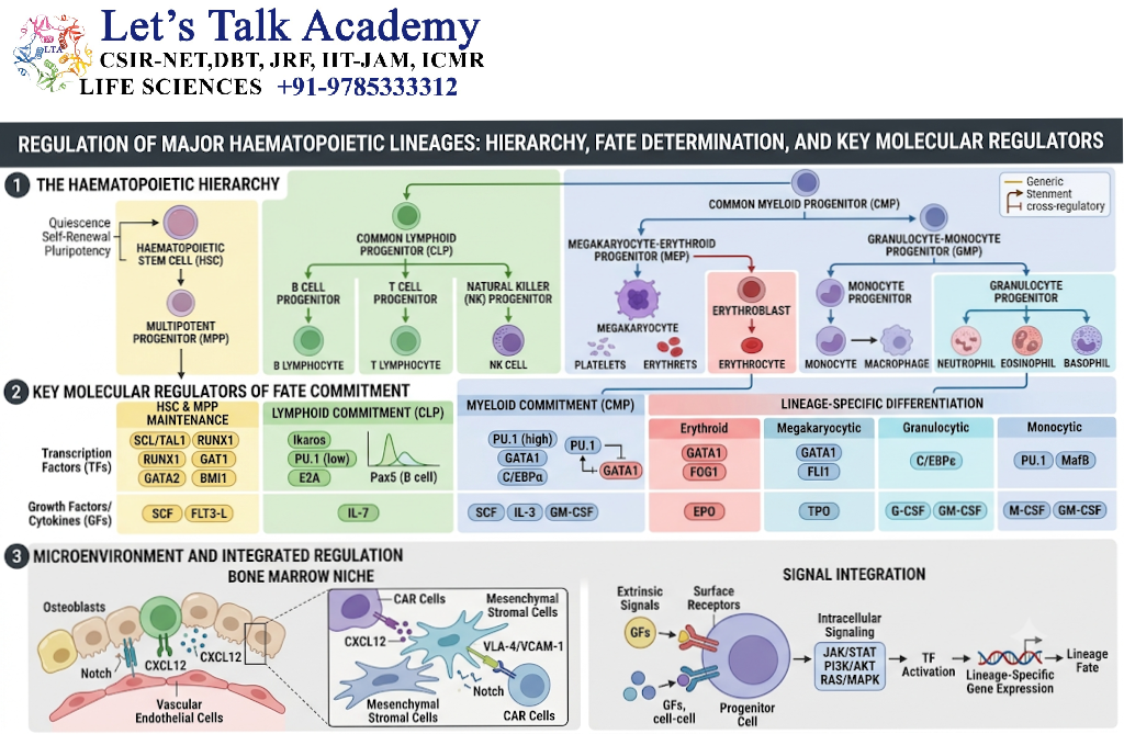
Haematopoiesis is traditionally represented as a hierarchical system in which haematopoietic stem cells give rise to progressively more specialized progenitor and precursor cells. This model is useful for understanding blood cell development, although modern research indicates that the system is more flexible and interconnected than a simple, rigid tree.
The classical developmental sequence is:
Haematopoietic stem cell (HSC) → Multipotent progenitor (MPP) → Lineage-biased progenitors → Committed progenitors → Precursor cells → Mature blood cells
The HSC maintains the ability to self-renew and generate multiple blood cell lineages. As cells progress through differentiation, their developmental potential generally becomes more restricted.
4.1.1 Multipotent progenitors
Multipotent progenitors arise from HSCs and retain the ability to generate multiple blood cell types. However, compared with long-term HSCs, they possess limited or reduced long-term self-renewal capacity.
These cells serve as an important transitional population between stem cell maintenance and the production of specialized blood cells.
4.1.2 Lineage-biased progenitors
Some progenitor populations display a preference for particular developmental pathways. For example, certain progenitors may produce predominantly myeloid cells, while others show a greater tendency toward lymphoid differentiation.
Lineage bias does not necessarily represent an irreversible commitment. The degree of developmental flexibility depends on the progenitor population and the physiological context.
4.1.3 Committed progenitors
Committed progenitors have a more restricted developmental potential. They are primarily involved in the generation of specific blood cell lineages.
Examples include progenitors associated with erythroid–megakaryocytic development and progenitors that contribute to granulocyte and monocyte formation.
4.1.4 Precursor cells
Precursor cells are more differentiated cells that undergo several rounds of proliferation and maturation before becoming fully functional blood cells.
For example, erythroblasts develop through recognizable stages before producing mature erythrocytes, while granulocyte precursors undergo sequential morphological and functional changes during granulopoiesis.
4.2 Major Blood Cell Lineages
Blood cell formation is broadly divided into two major developmental branches: the myeloid and lymphoid lineages.
4.2.1 Myeloid lineage
The myeloid lineage produces several types of blood cells, including:
-
Erythrocytes.
-
Megakaryocytes and platelets.
-
Neutrophils.
-
Eosinophils.
-
Basophils.
-
Monocytes.
-
Many conventional dendritic cell populations.
The regulation of myeloid differentiation involves transcription factors such as PU.1, C/EBP family proteins, GATA factors, and other lineage-associated regulators.
4.2.2 Lymphoid lineage
The lymphoid lineage gives rise to:
-
B lymphocytes.
-
T lymphocytes.
-
Natural killer cells.
-
Certain innate lymphoid cell populations.
Lymphoid development is influenced by transcription factors and signaling pathways involving Ikaros family proteins, E-proteins, Notch signaling, and cytokines such as interleukin-7 in appropriate developmental contexts.
4.3 Simplified Model of Blood Cell Development
The following diagram represents a classical overview of haematopoietic differentiation.
Haematopoietic Cell Development
Haematopoietic stem cell (HSC)
Self-renewal and multilineage differentiation
Multipotent progenitor
Myeloid-associated pathways
Erythroid, megakaryocytic, granulocytic and monocytic development
Lymphoid-associated pathways
B-cell, T-cell, NK-cell and related development
Lineage-specific progenitors and precursor cells
Mature blood cells
Red blood cells, platelets and diverse immune cells
Figure 1. A simplified representation of the classical haematopoietic hierarchy. Actual lineage relationships are dynamic and may include alternative developmental routes.
5. Fundamental Principles of Haematopoiesis Regulation
5.1 Intrinsic and Extrinsic Regulation
The regulation of haematopoiesis occurs through the combined action of intrinsic and extrinsic mechanisms.
Intrinsic regulation refers to processes operating within haematopoietic cells. These include gene expression, transcription factor activity, chromatin remodeling, epigenetic modifications, metabolic control, cell-cycle regulation, and DNA repair.
Extrinsic regulation refers to signals originating outside the haematopoietic cell. These signals are provided by the bone marrow microenvironment, neighbouring cells, circulating hormones, cytokines, chemokines, and systemic physiological conditions.
Neither mechanism functions independently. Intrinsic regulatory networks determine how a cell interprets external signals, while external signals can alter the transcriptional and metabolic state of the cell.
5.2 Regulation of Stem Cell Quiescence
HSC quiescence is one of the most important mechanisms for maintaining long-term blood-forming capacity.
Quiescent HSCs divide infrequently compared with actively proliferating progenitor cells. This helps preserve the stem cell population and limits the risks associated with repeated cell division.
Several factors contribute to quiescence:
-
Transforming growth factor beta (TGF-β).
-
Stem cell factor (SCF), acting through c-Kit.
-
CXCL12–CXCR4 signaling.
-
Low metabolic activity in many HSC populations.
-
Regulation of reactive oxygen species.
-
Intracellular cell-cycle inhibitors.
-
Interactions with perivascular stromal cells and endothelial cells.
Quiescence is not permanent. When blood cell demand increases, HSCs can become activated and generate progeny. After the demand decreases, some cells may return to a more quiescent state.
5.3 Regulation of Stem Cell Self-Renewal
Self-renewal must be carefully controlled to maintain a sufficient HSC reserve without producing uncontrolled stem cell expansion.
Self-renewal depends on coordinated transcriptional and signaling programs. Important regulators include:
-
HOX family transcription factors.
-
GATA-2.
-
RUNX1.
-
TET family proteins.
-
Polycomb-associated regulatory complexes.
-
PI3K–AKT and mTOR signaling.
-
Wnt-related pathways, whose effects depend on cellular context and signal strength.
-
Notch signaling in specific developmental or experimental contexts.
Self-renewal is also influenced by the age of the organism, the condition of the bone marrow niche, and the history of previous haematopoietic stress.
5.4 Regulation of Differentiation
Differentiation is the process through which an undifferentiated cell develops into a specialized cell type.
During haematopoiesis, differentiation involves changes in gene expression, cellular morphology, metabolism, proliferation, and functional properties.
Lineage differentiation is controlled by the combined activity of transcription factors and extracellular signals. For example:
-
GATA-1 promotes erythroid and megakaryocytic developmental programs in appropriate cellular contexts.
-
PU.1 is an important regulator of myeloid and lymphoid-associated gene expression.
-
C/EBPα contributes to granulocytic differentiation.
-
FLI1 has important roles in megakaryocytic development.
-
Ikaros family proteins contribute to lymphoid differentiation.
-
Notch signaling is essential for important stages of T-cell development.
The outcome of differentiation depends on the concentration, timing, and combination of signals rather than on the presence of a single molecule alone.
5.5 Regulation of Cell Survival and Apoptosis
The haematopoietic system must remove cells that are damaged, unnecessary, or unable to complete their developmental programs.
Apoptosis is a regulated form of cell death that helps maintain cellular balance.
The survival of HSCs and progenitor cells depends on signals that prevent inappropriate activation of cell death pathways. These include growth factor signaling, intracellular anti-apoptotic proteins, and interactions with supportive cells.
The BCL-2 family of proteins is an important regulator of mitochondrial apoptosis. Pro-survival members help maintain cell viability, whereas pro-apoptotic proteins can activate the cell death pathway under appropriate conditions.
Excessive apoptosis may contribute to reduced blood cell production, while failure to eliminate abnormal cells may allow the accumulation of dysfunctional or malignant populations.
6. The Bone Marrow Microenvironment and Haematopoietic Stem Cell Niche
6.1 Definition of the Haematopoietic Stem Cell Niche
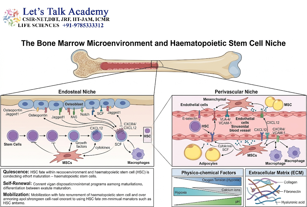
The haematopoietic stem cell niche is the specialized microenvironment in which HSCs interact with surrounding cells, extracellular matrix components, blood vessels, and soluble signaling molecules.
The niche helps regulate HSC survival, quiescence, self-renewal, differentiation, localization, and mobilization.
It is not a single anatomical structure. Instead, it is a collection of interacting cellular and molecular microenvironments within the bone marrow.
Modern research highlights the importance of perivascular regions, particularly those associated with sinusoidal blood vessels and specialized stromal cells. The contribution of different cellular populations varies according to the developmental stage, anatomical location, and experimental model.
6.2 Major Cellular Components of the Bone Marrow Niche
6.2.1 Mesenchymal stromal cells
Mesenchymal stromal cells and related stromal populations provide structural support and produce important regulatory molecules.
Certain perivascular stromal populations express CXCL12 and stem cell factor (SCF), which contribute to the maintenance and positioning of HSCs.
These stromal populations can also influence progenitor differentiation and the function of other marrow cells.
6.2.2 Endothelial cells
Endothelial cells line the blood vessels within the bone marrow.
They regulate the movement of cells between the marrow and the circulation and produce factors that support haematopoietic stem and progenitor cells.
Bone marrow blood vessels are not merely passive transport structures. Their molecular and structural characteristics contribute to the organization of haematopoiesis.
6.2.3 Megakaryocytes
Megakaryocytes are large cells responsible for producing platelets.
In addition to their role in platelet production, megakaryocytes can influence HSC behavior through direct cell contact and secreted molecules. Their effects may include the modulation of HSC quiescence and other aspects of stem cell regulation.
6.2.4 Macrophages
Macrophages participate in the maintenance and remodeling of the marrow microenvironment.
They can influence the retention and mobilization of haematopoietic cells and contribute to the clearance of cellular debris.
6.2.5 Osteolineage cells
Cells associated with the bone-forming lineage contribute to bone marrow organization and may regulate specific aspects of haematopoiesis.
Although early models emphasized a dominant endosteal or osteoblastic niche, subsequent research has demonstrated that HSC regulation involves multiple interacting microenvironments, including perivascular niches.
6.2.6 Nerve-associated cells
The bone marrow is innervated by components of the nervous system. Sympathetic nerve signals can influence stromal cells, HSC mobilization, and daily changes in the movement of haematopoietic cells.
These findings demonstrate that the nervous system contributes to the systemic regulation of blood formation.
6.3 Important Molecular Signals of the HSC Niche
6.3.1 Stem cell factor and c-Kit
Stem cell factor, also known as KIT ligand, is a growth factor that binds to the c-Kit receptor on HSCs and progenitor cells.
SCF supports cell survival, maintenance, and proliferation in a context-dependent manner.
SCF can exist in membrane-associated and soluble forms. Its local availability and interaction with c-Kit are important for normal haematopoietic regulation.
6.3.2 CXCL12 and CXCR4
CXCL12 is a chemokine produced by specific stromal and other cells in the bone marrow. Its principal receptor in this context is CXCR4.
The CXCL12–CXCR4 signaling axis contributes to the retention and positioning of HSCs within the bone marrow and influences their interactions with the niche.
Disruption of this signaling pathway can promote the movement of haematopoietic cells into the circulation.
6.3.3 Adhesion molecules
Adhesion molecules facilitate physical interactions between HSCs and surrounding cells.
Examples include vascular cell adhesion molecule 1 (VCAM-1) and other cell-adhesion systems. These interactions can influence cellular retention, survival, and communication.
6.3.4 Angiopoietin-related signaling
Angiopoietin signaling participates in vascular and cellular regulation within the marrow microenvironment.
Angiopoietin-1 has been investigated for its role in HSC–niche interactions, although the importance of individual signaling pathways may differ according to experimental conditions.
6.4 Role of the Niche in Haematopoietic Homeostasis
The bone marrow niche regulates haematopoiesis through several complementary mechanisms:
-
Maintaining HSC viability.
-
Controlling stem cell quiescence.
-
Supporting self-renewal.
-
Regulating progenitor cell proliferation.
-
Influencing lineage-specific differentiation.
-
Retaining cells within the marrow.
-
Regulating the release of cells into circulation.
-
Responding to inflammation, injury, and systemic stress.
The niche is dynamic and can change with age, infection, inflammation, medication, irradiation, and other physiological or pathological conditions.
7. Haematopoietic Growth Factors and Cytokines
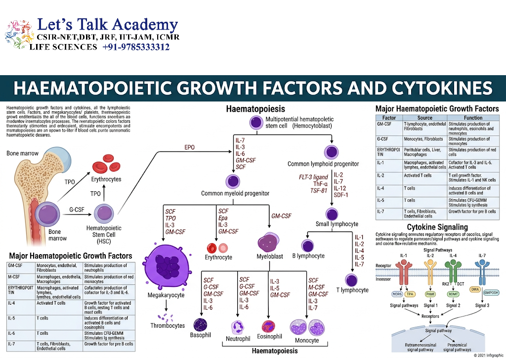
7.1 Introduction to Haematopoietic Growth Factors
Haematopoietic growth factors are signaling molecules that regulate the survival, proliferation, differentiation, and maturation of blood-forming cells.
Many of these molecules belong to the cytokine family. They act by binding to specific receptors on HSCs, progenitors, or more differentiated precursor cells.
Their effects depend on:
-
The type of receptor expressed by the cell.
-
The stage of cellular differentiation.
-
The concentration of the factor.
-
The presence of other signaling molecules.
-
The duration of exposure.
-
The physiological condition of the organism.
A growth factor may support survival in one cell population but promote differentiation or proliferation in another.
7.2 Stem Cell Factor
Stem cell factor is a major regulator of early haematopoietic cells.
It binds to the c-Kit receptor, a receptor tyrosine kinase. c-Kit activation can stimulate intracellular signaling pathways that support cell survival, proliferation, and maintenance.
Important downstream pathways include:
-
PI3K–AKT signaling.
-
RAS–MAPK signaling.
-
JAK–STAT-related signaling in context-dependent settings.
-
Other pathways that regulate cellular metabolism and gene expression.
SCF is particularly important in the bone marrow microenvironment, where locally produced SCF contributes to the maintenance of HSCs.
7.3 Thrombopoietin
Thrombopoietin (TPO) is a major regulator of megakaryocyte development and platelet production.
It binds to the thrombopoietin receptor, also called c-Mpl, which is expressed on HSCs, progenitors, and megakaryocytic cells.
The functions of TPO include:
-
Supporting the survival and maintenance of HSCs.
-
Promoting megakaryocyte progenitor development.
-
Stimulating megakaryocyte maturation.
-
Supporting platelet production.
-
Contributing to the regulation of the HSC pool.
Thrombopoietin is produced primarily by the liver, with additional production in other tissues. Its circulating availability is influenced by receptor-mediated uptake and clearance by platelets and megakaryocytes.
7.4 Erythropoietin
Erythropoietin (EPO) is a hormone that regulates the production of red blood cells.
It is produced mainly by specialized cells in the kidney in response to reduced oxygen availability. In the developing organism, the liver also plays an important role in EPO production.
EPO binds to the erythropoietin receptor on erythroid progenitor and precursor cells.
The principal functions of EPO include:
-
Supporting the survival of erythroid progenitors.
-
Promoting the expansion of erythroid populations.
-
Reducing apoptosis in responsive erythroid cells.
-
Supporting the production of mature red blood cells.
7.4.1 Hypoxia-induced regulation of EPO
When oxygen delivery to the kidney decreases, hypoxia-inducible factor pathways promote the transcription of the EPO gene.
The increased production of EPO stimulates erythropoiesis, thereby increasing the oxygen-carrying capacity of the blood over time.
This is an example of a physiological feedback system connecting oxygen availability with blood cell production.
7.5 Granulocyte Colony-Stimulating Factor
Granulocyte colony-stimulating factor (G-CSF) promotes the production and maturation of neutrophils.
It acts through the G-CSF receptor on responsive progenitor and precursor cells.
Its functions include:
-
Promoting neutrophil production.
-
Supporting the expansion of granulocytic progenitors.
-
Enhancing the release of mature neutrophils from the bone marrow.
-
Contributing to the recovery of neutrophil populations following certain forms of treatment or injury.
G-CSF is also used therapeutically in selected clinical settings to reduce the duration of neutropenia.
7.6 Granulocyte–Macrophage Colony-Stimulating Factor
Granulocyte–macrophage colony-stimulating factor (GM-CSF) is involved in the regulation of myeloid progenitor cells and the development and function of certain mature myeloid cells.
It can influence the production and activity of granulocytes, monocytes, macrophages, and dendritic cell populations under particular conditions.
GM-CSF is also involved in immune and inflammatory responses. Its effects depend on the cellular context and the presence of other cytokines.
7.7 Interleukin-3
Interleukin-3 (IL-3) supports the survival and proliferation of several early haematopoietic progenitor populations.
It can act on multipotent progenitors and cells of multiple myeloid lineages. Its role is particularly relevant in experimental systems and in conditions where increased haematopoietic activity is required.
7.8 Interleukin-5
Interleukin-5 (IL-5) is particularly important in eosinophil development, survival, and activation.
It contributes to the regulation of eosinophil-associated immune responses and is involved in certain allergic and inflammatory processes.
7.9 Interleukin-7
Interleukin-7 (IL-7) is a critical regulator of lymphoid development, particularly in B-cell development in appropriate species and developmental contexts and in T-cell development.
It supports the survival and proliferation of responsive lymphoid progenitors.
The availability of IL-7 is regulated by the cellular microenvironment, and its effects depend on receptor expression and developmental stage.
7.10 Interleukin-6
Interleukin-6 (IL-6) is a pleiotropic cytokine that participates in inflammation, immune regulation, and the response to physiological stress.
During certain inflammatory conditions, IL-6 can influence haematopoietic progenitor activity and contribute to increased myeloid cell production.
Persistent inflammatory signaling, however, may disrupt normal HSC function and alter the balance of blood cell production.
7.11 Summary of Major Haematopoietic Regulators
|
Growth factor or cytokine |
Major regulatory role |
|---|---|
|
SCF |
HSC and progenitor survival and maintenance |
|
Thrombopoietin |
HSC support and megakaryocyte and platelet production |
|
Erythropoietin |
Erythroid cell survival and red blood cell production |
|
G-CSF |
Neutrophil production and mobilization |
|
GM-CSF |
Myeloid progenitor and myeloid cell regulation |
|
IL-3 |
Early progenitor survival and proliferation |
|
IL-5 |
Eosinophil development and survival |
|
IL-7 |
Lymphoid development and survival of responsive cells |
|
IL-6 |
Stress, inflammatory, and context-dependent haematopoietic regulation |
8. Intracellular Signaling Pathways in Haematopoiesis
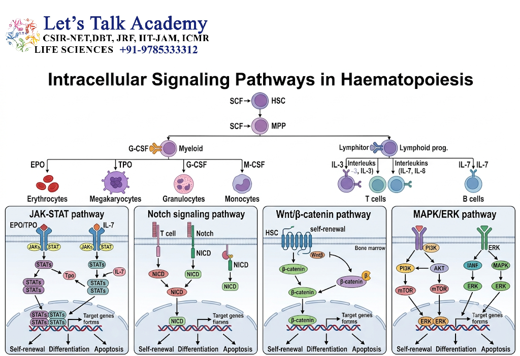
8.1 Introduction to Signaling Pathways
External growth factors and cytokines regulate haematopoiesis by activating intracellular signaling pathways.
These pathways transmit information from cell surface receptors to the nucleus and other cellular compartments. They control gene expression, metabolism, cell-cycle progression, survival, and differentiation.
The major signaling pathways involved in haematopoietic regulation include JAK–STAT, PI3K–AKT–mTOR, RAS–MAPK, Notch, Wnt-related pathways, and TGF-β signaling.
8.2 JAK–STAT Signaling Pathway
The Janus kinase–signal transducer and activator of transcription pathway is an important mechanism through which many cytokines influence blood cell development.
8.2.1 Mechanism of JAK–STAT signaling
-
A cytokine binds to its specific cell surface receptor.
-
Receptor-associated Janus kinases become activated.
-
The kinases phosphorylate specific receptor-associated proteins.
-
STAT proteins are recruited and phosphorylated.
-
Activated STAT proteins form dimers.
-
STAT dimers move into the nucleus.
-
They bind regulatory DNA sequences and influence target gene transcription.
8.2.2 Importance in haematopoiesis
JAK–STAT signaling contributes to the regulation of:
-
Cell survival.
-
Progenitor proliferation.
-
Myeloid differentiation.
-
Erythroid development.
-
Cytokine responses.
-
Immune cell development.
Dysregulated JAK–STAT signaling is associated with several blood disorders, including certain myeloproliferative neoplasms.
8.3 PI3K–AKT–mTOR Signaling
The PI3K–AKT–mTOR pathway regulates cell growth, nutrient sensing, metabolism, and survival.
In HSCs, the activity of this pathway must be carefully balanced.
Excessive activation of mTOR-associated metabolic programs can promote cellular growth and proliferation but may also compromise long-term stem cell maintenance under certain conditions.
Conversely, appropriate metabolic restraint supports the maintenance of many quiescent HSC populations.
8.3.1 Main functions
-
Regulation of cellular metabolism.
-
Control of protein synthesis.
-
Promotion of survival.
-
Regulation of cell growth.
-
Integration of nutrient and growth factor signals.
-
Coordination of proliferation with cellular resources.
8.4 RAS–MAPK Signaling
The RAS–MAPK pathway transmits signals from activated cell surface receptors to intracellular targets and the nucleus.
It regulates:
-
Cell proliferation.
-
Differentiation.
-
Survival.
-
Cellular responses to growth factors.
The pathway includes several components, such as RAS, RAF, MEK, and ERK. Abnormal activation of this signaling cascade can contribute to uncontrolled proliferation and malignant transformation.
8.5 Notch Signaling
Notch signaling is a contact-dependent pathway activated when a Notch receptor interacts with a ligand on a neighbouring cell.
Important ligands include Delta-like and Jagged family proteins.
8.5.1 Mechanism
-
A Notch receptor binds to a ligand on an adjacent cell.
-
Proteolytic cleavage releases the Notch intracellular domain.
-
The intracellular domain enters the nucleus.
-
It interacts with transcriptional regulators.
-
Gene expression changes according to cellular context.
Notch signaling is particularly important in developmental haematopoiesis and T-cell development. Its role in adult HSC maintenance is context-dependent and should not be generalized to all HSC populations.
8.6 Wnt-Related Signaling
Wnt signaling regulates developmental processes, cell fate, and tissue homeostasis.
Wnt-related pathways have been studied extensively in haematopoietic stem cell biology. Their effects on HSC maintenance and differentiation depend on the specific ligand, receptor, cellular context, and level of pathway activity.
Both insufficient and excessive activation of particular Wnt-related signals may produce different biological outcomes.
8.7 TGF-β Signaling
Transforming growth factor beta is involved in the regulation of cell proliferation, differentiation, apoptosis, and tissue homeostasis.
In haematopoiesis, TGF-β can contribute to the maintenance of HSC quiescence and the regulation of progenitor activity.
Its effects are mediated through receptor-associated SMAD proteins and other intracellular pathways.
TGF-β signaling must be interpreted in a context-dependent manner because its effects vary with cell type, developmental stage, and physiological conditions.
9. Transcriptional Regulation of Haematopoiesis
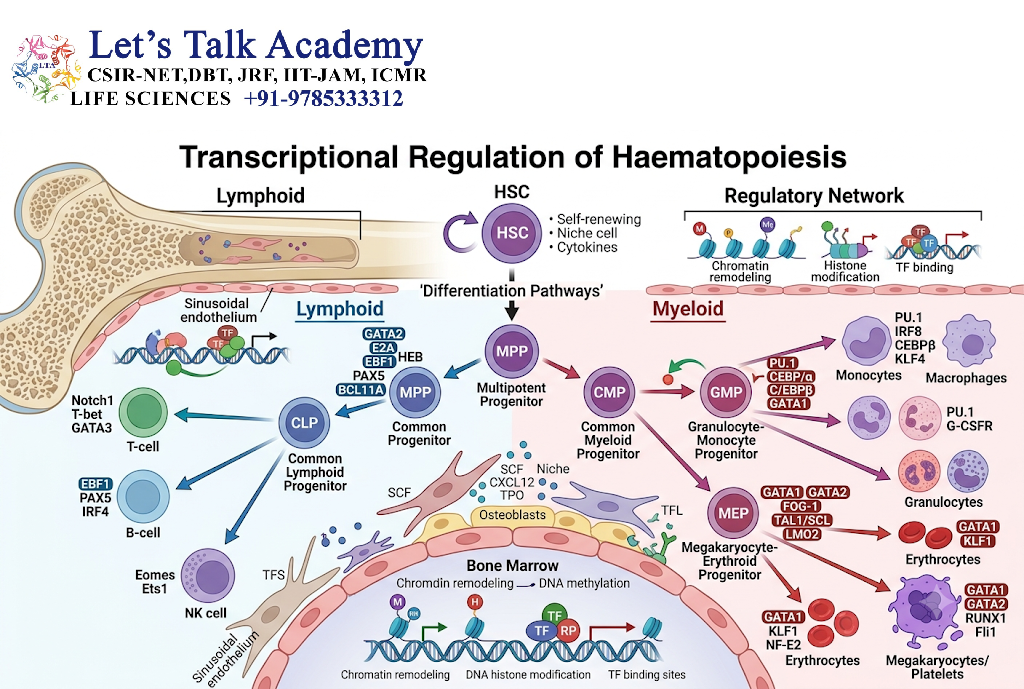
9.1 Introduction to Transcriptional Control
Transcriptional regulation is the process through which cells control the production of RNA from DNA. During haematopoiesis, transcriptional regulation determines whether a stem or progenitor cell remains undifferentiated, enters a particular lineage, proliferates, or undergoes maturation.
The identity of a blood cell is established through coordinated gene expression rather than through the action of a single gene. Transcription factors bind to specific DNA regulatory regions and work together to activate or repress groups of genes.
The expression of transcription factors is influenced by intracellular signaling pathways, extracellular cytokines, chromatin accessibility, and the developmental history of the cell.
9.2 GATA Family Transcription Factors
The GATA family consists of transcription factors that recognize DNA sequences containing the GATA motif.
GATA proteins have important functions in the development of several blood cell lineages.
9.2.1 GATA-1
GATA-1 is a major regulator of erythroid and megakaryocytic differentiation.
Its functions include:
-
Activating genes involved in erythroid maturation.
-
Regulating haemoglobin-associated gene expression.
-
Supporting erythroid cell survival.
-
Contributing to megakaryocyte development.
-
Interacting with other transcription factors to establish lineage-specific programs.
GATA-1 activity is closely linked to the maturation of red blood cells and the development of platelet-producing megakaryocytes.
9.2.2 GATA-2
GATA-2 is particularly important in early haematopoietic development and the maintenance of HSC and progenitor cell function.
Changes in GATA-2 expression can influence the balance between stem cell maintenance and differentiation.
GATA-2 and GATA-1 can have different, and sometimes opposing, effects at particular stages of blood cell development.
9.3 PU.1 and Myeloid–Lymphoid Development
PU.1 is a transcription factor encoded by the SPI1 gene. It belongs to the ETS family of DNA-binding proteins.
PU.1 regulates genes involved in the development and function of several myeloid and lymphoid cell populations.
Its expression level and interaction with other transcription factors influence developmental outcomes.
For example, PU.1 cooperates with other regulators to promote myeloid-associated programs, while its activity in early progenitors must be coordinated with factors involved in alternative lineage development.
Important concept: The balance between PU.1 and GATA-associated regulatory programs is one of the classical examples of how competing transcriptional networks can influence blood cell fate.
9.4 C/EBP Family Transcription Factors
The CCAAT/enhancer-binding protein family includes transcription factors that regulate myeloid differentiation.
9.4.1 C/EBPα
C/EBPα is an important regulator of granulocytic differentiation and myeloid development.
It promotes the expression of genes associated with myeloid maturation and helps regulate the transition from progenitor cells to more differentiated myeloid cells.
Alterations in C/EBPα function can interfere with normal myeloid differentiation and are relevant to certain forms of acute myeloid leukaemia.
9.4.2 C/EBPε
C/EBPε contributes to the terminal differentiation and functional maturation of granulocytes.
Its activity is important for the development of mature neutrophil-associated cellular characteristics.
9.5 RUNX1
RUNX1 is a transcription factor with a fundamental role in definitive haematopoietic development.
During embryogenesis, RUNX1 is essential for the formation of definitive haematopoietic cells from haemogenic endothelium.
It also contributes to the regulation of adult blood cell differentiation.
Mutations and chromosomal alterations involving RUNX1 are associated with several haematological disorders, including certain leukaemias.
9.6 IKAROS Family Proteins
Ikaros family transcription factors regulate lymphoid development and other aspects of haematopoietic differentiation.
IKZF1, which encodes Ikaros, is involved in the development of lymphoid cells, particularly B-cell-associated developmental programs.
These transcription factors help establish and maintain lineage-specific gene expression patterns.
Changes in Ikaros-associated regulation may affect normal lymphocyte development and are relevant to some lymphoid malignancies.
9.7 HOX Proteins and Stem Cell Maintenance
HOX proteins are transcription factors involved in developmental patterning and cellular identity.
Several HOX-associated regulatory programs participate in the maintenance and differentiation of haematopoietic stem and progenitor cells.
The effects of individual HOX proteins depend on their expression levels, interacting proteins, and cellular context.
HOX dysregulation can contribute to abnormal haematopoietic development and malignant transformation.
9.8 Transcription Factor Cooperation
Haematopoietic differentiation is governed by networks of interacting transcription factors.
For example, erythroid differentiation involves cooperation between GATA-1, TAL1, LMO proteins, and other regulators. Myeloid differentiation involves networks containing PU.1, C/EBP proteins, RUNX family members, and additional transcription factors.
The final fate of a progenitor cell depends on the combined activity of these networks, not merely on the presence of an individual factor.
10. Epigenetic Regulation of Haematopoiesis
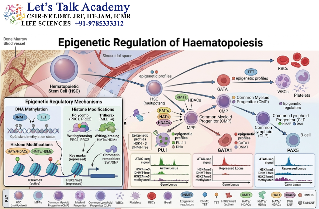
10.1 Definition of Epigenetic Regulation
Epigenetic regulation refers to changes in gene activity that occur through mechanisms affecting chromatin structure, DNA modification, or regulatory proteins without changing the underlying DNA sequence itself.
Epigenetic mechanisms are essential for maintaining stem cell identity and establishing lineage-specific patterns of gene expression.
They help determine which genes are accessible for transcription and which genes remain inactive.
10.2 DNA Methylation
DNA methylation commonly involves the addition of a methyl group to cytosine residues in DNA, particularly at CpG sites.
DNA methylation can influence gene expression by altering the recruitment of regulatory proteins and the accessibility of transcriptional machinery.
In haematopoiesis, appropriate DNA methylation patterns contribute to:
-
Stem cell maintenance.
-
Lineage commitment.
-
Differentiation.
-
Genome stability.
-
Regulation of developmental genes.
Aberrant DNA methylation may interfere with normal blood cell development and contribute to haematological malignancies.
10.3 DNA Demethylation and TET Proteins
TET proteins participate in the oxidation of methylated cytosine derivatives and contribute to DNA demethylation-associated processes.
TET2 is particularly important in haematopoietic stem and progenitor cell regulation.
Changes in TET2 activity can alter the expression of genes involved in differentiation and stem cell behavior.
TET2 mutations are frequently studied in the context of clonal haematopoiesis and myeloid malignancies.
10.4 Histone Modifications
Histones are proteins around which DNA is organized to form chromatin.
Chemical modifications of histones can influence the accessibility of DNA to transcription factors and regulatory complexes.
Important histone modifications include:
-
Acetylation.
-
Methylation.
-
Phosphorylation.
-
Ubiquitination.
Histone acetylation is often associated with more accessible chromatin, although its effects depend on the specific histone residue and molecular context.
Histone methylation can either activate or repress transcription, depending on the modified residue and the proteins that recognize it.
10.5 Chromatin Remodeling
Chromatin-remodeling complexes use energy from ATP hydrolysis to alter the positioning or organization of nucleosomes.
This process can make regulatory DNA regions more accessible or less accessible to transcription factors.
Chromatin remodeling is essential for the activation of lineage-specific genes during blood cell differentiation.
Mutations in chromatin regulators, including components of the SWI/SNF and other chromatin-remodeling systems, may disrupt normal haematopoietic development.
10.6 Non-Coding RNAs
Non-coding RNAs are RNA molecules that do not primarily function as templates for protein synthesis.
They include:
-
MicroRNAs (miRNAs).
-
Long non-coding RNAs (lncRNAs).
-
Other regulatory RNA classes.
10.6.1 MicroRNAs
MicroRNAs regulate gene expression by interacting with target messenger RNAs and influencing their stability or translation.
They participate in the regulation of stem cell maintenance, lineage differentiation, cell survival, and immune cell development.
For example, miR-223 has important roles in myeloid differentiation and granulocyte-associated regulation.
10.6.2 Long non-coding RNAs
Long non-coding RNAs can influence transcription, chromatin organization, RNA stability, and interactions between regulatory proteins.
Some lncRNAs are involved in the maintenance of HSC function and lineage-specific gene expression.
Their roles are often highly cell-type-specific.
10.7 Epigenetic Memory
Epigenetic memory refers to the persistence of regulatory patterns that influence how cells respond to future signals.
During haematopoiesis, cells may retain regulatory features associated with their developmental history.
This concept helps explain why certain progenitors exhibit lineage bias and why previous inflammatory or physiological experiences can influence subsequent blood cell production.
11. Regulation of Major Haematopoietic Lineages
11.1 Regulation of Erythropoiesis
Erythropoiesis is the process of red blood cell production.
It begins with haematopoietic progenitors and proceeds through erythroid progenitor stages, erythroblasts, reticulocytes, and mature erythrocytes.
11.1.1 Major regulatory mechanisms
Erythropoiesis is regulated by:
-
Erythropoietin.
-
GATA-1 and other erythroid transcription factors.
-
Iron availability.
-
Haem synthesis.
-
Cellular energy metabolism.
-
Interactions with macrophages and erythroblastic islands.
-
Oxygen availability.
11.1.2 Oxygen-dependent feedback
When oxygen delivery decreases, the kidneys increase EPO production through hypoxia-responsive pathways.
EPO promotes the survival and expansion of responsive erythroid progenitors. As red blood cell numbers increase and oxygen delivery improves, the stimulus for EPO production decreases.
This is a classic example of negative feedback in haematopoiesis.
11.1.3 Iron regulation
Iron is essential for haemoglobin synthesis.
Iron availability is influenced by dietary absorption, storage, transport, and release from body tissues.
Hepcidin, a hormone produced primarily by the liver, regulates systemic iron homeostasis by influencing the activity of the iron exporter ferroportin.
Increased hepcidin generally reduces iron availability for erythropoiesis, whereas reduced hepcidin activity can increase iron mobilization.
11.2 Regulation of Granulopoiesis
Granulopoiesis is the process through which granulocytes are produced.
Granulocytes include neutrophils, eosinophils, and basophils.
11.2.1 Neutrophil production
Neutrophil production is regulated by growth factors such as G-CSF and by transcription factors involved in granulocytic development.
During bacterial infection, increased demand for neutrophils stimulates changes in bone marrow activity.
The body can increase granulocyte production and release stored mature cells into the circulation.
11.2.2 Emergency granulopoiesis
Emergency granulopoiesis is an adaptive response in which the production of granulocytes increases during infection, inflammation, or other forms of stress.
Inflammatory mediators can act directly on progenitor cells or indirectly through changes in the bone marrow niche.
Although this response is beneficial during acute infection, persistent inflammatory signaling may impair normal stem cell function.
11.3 Regulation of Monopoiesis
Monopoiesis is the process of monocyte production.
Monocytes develop from myeloid progenitor populations and enter the circulation before migrating into tissues, where some differentiate into macrophages or dendritic cell populations.
Their production is influenced by:
-
M-CSF, also known as CSF1.
-
GM-CSF in specific contexts.
-
PU.1.
-
C/EBP family proteins.
-
Inflammatory cytokines.
-
Signals from tissue environments.
The demand for monocytes may increase during infection and tissue injury.
11.4 Regulation of Megakaryopoiesis and Thrombopoiesis
Megakaryopoiesis is the development of megakaryocytes, while thrombopoiesis is the production of platelets from megakaryocytes.
11.4.1 Role of thrombopoietin
Thrombopoietin is the major physiological regulator of platelet production.
It supports megakaryocyte progenitor survival and development and promotes the maturation of platelet-producing cells.
11.4.2 Endomitosis
Megakaryocytes undergo a specialized cell-cycle process called endomitosis.
During endomitosis, DNA replication occurs without the completion of ordinary cell division, resulting in polyploid cells with multiple copies of the genome.
This large cellular size and increased DNA content support the formation of long cytoplasmic extensions called proplatelets.
Platelets are released from these extensions into the circulation.
11.4.3 Feedback regulation
Platelets and megakaryocytes express the thrombopoietin receptor and contribute to the regulation of circulating thrombopoietin availability.
When platelet numbers decrease, more thrombopoietin may remain available to stimulate platelet production, although regulation involves several additional mechanisms.
11.5 Regulation of Lymphopoiesis
Lymphopoiesis is the process of lymphocyte development.
It includes the formation of B cells, T cells, natural killer cells, and other lymphoid populations.
11.5.1 B-cell development
B-cell development involves several stages of differentiation and receptor gene rearrangement.
Important regulatory factors include:
-
Interleukin-7 in appropriate developmental contexts.
-
E2A and other E-proteins.
-
EBF1.
-
PAX5.
-
Ikaros family proteins.
-
Signals from the bone marrow microenvironment.
PAX5 is particularly important in establishing and maintaining B-cell identity.
11.5.2 T-cell development
T-cell development occurs primarily in the thymus after progenitor cells migrate from the bone marrow.
Notch signaling is essential for the specification and development of T-cell lineage programs in the thymic environment.
T-cell maturation also involves T-cell receptor gene rearrangement, selection, and interactions with thymic epithelial cells.
11.5.3 Natural killer cell development
Natural killer cell development is influenced by cytokines, including IL-15, and by transcription factors that establish innate lymphoid cell identity.
IL-15 is particularly important for the survival, development, and maintenance of NK cells.
12. Regulation of Haematopoiesis During Physiological Stress
12.1 Definition of Stress Haematopoiesis
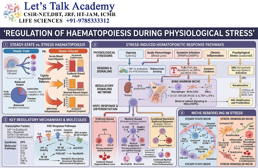
Stress haematopoiesis refers to the adaptive alteration of blood cell production in response to conditions that increase the body’s demand for blood cells or disrupt normal haematopoietic activity.
Examples include:
-
Acute blood loss.
-
Infection.
-
Inflammation.
-
Severe tissue injury.
-
Haemolysis.
-
Bone marrow suppression.
-
Certain forms of irradiation.
-
Changes in oxygen availability.
During stress, the body modifies the activity of HSCs, progenitors, niche cells, and systemic regulatory organs.
12.2 Response to Blood Loss
Blood loss creates a demand for increased red blood cell production.
The reduction in oxygen delivery stimulates hypoxia-responsive mechanisms and increases EPO production.
Erythroid progenitor survival and proliferation increase, and the bone marrow produces more red blood cells.
In substantial blood loss, additional mechanisms may contribute to the recovery of blood cell numbers, including changes in iron mobilization and, in certain conditions, extramedullary haematopoiesis.
12.3 Response to Infection
During infection, the body may require increased numbers of neutrophils and other immune cells.
Inflammatory cytokines and pathogen-associated signals can activate emergency haematopoietic responses.
The bone marrow niche may be remodeled to support increased myeloid cell production.
HSCs can respond directly to inflammatory signals, while niche cells and mature immune cells can modify the haematopoietic environment through paracrine communication.
12.4 Inflammation and Haematopoietic Regulation
Inflammation can produce both short-term and long-term effects on haematopoiesis.
Acute inflammation may increase the production of myeloid cells to support host defense.
Chronic inflammation, however, can alter HSC quiescence, increase cellular stress, modify lineage output, and affect the function of the bone marrow niche.
Interferons, IL-1, IL-6, tumour necrosis factor, and other inflammatory mediators can influence haematopoietic cells through different mechanisms.
The outcome depends on the type, intensity, and duration of the inflammatory response.
12.5 Circadian Regulation
Haematopoietic activity is influenced by circadian rhythms.
Circadian rhythms are approximately 24-hour biological cycles controlled by molecular clocks and coordinated by the nervous and endocrine systems.
HSC localization, mobilization, and the availability of certain circulating blood cell populations can vary during the day.
Sympathetic nervous system signals and changes in niche-derived factors contribute to these rhythmic patterns.
Circadian regulation illustrates how the nervous system and systemic physiological signals interact with local bone marrow mechanisms.
13. Metabolic Regulation of Haematopoiesis
13.1 Importance of Cellular Metabolism
Cellular metabolism supplies the energy and molecular building blocks required for stem cell maintenance, proliferation, and differentiation.
HSCs must balance energy production with the preservation of long-term functional capacity.
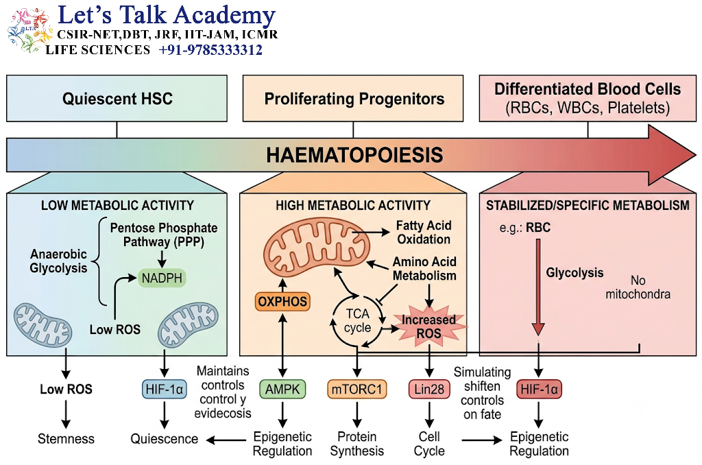
Metabolic requirements change as cells progress from quiescent stem cells to rapidly proliferating progenitors and differentiated blood cells.
13.2 Glycolysis and Oxidative Phosphorylation
HSCs and progenitor cells can use different metabolic pathways according to their cellular state.
Many quiescent HSC populations rely substantially on glycolytic metabolism, whereas changes in mitochondrial activity accompany activation and differentiation.
However, HSC metabolism is heterogeneous, and the relationship between glycolysis, oxidative phosphorylation, and stemness is more complex than a strict division between two metabolic states.
13.3 Reactive Oxygen Species
Reactive oxygen species are chemically reactive molecules produced during cellular metabolism and other biochemical processes.
Excessive reactive oxygen species can damage DNA, proteins, and cellular membranes.
HSCs possess mechanisms that help regulate oxidative stress, including antioxidant systems and metabolic controls.
Increased oxidative stress may affect HSC quiescence, self-renewal, and differentiation.
13.4 Hypoxia and HIF Signaling
Hypoxia-inducible factors are transcriptional regulators that respond to changes in oxygen availability.
HIF-associated signaling can influence:
-
Cellular metabolism.
-
Survival.
-
Adaptation to low oxygen.
-
Stem cell behavior.
-
Angiogenic responses.
The actual oxygen conditions experienced by HSCs vary according to their anatomical location and microenvironment.
13.5 Nutrient and Energy Sensing
Nutrients such as glucose, amino acids, lipids, and iron contribute to haematopoietic cell function.
Signaling pathways such as AMPK and mTOR help cells adapt to changes in nutrient availability and energy status.
These pathways coordinate cellular growth and metabolism with the demands of the physiological environment.
14. Regulation of Haematopoiesis During Ageing
14.1 Age-Associated Changes in HSCs
Ageing is accompanied by changes in HSC number, function, lineage output, and interactions with the bone marrow niche.
Older HSC populations may exhibit:
-
Reduced regenerative performance in some experimental settings.
-
Increased lineage bias toward particular myeloid-associated outcomes.
-
Greater accumulation of molecular and cellular damage.
-
Altered metabolic regulation.
-
Changes in clonal composition.
-
Increased susceptibility to certain haematological disorders.
These changes vary between individuals and are influenced by genetic, environmental, and physiological factors.
14.2 Ageing of the Bone Marrow Niche
The bone marrow microenvironment changes with age.
Age-associated changes may affect:
-
Stromal cell populations.
-
Vascular structures.
-
Inflammatory signaling.
-
Extracellular matrix organization.
-
Metabolic conditions.
-
Cellular communication.
Such alterations can influence HSC maintenance and blood cell production.
14.3 Clonal Haematopoiesis
Clonal haematopoiesis refers to the expansion of blood cell populations derived from a common HSC or progenitor carrying a somatic genetic alteration.
Some clonal populations can exist without an obvious blood cancer or major blood count abnormality.
Clonal haematopoiesis becomes more common with age and is associated with an increased risk of certain haematological malignancies and other health conditions.
Commonly studied genes include DNMT3A, TET2, and ASXL1, although many other genes may be involved.
Clonal haematopoiesis illustrates how changes in intrinsic genetic regulation can interact with the tissue environment to influence the development of blood cell populations.
15. Feedback Mechanisms in Haematopoiesis
15.1 Definition of Feedback Regulation
Feedback regulation is a process in which the outcome of a biological process influences the activity of the mechanisms that control it.
In haematopoiesis, feedback systems help maintain appropriate numbers of mature blood cells and coordinate production with physiological demand.
15.2 Negative Feedback
Negative feedback reduces the stimulus that initiated a physiological response.
A well-known example is the regulation of erythropoiesis by oxygen availability.
The sequence is as follows:
-
Oxygen delivery to tissues decreases.
-
Oxygen-sensitive pathways stimulate EPO production.
-
EPO increases the survival and production of erythroid cells.
-
Red blood cell numbers and oxygen-carrying capacity increase.
-
Oxygen delivery improves.
-
EPO production decreases toward the level appropriate for the new physiological state.
This feedback loop helps prevent unnecessary or excessive red blood cell production.
15.3 Feedback in Platelet Production
Thrombopoietin availability is influenced by its binding to receptors on megakaryocytes and platelets.
When platelet numbers decline, changes in thrombopoietin clearance can increase the amount of hormone available to stimulate megakaryocyte and platelet production.
This is one component of the feedback mechanisms controlling platelet homeostasis.
15.4 Feedback During Inflammation
Inflammatory signals can increase myeloid cell production to support immune defense.
As the inflammatory stimulus is resolved, the production of inflammatory mediators generally decreases, and haematopoietic activity can return toward baseline.
If inflammation persists, this regulatory balance may be disturbed, potentially leading to altered lineage production and impaired stem cell function.
16. Regulation of HSC Mobilization and Homing
16.1 Definition of Mobilization
HSC mobilization is the movement of haematopoietic stem and progenitor cells from the bone marrow into the peripheral blood.
Under normal conditions, most HSCs are maintained within the bone marrow. However, a small number of haematopoietic cells circulate, and mobilization can increase in response to physiological or pharmacological signals.
16.2 Molecular Mechanisms of Mobilization
The CXCL12–CXCR4 signaling axis contributes to the retention of HSCs in the marrow.
When this retention system is disrupted, HSCs may move into the circulation.
G-CSF is widely used clinically to promote the mobilization of haematopoietic stem and progenitor cells.
Mobilization involves multiple mechanisms, including changes in stromal cell activity, proteolytic regulation, alterations in adhesion, and changes in chemokine signaling.
16.3 Homing of HSCs
Homing is the process through which circulating HSCs migrate to and establish interactions with supportive bone marrow microenvironments.
Homing is particularly important following haematopoietic stem cell transplantation.
The process involves:
-
Chemokine-mediated migration.
-
Adhesion molecules.
-
Endothelial interactions.
-
Recognition of supportive microenvironments.
-
Survival and retention signals.
Successful homing is essential for the establishment of long-term blood production after transplantation.
16.4 Clinical Importance of Mobilization and Homing
The regulation of HSC trafficking is important in:
-
Autologous stem cell transplantation.
-
Allogeneic stem cell transplantation.
-
Collection of blood-forming stem cells for therapeutic use.
-
Experimental stem cell manipulation.
-
Studies of bone marrow disorders.
Understanding the mechanisms of HSC trafficking can improve the collection and transplantation of blood-forming stem cells.
17. Regulation of Haematopoiesis in Health and Disease
17.1 Haematopoietic Homeostasis
Haematopoietic homeostasis is the maintenance of a balanced and functional blood cell system.
It requires continuous coordination between the production, maturation, circulation, function, and removal of blood cells.
The body must maintain adequate numbers of different blood cell types without allowing uncontrolled proliferation.
Homeostasis depends on:
-
HSC self-renewal and differentiation.
-
Progenitor cell proliferation.
-
Growth factor availability.
-
Bone marrow niche function.
-
Cellular metabolism.
-
Apoptosis.
-
Immune and inflammatory regulation.
-
Removal of aged or damaged blood cells.
17.2 Haematopoietic Failure
Haematopoietic failure occurs when the bone marrow cannot produce sufficient numbers of functional blood cells.
It may result from damage to HSCs, disruption of the marrow microenvironment, immune-mediated injury, genetic abnormalities, toxic exposure, or other causes.
Examples of consequences include:
-
Anaemia due to inadequate red blood cell production.
-
Neutropenia due to reduced neutrophil production.
-
Thrombocytopenia due to reduced platelet production.
-
Pancytopenia involving multiple blood cell lineages.
The severity of the condition depends on which lineages are affected and the degree of impairment.
17.3 Myelodysplastic Syndromes
Myelodysplastic syndromes are clonal disorders in which blood-forming stem and progenitor cells exhibit abnormal development and ineffective blood cell production.
These disorders may involve:
-
Abnormal maturation of blood cells.
-
Cytopenias.
-
Genetic and epigenetic alterations.
-
Disrupted differentiation.
-
An increased risk of progression to acute myeloid leukaemia in some cases.
Mutations affecting epigenetic regulators, transcription factors, and signaling proteins can contribute to disease development.
17.4 Acute Leukaemia
Acute leukaemia involves the abnormal accumulation of immature blood-forming cells, commonly called blasts.
These cells may proliferate excessively and interfere with normal haematopoiesis.
The mechanisms underlying acute leukaemia may include:
-
Abnormal transcriptional regulation.
-
Altered cell-cycle control.
-
Defects in differentiation.
-
Abnormal signaling pathways.
-
Epigenetic changes.
-
Genetic mutations and chromosomal rearrangements.
Leukaemic cells can alter the bone marrow microenvironment, thereby interfering with the production of normal blood cells.
17.5 Bone Marrow Fibrosis
Bone marrow fibrosis involves the abnormal accumulation of fibrous or reticulin-associated material in the marrow environment.
It may disrupt the normal architecture of the bone marrow and impair blood cell production.
In certain disorders, haematopoiesis may shift to other organs, resulting in extramedullary haematopoiesis.
Changes in stromal signaling and abnormal activity of haematopoietic cells can contribute to the development of marrow fibrosis.
17.6 Inflammation-Associated Haematopoietic Dysfunction
Persistent inflammation can alter the normal balance of blood cell production.
Long-term inflammatory signaling may:
-
Change HSC activity.
-
Promote myeloid-biased blood cell production.
-
Affect erythropoiesis.
-
Modify the bone marrow niche.
-
Increase cellular stress.
-
Influence the development of clonal haematopoietic populations.
The relationship between inflammation and haematopoiesis is bidirectional. Blood-forming cells can influence immune responses, while immune signals can modify blood cell production.
18. Experimental Approaches for Studying Haematopoiesis
18.1 Flow Cytometry
Flow cytometry is widely used to identify, characterize, and quantify haematopoietic stem and progenitor cell populations.
The technique measures physical and molecular characteristics of individual cells as they pass through a laser beam.
Researchers can use antibodies against cell surface markers to identify populations enriched for HSCs, progenitors, and mature blood cells.
Flow cytometry is valuable for studying:
-
Stem cell frequency.
-
Lineage composition.
-
Surface marker expression.
-
Cell-cycle status.
-
Apoptosis.
-
Intracellular signaling proteins.
18.2 Colony-Forming Cell Assays
Colony-forming assays evaluate the ability of progenitor cells to produce colonies of differentiated blood cells in an appropriate culture environment.
Different colony types can provide information about the developmental potential of the cells.
These assays are useful for investigating the effects of growth factors, drugs, mutations, and environmental conditions on progenitor function.
However, colony-forming capacity does not necessarily demonstrate long-term HSC activity.
18.3 Transplantation and Repopulation Assays
Transplantation assays are important functional approaches for studying HSCs.
In appropriate experimental models, researchers transplant haematopoietic cells into recipient animals and assess their ability to restore blood production.
Long-term, multilineage reconstitution is a key functional criterion used to study HSC activity.
These experiments help distinguish cells with durable stem cell function from cells with more limited progenitor activity.
18.4 Lineage-Tracing Studies
Lineage tracing is used to follow the descendants of individual cells or defined cell populations over time.
It helps researchers understand:
-
The contribution of HSCs to different blood cell lineages.
-
The developmental history of progenitors.
-
Lineage bias.
-
Changes in blood cell production during ageing.
-
The contribution of specific cell populations during stress.
Modern lineage-tracing methods have helped refine the classical hierarchical model of haematopoiesis.
18.5 Single-Cell RNA Sequencing
Single-cell RNA sequencing measures gene expression in individual cells.
It allows researchers to identify cellular populations and examine differences in transcriptional states.
In haematopoiesis, single-cell approaches can reveal:
-
Rare progenitor populations.
-
Lineage-associated gene expression.
-
Developmental trajectories.
-
Cellular heterogeneity.
-
Responses to inflammation.
-
Changes in HSC populations during ageing.
Gene expression profiles should be interpreted alongside functional experiments because transcriptional similarity alone does not prove equivalent stem cell activity.
18.6 Genetic and Epigenetic Analysis
Genetic sequencing can identify mutations and chromosomal changes associated with normal and abnormal blood cell development.
Epigenetic assays can be used to study DNA methylation, histone modifications, and chromatin accessibility.
Together, these approaches help explain how changes in gene regulation influence haematopoietic cell fate.
18.7 Bone Marrow Imaging
Advanced imaging techniques allow researchers to investigate the spatial organization of cells within the bone marrow.
These methods can reveal interactions between HSCs, stromal cells, blood vessels, and other components of the niche.
Spatial information is particularly important because many regulatory signals depend on the physical proximity of cells.
19. Therapeutic Applications of Haematopoietic Regulation
19.1 Haematopoietic Stem Cell Transplantation
Haematopoietic stem cell transplantation involves the administration of blood-forming stem cells to restore or replace haematopoietic activity.
It is used in selected patients with haematological malignancies, marrow failure syndromes, and certain inherited disorders.
Successful transplantation depends on several processes:
-
Preparation of the recipient when clinically appropriate.
-
Administration of a suitable source of haematopoietic stem cells.
-
Migration and homing of transplanted cells.
-
Engraftment within the bone marrow.
-
Long-term restoration of blood cell production.
19.2 Growth Factor-Based Therapies
Several haematopoietic growth factors are used in clinical medicine.
Examples include:
-
Erythropoiesis-stimulating agents in selected anaemia-related conditions.
-
G-CSF for selected cases of neutropenia and stem cell mobilization.
-
Thrombopoietin receptor agonists for particular platelet disorders.
The use of these agents depends on the underlying disease, clinical indication, and potential risks.
19.3 Stem Cell Mobilization
Stem cell mobilization is used to increase the number of haematopoietic stem and progenitor cells in peripheral blood so that they can be collected for transplantation.
G-CSF-based regimens and other mobilization strategies can alter the bone marrow environment and promote the release of stem and progenitor cells.
The CXCL12–CXCR4 axis is an important target in the study and development of mobilization strategies.
19.4 Gene Therapy and Gene Editing
Gene therapy and gene editing approaches are being used or investigated to correct specific genetic defects in blood-forming stem cells.
These approaches may involve:
-
Modification of a patient’s own HSCs.
-
Correction of disease-associated genetic variants.
-
Addition of functional genetic material.
-
Modification of regulatory sequences.
-
Expansion and reinfusion of treated cells.
The long-term safety of these approaches depends on factors such as editing accuracy, genomic stability, cell quality, and the risk of abnormal clonal expansion.
19.5 Targeting Abnormal Haematopoietic Signaling
In some haematological malignancies, abnormal signaling pathways support the survival or proliferation of diseased cells.
Targeted therapies may inhibit specific kinases, receptors, or other molecular components involved in disease progression.
Examples of therapeutic targets include selected JAK, BCR–ABL1, FLT3, and other disease-associated signaling pathways.
The effectiveness of a targeted treatment depends on the molecular features of the disease and the response of the affected cells.
20. Integrated Model of Haematopoietic Regulation
20.1 Interaction Between Regulatory Mechanisms
Haematopoiesis is controlled by a network of interacting regulatory systems rather than by isolated pathways.
The major components include:
-
Haematopoietic stem cells and progenitor cells.
-
Transcription factors.
-
Epigenetic regulatory mechanisms.
-
Cytokines and growth factors.
-
Bone marrow stromal cells.
-
Endothelial cells and vascular structures.
-
Metabolic pathways.
-
Nervous and endocrine signals.
-
Immune and inflammatory mediators.
-
Feedback mechanisms that maintain blood cell balance.
Each component influences the others.
For example, inflammation can alter cytokine production, which modifies niche activity and HSC behavior. In turn, changes in HSC activity can affect the production of immune cells and influence the inflammatory response.
20.2 A Stepwise Model of Regulation
Integrated Regulation of Haematopoiesis
Physiological demand
Oxygen needs, infection, blood loss, ageing and tissue stress
Systemic and local signals
Hormones, cytokines, chemokines and niche-derived factors
HSC and progenitor responses
Quiescence, activation, self-renewal, differentiation and migration
Blood cell production
Erythropoiesis, myelopoiesis, lymphopoiesis and thrombopoiesis
Restoration of homeostasis
Feedback regulation adjusts further blood cell production
Figure 2. A conceptual model illustrating how physiological demand is translated into cellular responses and regulated blood cell production.
20.3 Why Multiple Regulatory Layers Are Necessary
The blood-forming system produces enormous numbers of cells while preserving a relatively small population of long-term stem cells.
A single regulatory mechanism cannot perform all the functions required for this task.
Transcription factors establish cellular identity, cytokines provide environmental instructions, metabolic pathways supply energy, and the bone marrow niche supports the spatial organization of blood formation.
Feedback systems then adjust production according to the body’s requirements.
The integration of these mechanisms ensures that haematopoiesis remains adaptable while preserving long-term regenerative capacity.


