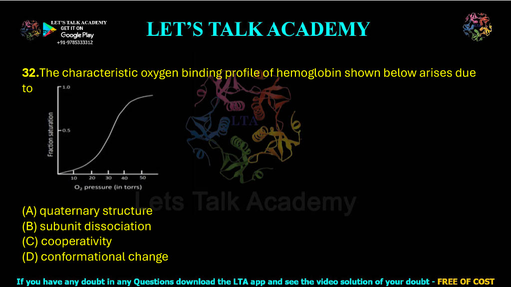
32.The characteristic oxygen binding profile of hemoglobin shown below arises due to the .
(A) quaternary structure
(B) subunit dissociation
(C) cooperativity
(D) conformational change
Hemoglobin Oxygen Binding Curve Explained | Why Hemoglobin Shows a Sigmoidal Oxygen Binding Curve?
Correct Answer
(C) Cooperativity
Introduction
Hemoglobin is the principal oxygen-transport protein present in the red blood cells of vertebrates. Its primary function is to bind oxygen efficiently in the lungs, where oxygen concentration is high, and release it in peripheral tissues, where oxygen demand is greater. Unlike myoglobin, which displays a simple hyperbolic oxygen-binding curve, hemoglobin exhibits a sigmoidal (S-shaped) oxygen saturation curve. This unique binding behavior enables hemoglobin to load oxygen rapidly in the lungs and unload it efficiently in actively respiring tissues.
The sigmoidal nature of the hemoglobin oxygen-binding curve is one of the most important concepts in biochemistry and physiology. It reflects the phenomenon of positive cooperativity, whereby the binding of one oxygen molecule increases the affinity of the remaining subunits for oxygen.
Understanding the Concept Behind the Question
The graph shown in the question is the classic sigmoidal oxygen saturation curve of hemoglobin. The S-shaped profile indicates that oxygen does not bind independently to each subunit.
Instead, when the first oxygen molecule binds to one subunit, it induces structural changes that increase the oxygen affinity of the remaining three subunits. As additional oxygen molecules bind, oxygen binding becomes progressively easier.
This phenomenon is known as positive cooperativity.
At low oxygen pressure, oxygen binding is relatively slow because hemoglobin is predominantly in the T (tense) state, which has low affinity for oxygen. As oxygen begins to bind, hemoglobin gradually converts into the R (relaxed) state, which has a much higher affinity for oxygen. This transition produces the characteristic S-shaped binding curve.
Therefore, the sigmoidal oxygen-binding profile arises because of cooperative binding.
Hence, Option (C) is the correct answer.
Why Option (A) Is Incorrect
Quaternary Structure
Hemoglobin indeed possesses a quaternary structure, consisting of two α-subunits and two β-subunits.
This quaternary organization is essential because cooperativity cannot occur in a single-subunit protein. However, the question asks what directly produces the characteristic sigmoidal oxygen-binding curve.
Although quaternary structure provides the structural basis for cooperative interactions, the actual reason for the sigmoidal curve is cooperativity, not merely the presence of multiple subunits.
Therefore,
Option (A) is incorrect.
Why Option (B) Is Incorrect
Subunit Dissociation
During normal oxygen binding, the four hemoglobin subunits remain associated throughout the entire process.
Oxygen binding does not cause the α and β subunits to separate. Instead, only subtle changes occur in their relative positions, allowing communication between subunits.
Since hemoglobin functions as an intact tetramer during oxygen transport, subunit dissociation is not responsible for the sigmoidal binding curve.
Therefore,
Option (B) is incorrect.
Why Option (C) Is Correct
Cooperativity
Cooperativity refers to the phenomenon in which binding of a ligand to one subunit influences ligand binding at the remaining subunits.
In hemoglobin:
- Binding of the first oxygen molecule slightly increases affinity.
- Binding of the second oxygen molecule becomes easier.
- Binding of the third and fourth oxygen molecules occurs even more readily.
This sequential increase in affinity produces the characteristic sigmoidal oxygen-binding curve.
The molecular basis of this phenomenon is the transition between the T-state (low affinity) and the R-state (high affinity).
Because each oxygen molecule facilitates binding of the next oxygen molecule, hemoglobin displays positive cooperativity.
Therefore,
Option (C) is correct.
Why Option (D) Is Incorrect
Conformational Change
Hemoglobin certainly undergoes conformational changes during oxygen binding. Specifically, oxygen binding shifts the protein from the T-state to the R-state.
However, these conformational changes are the mechanism through which cooperativity is achieved rather than the phenomenon responsible for the characteristic binding profile.
The question asks why the oxygen-binding curve is sigmoidal.
The accepted biochemical answer is positive cooperativity.
Therefore,
Option (D) is incorrect.
What Is Positive Cooperativity?
Positive cooperativity occurs when the binding of one ligand increases the affinity of the remaining binding sites for additional ligand molecules.
For hemoglobin:
First O₂ binds
↓
T-state begins changing
↓
Affinity increases
↓
Second O₂ binds more easily
↓
Third and fourth O₂ bind even faster
This sequential increase in affinity creates the characteristic S-shaped (sigmoidal) oxygen saturation curve.
T-State and R-State of Hemoglobin
Hemoglobin exists in two major conformations.
T-State (Tense State)
- Low oxygen affinity
- Predominates in peripheral tissues
- Stabilized by 2,3-BPG, H⁺, and CO₂
R-State (Relaxed State)
- High oxygen affinity
- Predominates in the lungs
- Stabilized by oxygen binding
The transition from the T-state to the R-state is responsible for cooperative oxygen binding.
Hemoglobin vs Myoglobin
| Feature | Hemoglobin | Myoglobin |
|---|---|---|
| Number of Subunits | 4 | 1 |
| Oxygen Binding Curve | Sigmoidal | Hyperbolic |
| Cooperativity | Present | Absent |
| Primary Function | Oxygen transport | Oxygen storage |
| Allosteric Regulation | Yes | No |
This comparison is extremely important in competitive examinations.
Biological Importance of Cooperativity
Cooperative binding enables hemoglobin to function as an exceptionally efficient oxygen transporter. In the lungs, where oxygen pressure is high, hemoglobin rapidly binds oxygen because the R-state is favored. In actively metabolizing tissues, where oxygen pressure is lower, hemoglobin readily releases oxygen because the T-state becomes more stable.
Without cooperativity, oxygen loading in the lungs and unloading in tissues would be far less efficient. Thus, positive cooperativity is a remarkable evolutionary adaptation that optimizes oxygen transport throughout the body.
Common Mistakes in Competitive Examinations
Many students choose quaternary structure because they know hemoglobin contains four subunits. Although quaternary structure is necessary for cooperative interactions, it does not directly explain the sigmoidal binding curve. The immediate cause is positive cooperativity.
Another common mistake is selecting conformational change. While the T-to-R transition is the structural mechanism underlying cooperativity, the accepted biochemical term describing the binding behavior is cooperativity.
Students should always remember that sigmoidal curve = positive cooperativity.
High-Yield Points
- Hemoglobin contains four subunits.
- Oxygen binding curve is sigmoidal.
- Sigmoidal curve indicates positive cooperativity.
- Myoglobin exhibits a hyperbolic oxygen-binding curve.
- Oxygen binding shifts hemoglobin from T-state → R-state.
- Cooperative binding improves oxygen transport efficiency.
Frequently Asked Questions
Why is the hemoglobin oxygen-binding curve sigmoidal?
Because oxygen binding to one subunit increases the affinity of the remaining subunits through positive cooperativity.
Why doesn’t myoglobin show a sigmoidal curve?
Myoglobin contains only one polypeptide chain, so there are no neighboring subunits with which to exhibit cooperative interactions. Therefore, its oxygen-binding curve is hyperbolic.
Is quaternary structure important?
Yes. Quaternary structure provides the structural framework required for cooperativity, but the direct reason for the sigmoidal oxygen-binding profile is positive cooperativity.
Key Takeaways
The characteristic sigmoidal oxygen-binding curve of hemoglobin arises because of positive cooperativity among its four subunits. Binding of the first oxygen molecule induces a transition from the low-affinity T-state to the high-affinity R-state, making it progressively easier for additional oxygen molecules to bind. Although hemoglobin’s quaternary structure and conformational changes are essential for this process, the phenomenon responsible for the S-shaped oxygen-binding profile is cooperativity. This mechanism allows efficient oxygen loading in the lungs and oxygen release in peripheral tissues, making hemoglobin an exceptionally effective oxygen transport protein.
Final Answer
Correct Option: (C) Cooperativity
Explanation
The sigmoidal oxygen-binding curve of hemoglobin is produced by positive cooperativity, in which the binding of one oxygen molecule increases the affinity of the remaining subunits for oxygen. This cooperative behavior results from oxygen-induced T-state to R-state conformational transitions within the hemoglobin tetramer. Although the protein’s quaternary structure is necessary for cooperativity and conformational changes occur during oxygen binding, the characteristic S-shaped binding profile specifically arises because of cooperative binding. Therefore, the correct answer is Option (C) – Cooperativity.


